. 2003 Apr 1;548(Pt 1):53-69.
doi: 10.1113/jphysiol.2002.027854. Epub 2003 Feb 7.

Distinct contributions of small and large conductance Ca2+-activated K+ channels to rat Purkinje neuron function

Affiliations

Distinct contributions of small and large conductance Ca2+-activated K+ channels to rat Purkinje neuron function

Jeremy R Edgerton et al. J Physiol. .

Abstract

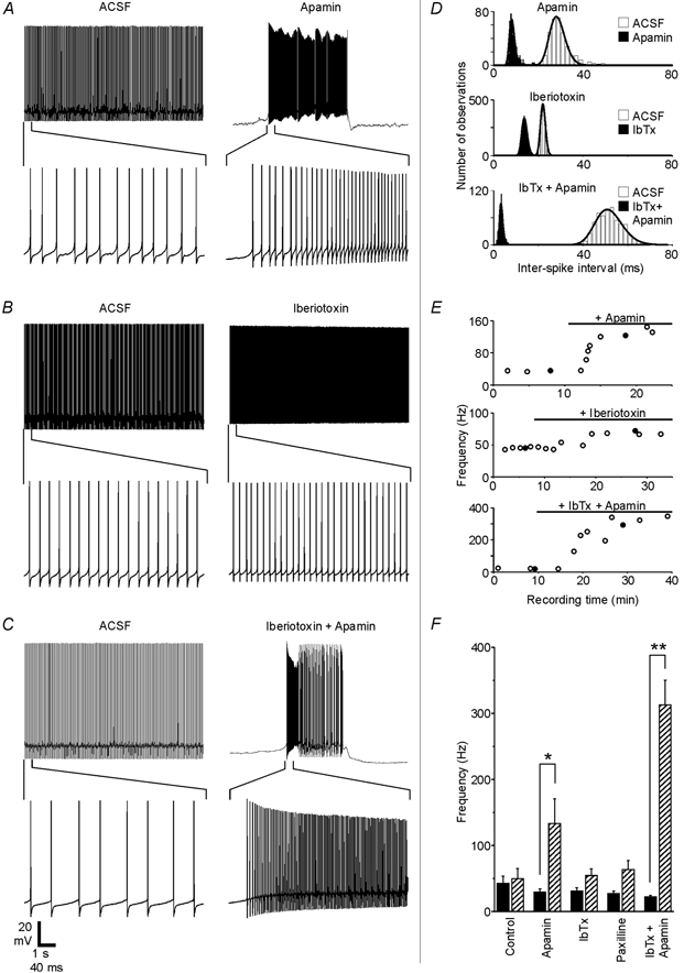
The cerebellum is important for many aspects of behaviour, from posture maintenance and goal-oriented reaching movements to timing tasks and certain forms of learning. In every case, information flowing through the cerebellum passes through Purkinje neurons, which receive input from the two primary cerebellar afferents and generate continuous streams of action potentials that constitute the sole output from the cerebellar cortex to the deep nuclei. The tonic firing behaviour observed in Purkinje neurons in vivo is maintained in brain slices even when synaptic inputs are blocked, suggesting that Purkinje neuron activity relies to a significant extent on intrinsic conductances. Previous research has suggested that the interplay between Ca2+ currents and Ca2+-activated K+ channels (KCa channels) is important for Purkinje cell activity, but how many different KCa channel types are present and what each channel type contributes to cell behaviour remains unclear. In order to better understand the ionic mechanisms that control the behaviour of these neurons, we investigated the effects of different Ca2+ channel and KCa channel antagonists on Purkinje neurons in acute slices of rat cerebellum. Our data show that Ca2+ entering through P-type voltage-gated Ca2+ channels activates both small-conductance (SK) and large-conductance (BK) KCa channels. SK channels play a role in setting the intrinsic firing frequency, while BK channels regulate action potential shape and may contribute to the unique climbing fibre response.


Figures

Figure 1
Figure 1. Current clamp recordings from Purkinje neurons in cerebellar slices demonstrate both Na+ and Ca2+ action potentials
Right-hand traces are at expanded time scales. A, under control conditions, injection of 0.5 nA depolarizing current through the recording pipette evoked bursts of Na+ spikes (arrows) interrupted by single Ca2+ spikes (▵). B, when TTX was added to the bath solution, the same cell fired only Ca2+ spikes. Each spike was followed by a deep after-hyperpolarization (AHP). C, recordings from a different cell demonstrate Na+ spikes in the absence of Ca2+ spikes due to the block of Ca2+ channels with 100 μm bath-applied cadmium chloride.
Figure 2
Figure 2. The spontaneous firing pattern of Purkinje neurons is Ca2+ dependent
A, current clamp recordings showing the spontaneous firing pattern of a Purkinje neuron under control conditions (left trace), when Ca2+ channels were blocked by cadmium chloride (middle trace) and following removal of the cadmium by perfusion (right trace). B, inter-spike interval (ISI) distributions and log-normal fits are shown for each trace in panel A. Firing frequency components were determined from the peaks of the fits. For the ACSF trace, the peak was at 20 ms, corresponding to a firing frequency of 50 Hz. For the cadmium trace, three peaks could be discerned, corresponding to frequencies of 161, 88 and 56 Hz. After the wash period, the distribution again showed a single peak corresponding to a firing frequency of 45 Hz. C and D, recordings and ISI distributions from a different Purkinje neuron show how the firing pattern and frequency changed as 10 mm BAPTA diffused into the cell from the patch pipette. A few seconds after the whole-cell configuration was established, the cell fired at 84 Hz (left trace). After only 30 s, the firing pattern was irregular, displaying both Na+ and Ca2+ spikes, with an intra-burst firing frequency of 293 Hz (middle trace). An additional 2.5 min of BAPTA diffusion time had little effect on the intra-burst firing frequency, increasing it to 296 Hz (right trace). E, firing rates (means ±s.e.m.) are shown for each condition. Black bars represent the baseline firing rate for each group while hatched bars represent firing rates under treatment conditions. BAPTA increased the firing rate from 174 ± 40 to 534 ± 70 Hz as it diffused into the cell (n = 5), while cadmium changed the firing rate from 53 ± 11 to 187 ± 26 Hz (n = 4). The firing rate of the control group remained stable over time, changing from 41 ± 16 initially to 47 ± 19 Hz after 10 min in ACSF (n = 6). The BAPTA group differed significantly from the control group both in its baseline rate (P < 0.0001) and in the difference between the treatment condition and the baseline (P < 0.0001).
Figure 3
Figure 3. Ca2+-dependent conductances contribute to the AHPs following Na+ and Ca2+ spikes
A, the Na+ spike AHP requires Ca2+ influx. Left panel, reducing Ca2+ influx with 100 μm cadmium chloride (thick trace) attenuated the Na+ spike AHP without affecting spike repolarization. Right panel, when spike repolarization was first slowed with 100 μm 4-AP (thick continuous trace), Ca2+-dependent conductances became important for repolarization; if they were subsequently blocked with 100 μm cadmium added in the presence of 4-AP (dotted trace), spike repolarization was significantly delayed. The inset shows the same traces at an expanded scale (scale bar = 25 mV × 0.5 ms for inset). The vertical line in the inset is 0.4 ms after the beginning of the spike. B, the Ca2+ spike AHP involves Ca2+-activated conductances. Ca2+ spikes were evoked using the same protocol as in Fig. 1 A but with 10 mm BAPTA in the pipette solution. Representative Ca2+ spikes are shown from recordings taken immediately after establishing the whole-cell configuration (thin traces) and 12 min later, when BAPTA had diffused from the patch pipette into the cell (thick traces). The expanded traces in the right-hand panel show the effect of BAPTA on the AHP at different time points. At 1 ms after the peak of the spike (dashed line), there was no difference between the two conditions. However, the AHP of the later trace ended within about 3 ms of the spike peak (dotted line), while the control trace AHP showed a slower component that was still approaching its minimum value at 5 ms after the spike peak (continuous line). At the 5 ms time point, the baseline condition trace was 14 mV more negative than the trace recorded later.
Figure 4
Figure 4. SK and BK channels regulate Purkinje cell firing patterns
A-C, recordings from three different Purkinje neurons show how spontaneous firing was affected by 200 nm apamin, 100 nm iberiotoxin or the combination of the two blockers. The same scale bar applies to each set of traces. D, ISI distributions and fits are shown for the traces in A-C. E, spontaneous firing frequency plotted as a function of recording time for each experiment. •, the traces shown in A-C. Black horizontal bars show when toxins were present in the bath solution. Response delays partly reflect the time needed for residual control solution to empty out of the perfusion tubing and inline solution heater. F, the effects of specific KCa channel blockers on the firing frequency are summarized. Black bars show the baseline frequency for each group (means ±s.e.m.), while hatched bars show the frequency in the presence of the blocker. The firing frequency increased from 29 ± 5 to 133 ± 37 Hz with the application of apamin (n = 7 cells), from 29 ± 5 to 50 ± 12 Hz with iberiotoxin (n = 6), from 27 ± 4 to 63 ± 14 Hz with 1 μm paxilline (n = 5) and from 22 ± 3 to 313 ± 37 Hz when both iberiotoxin and apamin were applied together (n = 5). For the control group, the firing rate increased from 41 ± 16 initially to 47 ± 19 Hz after 10 min of control recording time (n = 6). Apamin had a significant effect compared to the control (P < 0.05), and the simultaneous application of iberiotoxin and apamin differed significantly from both the control group (P < 0.0001) and from the apamin group (P < 0.01). When applied alone, neither iberiotoxin nor paxilline had a statistically significant effect compared to the control group (P > 0.5).
Figure 5
Figure 5. BK channels are major contributors to the Na+ spike AHP
A-D, spontaneous Na+ spikes were recorded in ACSF (thin traces), then in the presence of one or more channel blockers (thick traces). Each trace is an average of 10 action potentials except the thick trace in panel D, which shows a single spike burst characteristic of spontaneous activity in this condition. E, spontaneous Na+ spikes were recorded in ACSF while 10 mm BAPTA diffused into the cell from the pipette. The thin trace is an average of 10 action potentials recorded immediately after establishing the whole-cell configuration. The thick trace is a representative action potential burst recorded 3 min later. F, Na+ spike AHP amplitude was quantified as the difference between the initial voltage and the voltage 1 ms after the peak. G, the degree to which each treatment reduced the Na+ spike AHP amplitude is summarized for all groups (means ±s.e.m.). Apamin did not significantly affect the AHP, reducing it by only 2 ± 5 % (n = 5 cells). Iberiotoxin reduced the AHP by 53 ± 6 % (n = 7), paxilline by 57 ± 6 % (n = 5), cadmium by 71 ± 14 % (n = 5), BAPTA by 148 ± 29 % (n = 5) and the combination of iberiotoxin and apamin reduced the Na+ spike AHP by 106 ± 17 % (n = 5). All treatments except apamin differed significantly (P < 0.05) from the control, in which the AHP decreased in amplitude by 0 ± 9 % during 10 min in ACSF (n = 7).
Figure 6
Figure 6. Both BK and SK channels contribute to the Ca2+ spike AHP
A, three traces from the same cell show Ca2+ spikes evoked under control conditions (left trace), in the presence of apamin (middle trace) and in the combined presence of iberiotoxin and apamin (right trace). Spikes were evoked by applying a 2 nA depolarizing current step for 3 s. B, (upper panel) five sequential Ca2+ spikes from each trace in A (spikes indicated by brackets) are shown aligned to peak time. Control traces are black, apamin traces are blue and iberiotoxin + apamin traces are green. The lower panel shows one trace from each condition on an expanded time scale. By 3 ms after the peak of the spike (dashed line), the iberiotoxin + apamin trace was 20 mV more depolarized than the apamin-only trace. C, iberiotoxin by itself (100 nm, red traces) increased the peak amplitude of Ca2+ spikes and reduced the AHP compared to control (black traces), but the subsequent addition of apamin to the iberiotoxin-containing bath (green traces) further attenuated the AHP (same scale as the upper traces in B.) D1, for all cells, Ca2+ spike AHP amplitudes were measured as the difference between the membrane potential 3 ms before the peak and the membrane potential 3 ms after the peak (when control spikes typically reached minimum voltages). Measurements are compared before and after the addition of a blocker or, in the case of the control group, before and after a 10 min time period. The effect of the different treatments is expressed as the change in AHP amplitude as a percentage of the initial AHP amplitude (100 × (treatment – control)/control). For the control group, the AHP amplitude changed by 0 ± 4 % (n = 5) over the 10 min recording period. Apamin increased the AHP amplitude by 10 ± 9 % (n = 4), but this did not differ significantly from the control group (P > 0.3). Iberiotoxin reduced the AHP amplitude by 31 ± 4 % (n = 5), while the combination of iberiotoxin and apamin reduced the AHP by 45 ± 14 % (n = 4), and both of these differed significantly from the control group (P < 0.05). D2, when BK channels were first blocked with iberiotoxin, the subsequent addition of apamin caused a further reduction of the AHP amplitude by 18 ± 5 % (n = 3) compared to iberiotoxin alone (green and black hatched bar), which was a significant effect (P < 0.05).
Figure 7
Figure 7. P-type voltage-gated Ca2+ channels contribute to the activity of both BK and SK channels
A and B, application of the P-type Ca2+ channel blocker ω-Aga-IVA (100 nm) caused a Purkinje neuron to switch from a regular spontaneous firing pattern at a frequency of 40 Hz to a bursting pattern with an intra-burst frequency of 259 Hz. In contrast to cadmium experiments, Ca2+ spikes could still be observed in this condition (arrows in A). C, the AHP of spontaneous Na+ spikes was reduced in the presence of ω-Aga-IVA (thick trace, representative action potential from the beginning of a burst) compared to Na+ spikes recorded from the same neuron before the addition of the blocker (thin trace, average of 10 sequential action potentials in ACSF). D, the Na+ spike AHP was measured using the same method as in Fig. 5 F. ω-Aga-IVA significantly reduced the AHP by 54 ± 12 % (n = 4 cells, P < 0.05), while neither 10 μm nifedipine (8 ± 4 % increase in AHP, n = 3 cells) nor 200 nm SNX-482 (6 ± 20 % increase in AHP, n = 3 cells) had a significant effect. E, ω-Aga-IVA significantly increased the spontaneous firing frequency of Purkinje neurons from their baseline rate of 55 ± 11 to 201 ± 48 Hz in the presence of the blocker (n = 5, P < 0.05). The spontaneous firing rate was not affected by nifedipine (36 ± 9 Hz baseline, 33 ± 9 Hz in nifedipine, n = 3 cells) or SNX-482 (41 ± 26 Hz baseline, 41 ± 18 Hz in SNX-482, n = 3 cells).

Similar articles

Cited by

References

    1. Armstrong DM, Rawson JA. Activity patterns of cerebellar cortical neurones and climbing fibre afferents in the awake cat. J Physiol. 1979;289:425–448. - PMC - PubMed
    1. Blatz AL, Magleby KL. Single apamin-blocked Ca-activated K+ channels of small conductance in cultured rat skeletal muscle. Nature. 1986;323:718–720. - PubMed
    1. Chung YH, Shin C, Park KH, Cha CI. Immunohistochemical study on the distribution of neuronal voltage-gated calcium channels in the rat cerebellum. Brain Res. 2000;865:278–282. - PubMed
    1. Cingolani LA, Gymnopoulos M, Boccaccio A, Stocker M, Pedarzani P. Developmental regulation of small-conductance Ca2+-activated K+ channel expression and function in rat Purkinje neurons. J Neurosci. 2002;22:4456–4467. - PMC - PubMed
    1. Crepel F, Penit-Soria J. Inward rectification and low threshold calcium conductance in rat cerebellar Purkinje cells. An in vitro study. J Physiol. 1986;372:1–23. - PMC - PubMed

Publication types

MeSH terms

LinkOut - more resources