. 2003 Feb 18;100(4):1849-54.
doi: 10.1073/pnas.0430327100. Epub 2003 Feb 7.

Defective presentation of the CD1d1-restricted natural Va14Ja18 NKT lymphocyte antigen caused by beta-D-glucosylceramide synthase deficiency

Affiliations

Defective presentation of the CD1d1-restricted natural Va14Ja18 NKT lymphocyte antigen caused by beta-D-glucosylceramide synthase deficiency

Aleksandar K Stanic et al. Proc Natl Acad Sci U S A. .

Abstract

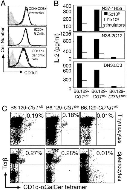
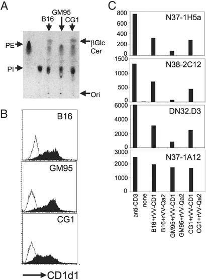
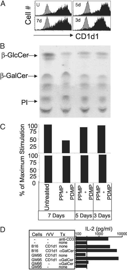
Va14Ja18 natural T (NKT) cells play an immunoregulatory role, which is controlled by a self glycolipid(s) presented by CD1d. Although the synthetic antigen alpha-D-galactosylceramide (alpha-D-GalCer) stimulates all Va14Ja18 NKT cells, alpha-anomeric D-glycosylceramides are currently unknown in mammals. We have used beta-D-GalCer-deficient mice and beta-D-glucosylceramide (beta-D-GlcCer)-deficient cells to define the chemical nature of a natural NKT cell antigen. beta-D-GalCer-deficient mice exhibit normal NKT cell development and function, and cells from these animals potently stimulate NKT hybridomas. In striking contrast, the same hybridomas fail to react to CD1d1 expressed by a beta-D-GlcCer-deficient cell line. Importantly, human beta-D-GlcCer synthase cDNA transfer, and hence the biosynthesis of beta-D-GlcCer, restores the recognition of mutant cells expressing CD1d1 by the Va14Ja18 NKT hybridomas. Additionally, suppression of beta-D-GlcCer synthesis inhibits antigen presentation to Va14Ja18 NKT cells. The possibility that beta-D-GlcCer itself is the natural NKT cell antigen was excluded because it was unable to activate NKT hybridomas in a cell-free antigen-presentation assay. These findings suggest that beta-D-GlcCer may play an important role in generating and/or loading a natural Va14Ja18 NKT antigen.


Figures

Figure 1
Figure 1
Expression of functional CD1d1 and development of Va14Ja18 NKT cells in β-D-GalCer synthase-null mice. (A) CD1d1 expression in CD4+8+ thymocytes, B220+ B lymphocytes, and CD11c+ dendritic cells was monitored by flow cytometry after staining with biotinylated CD1-specific mAb 1B1. Note that cells from B6.129-Cgt+/0 and B6.129-Cgt0/0 mice express equal levels of CD1d1, and hence the two histograms overlap. The filled gray histogram represents CD1d1 expression in B6.129-CD1d10/0-deficient cells. (B) The functional status of CD1d1 expressed by β-D-GalCer-containing and -deficient cells was determined in the stimulator–responder coculture system. IL-2 secreted (an indicator of T cell activation) from 5 × 10⁴ hybridoma cells stimulated with 1–5 × 10⁵ wild-type and mutant thymocytes was monitored by ELISA. (C) Dot plots demonstrating the percentage of Va14Ja18 NKT lymphocytes within the thymus and spleen of B6.129-Cgt0/+, B6.129-Cgt0/0, and B6.129-CD1d10/0 mice. Mononuclear cells were stained with Tcrβ-specific H57–597-phycoerythrin mAb and CD1d1-α-D-GalCer tetramer-allophycocyanin. Results are representative of at least three experiments involving at least six mice in each group.
Figure 2
Figure 2
Defective presentation of the natural Va14Ja18 NKT cell antigen by CD1d1 expressed by β-D-GlcCer synthase-deficient GM95 cells. (A) Total cellular lipids were extracted from B16, GM95, and CG1 cells, separated by TLC, and visualized by charring. Commercial β-D-GlcCer was separated alongside as a standard. PE, phosphatidylethanolamine. (B) The expression of CD1d1 after infection with rVV-CD1d1 (filled histogram) or rVV-Qa2 (open histogram) was monitored after staining with biotinylated-1B1 as in Fig. 1A. (C) Mouse CD1 was expressed in the three cell lines by recent infection with rVV-CD1d1. The ability of Va14Ja18-positive (N37–1H5a, N38–2C12, and DN32.D3) and -negative (N37–1A12) NKT cell hybridomas to recognize CD1d1 expressed by β-D-GlcCer-containing and -deficient cells was determined in the stimulator–responder coculture system described for Fig. 1B. Results are representative of at least three experiments.
Figure 3
Figure 3
CD1d1 molecules expressed by β-D-GlcCer synthase-deficient cells preserve structural and functional integrity. (A) CD1d levels on uninfected or rVV-CD1d1-infected B16 cells treated for the indicated times with 2.5 μM PPMP and 10 μM PDMP were determined as in Fig. 1A. (B) β-D-GlcCer levels of PPMP+PDMP-treated B16 cells were evaluated by TLC. (C) The ability of Va14Ja18 (Upper, N37–1H5a; N38–2C12 and DN32-D3 not shown) and non-Va14 (Lower, N37–1A12) NKT cell hybridomas to recognize CD1d1 expressed by PPMP+PDMP-treated B16 cells was determined in the stimulator–responder coculture system. (D) Uninfected or recent rVV-CD1d1-infected B16 and GM95 were pulsed with vehicle or 0.5 μM α-D-GalCer for 4 h and washed extensively to remove unbound antigen. The ability of these cells to stimulate N38–2C12 Va14Ja18 NKT hybridoma was tested as described for Fig. 1B. Tx, treatment. Results are representative of three experiments.
Figure 4
Figure 4
Normal CD1d1 trafficking through late endosomes/lysosomes in β-D-GlcCer synthase-deficient cells. (A) Internalization of wild-type CD1d1 and CD1d1Δcyt expressed by GM95 and CG1 was monitored by using biotin labeling of cell-surface molecules as described in Materials and Methods. H2b class I and transferrin receptor were used as negative and positive controls (data not shown). (B) The traffic of CD1d1 through the low-pH endosomal/lysosomal compartment was monitored by the colocalization of mouse CD1 with LAMP-1 by using confocal microscopy. Wild-type CD1d1 or CD1d1Δcyt was expressed by infection of B16, GM95, and CG1 with the corresponding rVV. CD1d1 was detected 8 h after viral transduction with a specific mAb 1H6 (green) and LAMP-1 with 1D4B (red). (C) Recycling of wild-type CD1d1 or CD1d1Δcyt expressed by GM95 and CG1 was monitored after biotin labeling of cell-surface molecules as described in Materials and Methods. Results are representative of three (A and C) or four (B) independent experiments.
Figure 5
Figure 5
Specific cell-free recognition of CD1d1-α-D-GalCer by Va14Ja18 NKT hybridomas. (A) Microtiter wells were coated with 5 μg/ml sCD1d1, blocked with fetal bovine serum, and pulsed with the indicated lipids. Increasing concentrations of α-D-GalCer or a fixed concentration of PI, β-D-GalCer, or β-D-GlcCer was used. Cell-free presentation of lipids to NKT hybridomas by plate-bound sCD1d1 was monitored. (B) Plate-bound sCD1d1 was first pulsed with 0.0567 μM α-D-GalCer, and then increasing concentrations of PI or β-d-GlcCer were added. The ability of PI, β-D-GlcCer, and p99, a hydrophobic peptide known to bind CD1d1, to inhibit the presentation of α-D-GalCer was tested by using Va14Ja18 NKT hybridomas. In all assays, IL-2 secretion indicated the recognition of antigen by the hybridomas. Results are representative of at least three experiments.

References

    1. Joyce S. Cell Mol Life Sci. 2001;58:442–469. - PMC - PubMed
    1. Grakoui A, Bromley S K, Sumen C, Davis M M, Shaw A S, Allen P M, Dustin M L. Science. 1999;285:221–227. - PubMed
    1. Krummel M, Wulfing C, Sumen C, Davis M M. Philos Trans R Soc London B. 2000;355:1071–1076. - PMC - PubMed
    1. Bendelac A, Lantz O, Quimby M E, Yewdell J W, Bennink J R, Brutkiewicz R R. Science. 1995;268:863–865. - PubMed
    1. Brossay L, Chioda M, Burdin N, Koezuka Y, Casorati G, Dellabona P, Kronenberg M. J Exp Med. 1998;188:1521–1528. - PMC - PubMed

Publication types

LinkOut - more resources