. 2003 Feb 17;160(4):529-39.
doi: 10.1083/jcb.200210095. Epub 2003 Feb 10.

Substrate-specific function of the translocon-associated protein complex during translocation across the ER membrane

Affiliations

Substrate-specific function of the translocon-associated protein complex during translocation across the ER membrane

Ryen D Fons et al. J Cell Biol. .

Abstract

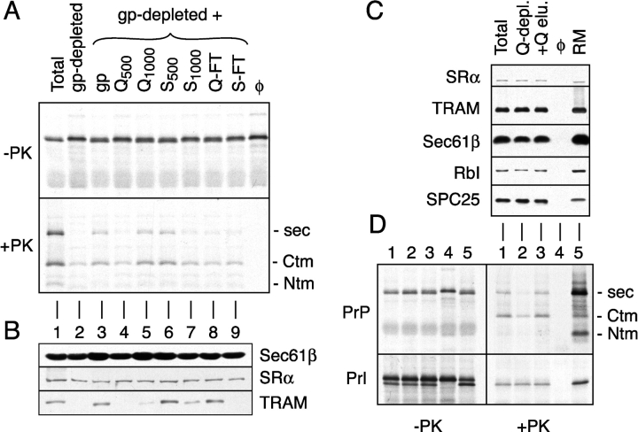
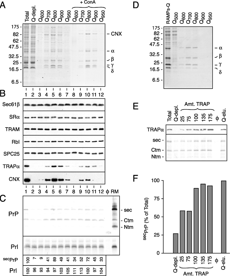
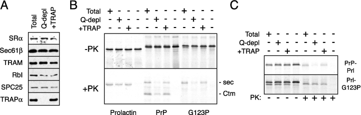
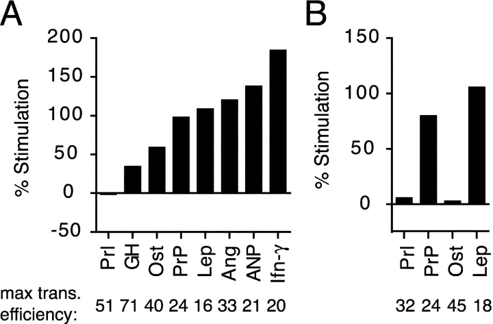
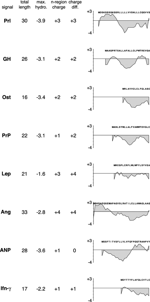
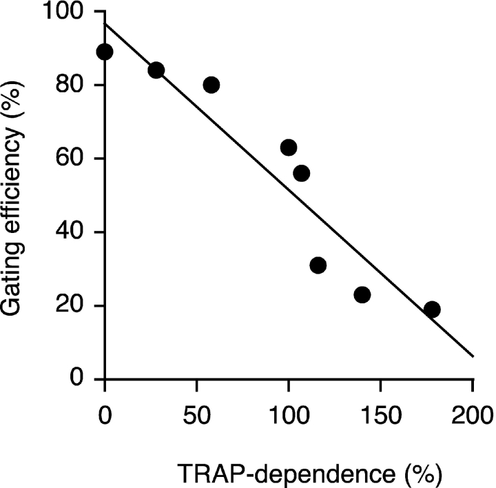
Although the transport of model proteins across the mammalian ER can be reconstituted with purified Sec61p complex, TRAM, and signal recognition particle receptor, some substrates, such as the prion protein (PrP), are inefficiently or improperly translocated using only these components. Here, we purify a factor needed for proper translocation of PrP and identify it as the translocon-associated protein (TRAP) complex. Surprisingly, TRAP also stimulates vectorial transport of many, but not all, other substrates in a manner influenced by their signal sequences. Comparative analyses of several natural signal sequences suggest that a dependence on TRAP for translocation is not due to any single physical parameter, such as hydrophobicity of the signal sequence. Instead, a functional property of the signal, efficiency of its post-targeting role in initiating substrate translocation, correlates inversely with TRAP dependence. Thus, maximal translocation independent of TRAP can only be achieved with a signal sequence, such as the one from prolactin, whose strong interaction with the translocon mediates translocon gating shortly after targeting. These results identify the TRAP complex as a functional component of the translocon and demonstrate that it acts in a substrate-specific manner to facilitate the initiation of protein translocation.


Figures

Figure 1.
Figure 1.
Detection and fractionation of a translocation accessory factor activity. (A) Analysis of PrP translocation activity in fractionated proteoliposomes. A glycoprotein-depleted detergent extract was mixed with either buffer (lane 2), total glycoproteins (lane 3), or ion exchange fractions of total glycoproteins (lanes 4–9). Proteoliposomes were prepared from each mixture and assayed for their ability to translocate PrP. Shown are translation products before and after digestion with PK. The positions of protease-protected fragments of PrP corresponding to the secPrP, CtmPrP, and NtmPrP forms (Hegde et al., 1998a) are indicated to the right of the autoradiograph. Q-FT and S-FT indicate flowthrough fractions after binding at 200 mM KAc to Q- and S-sepharose, respectively. Fractions resulting from sequential elution of these resins with either 500 mM or 1,000 mM KAc are indicated with a subscript. For comparison, shown are translocation reactions lacking membranes (last lane) and containing proteoliposomes reconstituted from a total unfractionated detergent extract (lane 1). (B) Immunoblots of each of the proteoliposomes from panel A with antibodies against Sec61β, SRα, and TRAM. (C) Proteoliposomes were prepared from a total detergent extract, a detergent extract after depletion of proteins that bind to Q-sepharose (Q-depl.), and a Q-depleted extract replenished with the protein eluted from Q-sepharose (+Q-elu.). Aliquots of each proteoliposome preparation, along with the starting RMs, were immunoblotted with antibodies against the indicated proteins. (D) Translocation of PrP and Prl into the proteoliposomes from panel C. Aliquots of the translation products before and after digestion with PK are shown on the left and right, respectively. Lane 4 is a translocation reaction lacking membranes. The topologic forms of PrP are indicated to the right of the autoradiograph.
Figure 2.
Figure 2.
Purification of TrAF and identification as the TRAP complex. (A) Shown is a Coomassie blue–stained gel of fractions resulting from the separation of membrane proteins by ion exchange and ConA chromatography. The positions of CNX and the α through δ subunits of the TRAP complex are indicated. (B) Immunoblots against various proteins in proteoliposomes prepared using the fractions in panel A. Lanes 1 and 2 contain proteoliposomes reconstituted from the total and Q-depleted detergent extracts, respectively. Lanes 3–12 contain proteoliposomes coreconstituted with the Q-depleted detergent extract plus the respective individual fractions in lanes 3–12 of A. (C) Translocation assays of PrP and Prl using proteoliposomes from panel B. Control reactions lacking membranes or containing RMs are also shown for comparison. Only the translocated material, remaining after digestion of the translation reactions with PK, is shown. The efficiencies of secPrP and Prl translocation, relative to the unfractionated proteoliposomes in lane 1, are shown below the autoradiograph. (D) Purification of the TRAP complex from the RAMP fraction. Shown is the Coomassie blue–stained gel containing the final fractions of the purification. (E) A Q-depleted detergent extract was replenished with varying concentrations of RAMP-purified TRAP or total Q eluate and reconstituted into proteoliposomes. As a control, an unfractionated detergent extract was also reconstituted in parallel. Shown in the top panel is an immunoblot against TRAPα of the different proteoliposomes. The amount of TRAP, as a percent of that found in the unfractionated proteoliposomes, is indicated above the blot. The bottom panel shows the assay for PrP translocation into these proteoliposomes. Only the translocated products remaining after protease digestion are shown. (F) The extent of secPrP translocation in the assay from panel E was quantitated and plotted as a bar graph. The amount of translocation in the Q-depleted proteoliposomes replenished with the total Q-eluate fraction was defined as 100%.
Figure 3.
Figure 3.
Dependence on TRAP for translocation is influenced by the signal sequence. (A) Immunoblots against various proteins of proteoliposomes prepared from an unfractionated detergent extract (Total), a Q-depleted detergent extract, and a Q-depleted extract replenished with RAMP-purified TRAP at a level comparable to that in the unfractionated extract. (B) Translocation of Prl, PrP, and PrP(G123P) into the proteoliposomes from panel A, or a control reaction lacking proteoliposomes. Aliquots of the translation reaction before (top) and after (bottom) digestion with PK are shown. (C) Prl–G123P (which contains the signal sequence of Prl fused to the mature domain of PrP[G123P]) and PrP–Prl (containing the signal of PrP fused to the mature domain of Prl) were assayed for their TRAP dependence, as in B.
Figure 4.
Figure 4.
Signal sequences from different substrates vary in their TRAP dependence. (A) Constructs containing the signal sequences of various mammalian proteins fused to PrP (Kim et al., 2002) were tested for their translocation into Q-depleted proteoliposomes lacking or containing RAMP-purified TRAP (as in Fig. 3 A). To facilitate direct comparisons, all constructs were analyzed in parallel using the same batch of proteoliposomes.The percent increase in overall translocation into the TRAP-containing proteoliposomes, relative to the membranes lacking TRAP, is plotted. The maximal overall translocation efficiency for each substrate (as a percent of total synthesized translation product) into proteoliposomes containing TRAP is indicated below the graph. The sequences and properties of the signals used in this experiment are depicted in Fig. 5. (B) The indicated signal sequences fused to the mature domain of bovine Prl were analyzed for their degree of dependence on TRAP for translocation, as in A.
Figure 5.
Figure 5.
Physical properties of signal sequences that differ in their TRAP dependence. Shown are several parameters for eight signal sequences (human Prl, pig GH, rat Ost, hamster PrP, pig leptin [Lep], human angiotensinogen [Ang], pig atrial naturetic peptide [ANP], and pig interferon-γ [Ifn-γ]). Hydropathy was determined by the method of Kyte and Doolittle (1982) using a window of seven residues. In calculating the net charge of the n-region (the domain preceding the hydrophobic core) or the charge difference flanking the hydrophobic domain, the amino terminus, lysine, and arginine were each taken to contribute a net +1 charge, whereas aspartate and glutamate were taken to contribute a net −1 charge. All of the hydropathy plots are shown on the same scale to allow direct comparisons to be made.
Figure 6.
Figure 6.
Correlation of a signal's TRAP dependence and post-targeting gating function. The eight signal sequences from Fig. 5 are plotted on a graph that shows their relative dependence on TRAP for translocation on the x axis and their post-targeting gating activity on the y axis. Each signal sequence's gating activity was taken from data reported in previously published work (Kim et al., 2002). In this study, gating was defined as a signal's ability to initiate substrate translocation at the translocon and was measured based on an assay using the topology of PrP as a reporter of the efficiency of NH2-terminal translocation (Kim et al., 2002).
Figure 7.
Figure 7.
Functional analysis of signal sequence–translocon interactions. (A) Translocation intermediates of 30, 50, or 70 residues beyond the signal sequence of either Prl or PrP–Prl were prepared and examined by protease protection and salt-resistant binding assays. Equal aliquots of untreated, PK-digested, and salt-resistant samples are shown for each intermediate. In addition, the full-length proteins were analyzed in parallel. (B) Translocation intermediates at 56 residues beyond the signal of either Prl or PrP–Prl were examined for protease protection, salt-resistant binding, and ability to translocate upon release from the ribosome with puromycin. (C) Shown is a schematic diagram of the experimental protocol and interpretation of the experiment in B. Nascent chains that are protected from protease digestion appear to translocate into the lumen (with concomitant signal sequence cleavage) upon release with puromycin, whereas protease-accessible nascent chains slip into the cytosol upon puromycin release. (D) Translocation intermediates of 101 residues beyond the signal sequence were examined for cytosolic accessibility, salt-resistant binding, and translocation, as in B. Each substrate is the mature region of PrP containing the indicated signal sequences. The positions of the precursor and processed (i.e., signal cleaved) forms for each substrate are indicated to the left. Also indicated is the position of the COOH-terminal fragment (CTF) that represents the segment of the nascent chain within the ribosome and, hence, is protected from protease digestion. The percent of translocon-bound chains that are translocated (and, hence, protease protected) upon puromycin release was calculated by dividing the amount of substrate in lane 7 by that in lane 4, and is indicated to the right of each autoradiograph.
Figure 8.
Figure 8.
Modulation of TRAP dependence by changing signal–translocon interactions. (A) Sequences of the wild-type, N7 mutant, and N9 mutant signal sequences of PrP. The mutated residue is indicated in bold. (B) The N7 and N9 signal sequences fused to Prl were assayed for their dependence on TRAP for translocation, as in Fig. 3 B. Control reactions containing RMs or lacking membranes are also shown for comparison.

References

    1. Andrews, D.W., and A.E. Johnson. 1996. The translocon: more than a hole in the ER membrane? Trends Biochem. Sci. 21:365–369. - PubMed
    1. Beckmann, R., D. Bubeck, R. Grassucci, P. Penczek, A. Verschoor, G. Blobel, and J. Frank. 1997. Alignment of conduits for the nascent polypeptide chain in the ribosome-Sec61 complex. Science. 278:2123–2126. - PubMed
    1. Crowley, K.S., S. Liao, V.E. Worrell, G.D. Reinhart, and A.E. Johnson. 1994. Secretory proteins move through the endoplasmic reticulum membrane via an aqueous, gated pore. Cell. 78:461–471. - PubMed
    1. Gorlich, D., and T.A. Rapoport. 1993. Protein translocation into proteoliposomes reconstituted from purified components of the endoplasmic reticulum membrane. Cell. 75:615–630. - PubMed
    1. Gorlich, D., E. Hartmann, S. Prehn, and T.A. Rapoport. 1992. a. A protein of the endoplasmic reticulum involved early in polypeptide translocation. Nature. 357:47–52. - PubMed

Publication types

MeSH terms