. 2003 Feb 17;160(4):487-93.
doi: 10.1083/jcb.200209105. Epub 2003 Feb 10.

Cyclic AMP induces integrin-mediated cell adhesion through Epac and Rap1 upon stimulation of the beta 2-adrenergic receptor

Affiliations

Cyclic AMP induces integrin-mediated cell adhesion through Epac and Rap1 upon stimulation of the beta 2-adrenergic receptor

Savithri Rangarajan et al. J Cell Biol. .

Abstract

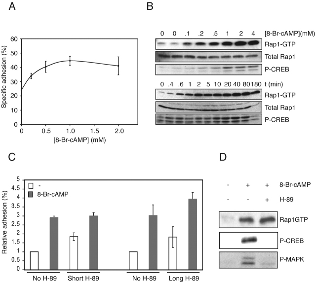
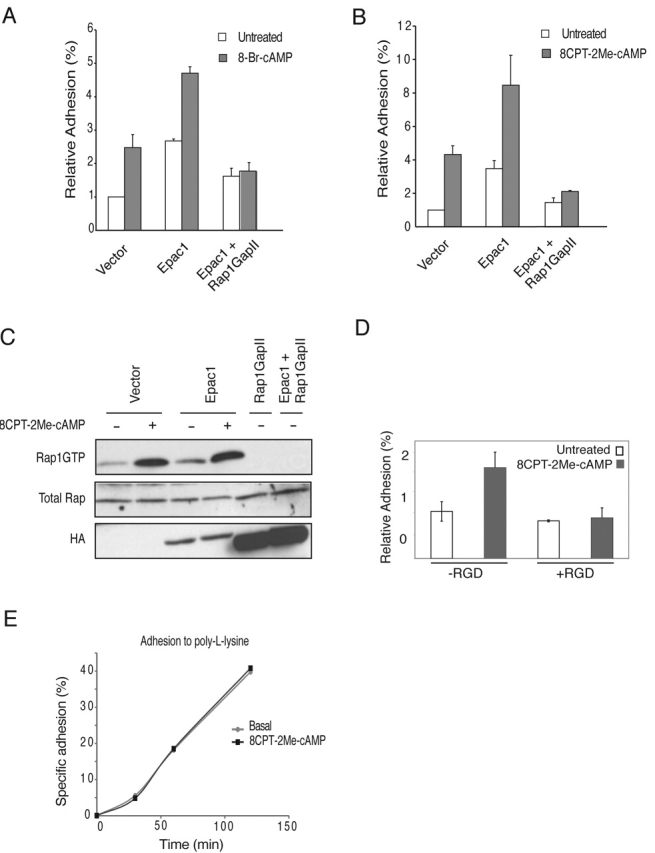
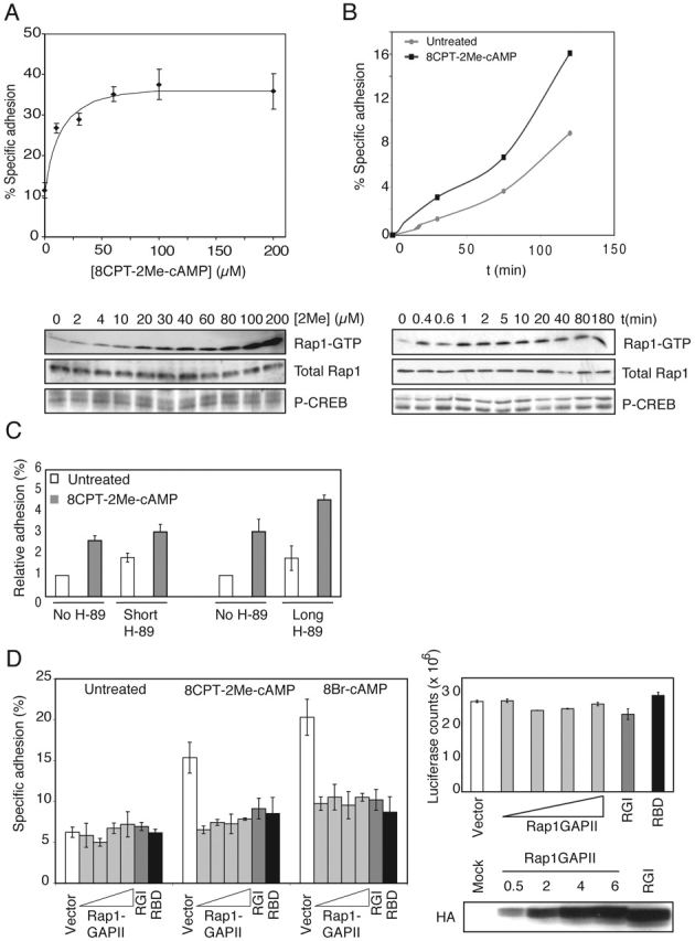
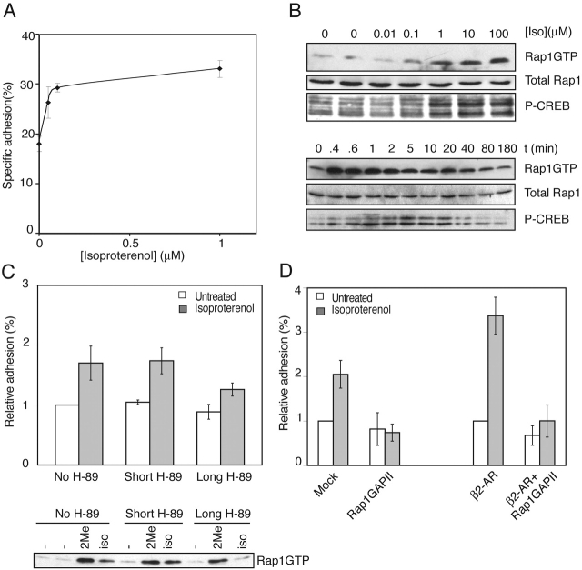
cAMP controls many cellular processes mainly through the activation of protein kinase A (PKA). However, more recently PKA-independent pathways have been established through the exchange protein directly activated by cAMP (Epac), a guanine nucleotide exchange factor for the small GTPases Rap1 and Rap2. In this report, we show that cAMP can induce integrin-mediated cell adhesion through Epac and Rap1. Indeed, when Ovcar3 cells were treated with cAMP, cells adhered more rapidly to fibronectin. This cAMP effect was insensitive to the PKA inhibitor H-89. A similar increase was observed when the cells were transfected with Epac. Both the cAMP effect and the Epac effect on cell adhesion were abolished by the expression of Rap1-GTPase-activating protein, indicating the involvement of Rap1 in the signaling pathway. Importantly, a recently characterized cAMP analogue, 8-(4-chloro-phenylthio)-2'-O-methyladenosine-3',5'-cyclic monophosphate, which specifically activates Epac but not PKA, induced Rap-dependent cell adhesion. Finally, we demonstrate that external stimuli of cAMP signaling, i.e., isoproterenol, which activates the G alpha s-coupled beta 2-adrenergic receptor can induce integrin-mediated cell adhesion through the Epac-Rap1 pathway. From these results we conclude that cAMP mediates receptor-induced integrin-mediated cell adhesion to fibronectin through the Epac-Rap1 signaling pathway.


Figures

Figure 1.
Figure 1.
cAMP induces cell adhesion to fibronectin in a PKA-independent manner. (A) Treatment with 8-Br-cAMP induces adhesion to fibronectin. Ovcar3 cells were transiently transfected with CMV-luciferase plasmid, and cells adhering to fibronectin (2 μg/ml) in the presence of increasing concentrations of 8-Br-cAMP were quantified as described in Materials and methods. (B) 8-Br-cAMP induces Rap1 activation. (Top) Ovcar3 cells were treated with increasing concentrations 8-Br-cAMP for 15 min. Cells were lysed, and equal amounts of cell lysate were analyzed for activation of Rap1 (top blot) and CREB (bottom blot). Total levels of Rap1 in cell lysates are shown (middle blot). (Bottom) Ovcar3 cells were treated with 1 mM 8-Br-cAMP for the indicated times. Cells were lysed as above and analyzed for activation of Rap1 (top blot) and CREB (bottom blot). Total Rap1 levels are shown (middle blot). (C) 8-Br-cAMP– induced adhesion is independent of PKA. Ovcar3 cells transiently transfected with CMV-luciferase plasmid were either preincubated at 37°C for 30 min with the PKA inhibitor H-89 (10 μM) 30 min before seeding onto the wells (Short), or H-89 was added 30 min before trypsinization and during the recovery period (Long) and seeded onto wells with or without 8-Br-cAMP. Cells were allowed to adhere for 1 h, and nonadherent cells were removed. The percentage of adherent cells was quantified and plotted relative to unstimulated cells (range from 2–10%). The plot shown is representative of two (long pretreatment) and five (short pretreatment) experiments each in triplicate. Error bars represent SD. (D) Activation of CREB and ERK but not Rap1 is blocked by H-89. Ovcar3 cells were pretreated with either H-89 or carrier for 30 min followed by stimulation with 8-Br-cAMP for 15 min. Cells were lysed, and equal amounts of cell lysates were incubated with precoupled GST-RalGDS-RBD, and activation of Rap1 was analyzed by immunoblotting using a Rap1 antibody. Phosphorylation of CREB and ERK was assayed by Western blotting using phospho-specific antibodies.
Figure 2.
Figure 2.
Overexpression of Epac1 increases cAMP-induced cell adhesion, which is Rap1GAPII sensitive. (A) 8-Br-cAMP-Epac1–induced cell adhesion is blocked by Rap1GAPII. Ovcar3 cells were transiently transfected with TK-luciferase plasmid and either mock DNA (vector), HA-tagged Epac1, or HA-tagged Rap1GAPII where indicated. Cells were stimulated with 8-Br-cAMP, and adhesion of cells to fibronectin was quantified. The percentage of adherent cells was plotted relative to unstimulated, mock-transfected cells. Representative data performed in triplicate are shown, and error bars represent SD. The experiments were repeated at least four times with identical results. (B) 8CPT-2Me-cAMP-Epac1–induced cell adhesion is blocked by Rap1GAPII. Ovcar3 cell were transiently transfected as above. Cells were stimulated with 8CPT-2Me-cAMP, and adhesion of cells to fibronectin was quantified. The percentage of adherent cells was plotted relative to unstimulated, mock-transfected cells. Representative data performed in triplicate are shown, and error bars represent SD. The experiments were repeated at least four times with identical results. (C) 8CPT-2Me-cAMP-Epac1–induced Rap1 activation is blocked by Rap1GAPII. Cells were treated with 50 μM 8CPT-2Me-cAMP for 15 min, lysed, and GTP-bound Rap1 levels were determined as described in Materials and methods (top). Rap1 protein levels were equal (middle), and expression of transfected proteins was confirmed with an anti-HA antibody (bottom). (D) A β1-integrin–blocking peptide containing the RGD sequence present in fibronectin inhibits 8CPT-2Me-cAMP–induced cell adhesion. Ovcar3 cells were pretreated for 20 min with RGD peptide (100 μM) where indicated and seeded in wells with or without 8CPT-2Me-cAMP. Cells were allowed to adhere for 1 h, and nonadherent cells were removed. The percentage of adherent cells was measured and plotted relative to unstimulated cells. Representative data from two experiments performed in triplicate are shown with error bars representing SD. (E) 8CPT-2Me-cAMP does not increase the rate of cell adhesion to poly- l-lysine. Ovcar3 cells were transfected with CMV-luciferase and seeded onto poly-l-lysine–coated plates. At various time points, nonadherent cells were removed and adherent cells were quantified. A representative experiment in triplicate is shown.
Figure 3.
Figure 3.
8CPT-2Me-cAMP induces cell adhesion via Epac and Rap1. (A) 8CPT-2Me-cAMP stimulates cell adhesion. (Top) Ovcar3 cells were transiently transfected with CMV- luciferase plasmid and treated with increasing concentrations of 8CPT-2Me-cAMP. Cells adhering to fibronectin (2 μg/ml) were quantified as described in Materials and methods. (Bottom) Ovcar3 cells were treated with increasing concentrations of 8CPT-2Me-cAMP for 15 min, and cells were lysed and analyzed for activation of Rap1 (top blot) and CREB (bottom blot). Total Rap1 levels are shown (middle blot). (B) 8CPT-2Me-cAMP increases the rate of cell adhesion. (Top) Ovcar3 cells were transfected with TK-luciferase and seeded onto fibronectin-coated plates. At various time points, nonadherent cells were removed, and adherent cells were quantified. (Bottom) cells were treated with 60 μM 8CPT-2Me-cAMP for the indicated times. Cells were lysed, and equal amounts of cell lysates were analyzed for activation of Rap1 (top blot) and CREB (bottom blot). Total levels of Rap1 in cell lysates are shown (middle blot). (C) Ovcar3 cells were pretreated with H-89 as described in the legend to Fig. 1 C and seeded onto wells in the absence or presence of 8CPT-2Me-cAMP (100 μM). Cells were allowed to adhere for 1 h, and nonadherent cells were removed. The percentage of adherent cells was quantified and plotted relative to unstimulated cells (range from 2–10%). The plot shown is representative of two (long pretreatment) and five (short pretreatment) experiments, each in triplicate. Error bars represent SD. (D) cAMP-induced adhesion to fibronectin is blocked by inhibitors of Rap1. (Left) Ovcar3 cells were transiently transfected with CMV-luciferase and either mock DNA, increasing concentrations of HA-Rap1GAP II (0.5, 1, 2, or 6 μg, respectively), HA-Rap1GAPI (6 μg), or HA-RBD of RalGDS (6 μg), respectively. Cells were treated with 8-Br-cAMP or 8CPT-2Me-cAMP, and adhesion to fibronectin (5 μg/ml) was determined and plotted relative to unstimulated, mock-transfected cells. Representative data from experiments performed in triplicate are shown with error bars representing SD. The experiments were repeated (Rap1GAPII, at least four times; Rap1GAPI and RBD, twice) with identical results. (Top right) Luciferase counts of total input cells per well in the above experiment are shown with error bars representing SD of triplicates. (Bottom left panel) Expression of HA-Rap1GAPs in the above experiment is shown.
Figure 4.
Figure 4.
Stimulation of the β2-AR with isoproterenol induces cell adhesion. (A) Isoproterenol induces adhesion to fibronectin. Ovcar3 cells transiently transfected with CMV- luciferase plasmid were treated with increasing concentrations of the β2-AR agonist isoproterenol, and cells adhering to fibronectin (2 μg/ml) were quantified as described in Materials and methods. (B) Isoproterenol induces activation of Rap1 and CREB. (Top) Ovcar3 cells were treated with increasing concentrations of isoproterenol for 5 min. Cells were lysed, and equal amounts of cell lysate were analyzed for activation of Rap1 (top) and CREB (bottom). Total levels of Rap1 in cell lysates are shown (middle blot). (Bottom) Cells were treated with 10 μM of isoproterenol for the indicated times. Cells were lysed, and equal amounts of cell lysate were analyzed for activation of Rap1 (top blot) and CREB (bottom blot). Total levels of Rap1 in cell lysates are shown (middle blot). (C) Isoproterenol-induced adhesion to fibronectin is independent of PKA. (Top) Ovcar3 cells were pretreated with H-89 as described in the legend to Fig. 1 C and seeded onto wells in the absence or presence of isoproterenol (100 μM). Cells were allowed to adhere for 1 h, and nonadherent cells were removed. The percentage of adherent cells was quantified and plotted relative to unstimulated cells (range from 2–10%). The plot shown is representative of two (long pretreatment) and four (short pretreatment) experiments, each in triplicate. Error bars represent SD. (Bottom) Cells were pretreated with either DMSO or H-89 for 30 min before trypsinization and during the recovery period (DMSO and long H-89 treatment, respectively) or during the last 30 min of recovery (short H-89 treatment). Then cells were stimulated with either 50 μM 8CPT-2Me-cAMP for 10 min or isoproterenol for 2 min, respectively. Cells were centrifuged, cell pellets were lysed, and equal amounts of cell lysate were incubated with precoupled GST-RalGDS-RBD, and activation of Rap1 was analyzed on Western blot using a Rap1 antibody. (D) Isoproterenol-induced adhesion to fibronectin is inhibited by Rap1GAPII. Ovcar3 cells were transfected with either mock DNA (Vector) or HA-Rap1GapII alone or in combination with a β2-AR expression vector where indicated. Adhesion of cells to fibronectin in the absence or presence of isoproterenol was quantified. The percentage of adherent cells was plotted relative to unstimulated, mock-transfected cells (range 5–15%). Summarizing data of four (for the left half of the plot) and two (for the right half of the plot) independent experiments performed in triplicate are shown with error bars representing SD.

References

    1. Arai, A., Y. Nosaka, E. Kanda, K. Yamamoto, N. Miyasaka, and O. Miura. 2001. Rap1 is activated by erythropoietin or interleukin-3 and is involved in regulation of β1 integrin-mediated hematopoietic cell adhesion. J. Biol. Chem. 276:10453–10462. - PubMed
    1. Bertoni, A., S. Tadokoro, K. Eto, N. Pampori, L.V. Parise, G.C. White, and S.J. Shattil. 2002. Relationships between Rap1b, affinity modulation of integrin α IIbβ 3, and the actin cytoskeleton. J. Biol. Chem. 277:25715–25721. - PubMed
    1. Bos, J.L., J. de Rooij, and K.A. Reedquist. 2001. Rap1 signalling: adhering to new models. Nat. Rev. Mol. Cell Biol. 2:369–377. - PubMed
    1. Brakebusch, C., D. Bouvard, F. Stanchi, T. Sakai, and R. Fassler. 2002. Integrins in invasive growth. J. Clin. Invest. 109:999–1006. - PMC - PubMed
    1. Buczek-Thomas, J.A., N. Chen, and T. Hasan. 1998. Integrin-mediated adhesion and signalling in ovarian cancer cells. Cell. Signal. 10:55–63. - PubMed

Publication types

MeSH terms