. 2003 Feb 18;100(4):1633-8.
doi: 10.1073/pnas.0437927100. Epub 2003 Feb 10.

Loading of the human 9-1-1 checkpoint complex onto DNA by the checkpoint clamp loader hRad17-replication factor C complex in vitro

Affiliations

Loading of the human 9-1-1 checkpoint complex onto DNA by the checkpoint clamp loader hRad17-replication factor C complex in vitro

Vladimir P Bermudez et al. Proc Natl Acad Sci U S A. .

Abstract

The human DNA damage sensors, Rad17-replication factor C (Rad17-RFC) and the Rad9-Rad1-Hus1 (9-1-1) checkpoint complex, are thought to be involved in the early steps of the DNA damage checkpoint response. Rad17-RFC and the 9-1-1 complex have been shown to be structurally similar to the replication factors, RFC clamp loader and proliferating cell nuclear antigen polymerase clamp, respectively. Here, we demonstrate functional similarities between the replication and checkpoint clamp loader/DNA clamp pairs. When all eight subunits of the two checkpoint complexes are coexpressed in insect cells, a stable Rad17-RFC/9-1-1 checkpoint supercomplex forms in vivo and is readily purified. The two individually purified checkpoint complexes also form a supercomplex in vitro, which depends on ATP and is mediated by interactions between Rad17 and Rad9. Rad17-RFC binds to nicked circular, gapped, and primed DNA and recruits the 9-1-1 complex in an ATP-dependent manner. Electron microscopic analyses of the reaction products indicate that the 9-1-1 ring is clamped around the DNA.


Figures

Figure 1
Figure 1
Purification of Rad17-RFC/9-1-1 checkpoint supercomplex. The checkpoint complexes were reconstituted in H5 cells by coinfection with: lane 1 (9-1-1 complex), three baculoviruses expressing Flag-Rad9, Hus1, and Rad1; lane 2 (Rad17-RFC), five baculoviruses expressing Flag-Rad17, p40, His-p38, p37, and p36; and lane 3 (supercomplex), eight baculoviruses expressing His-Rad17, p40, His-p38, p37, p36, Rad9, Flag-Hus1, and Rad1. Complexes were purified by chromatography with Ni-NTA and/or anti-Flag agarose as described in Materials and Methods, and proteins were visualized after SDS/PAGE by silver staining.
Figure 2
Figure 2
Nucleotide cofactor requirement for binding of Rad17-RFC to the 9-1-1 complex. (A) Coimmunoprecipitation with anti-Rad9 antibodies. One picomole each of purified Rad17-RFC and 9-1-1 complexes were incubated with or without ATP and immunoprecipitated with anti-Rad9 antibodies; the bound and unbound proteins were analyzed by Western blotting with anti-Flag antibodies. (B) Coimmunoprecipitation with anti-RFCp37 antibodies. Reaction mixtures (25 μl) containing 5 pmol of purified RFC or Rad17-RFC and 0.5 pmol of 32P-labeled 9-1-1 were incubated with or without 10 μM of the indicated nucleotide as described in Materials and Methods. Lo denotes 10% input, Pr denotes preimmune serum, and 37 denotes anti-RFCp37 serum used for immunoprecipitations. The bound proteins were eluted with SDS loading buffer, subjected to SDS/12% PAGE, and autoradiographed. (C) The 32P-labeled 9-1-1 bound to Rad-17RFC shown in B was quantitated by phosphorimaging analyses. (D) Analysis by glycerol gradient velocity sedimentation. Reaction mixtures (100 μl) containing purified Rad17-RFC (5 pmol) and 32P-labeled 9-1-1 (1 pmol) were incubated in the presence or absence of 10 μM indicated nucleotides. After incubation, mixtures were subjected to glycerol gradient centrifugation as described in Materials and Methods. Nucleotide additions were as follows: ⧫, no ATP; ◊, ATP; ●, ADP; ■, AMP; and ▴, adenosine 5′-[β,γ-imido]triphosphate (AMPPNP).
Figure 3
Figure 3
Interactions of RFC and Rad17-RFC with PCNA and the 9-1-1 checkpoint complex. Reaction mixtures (25 μl) containing purified Rad17-RFC or RFC (5 pmol) and 1 pmol of either 32P-labeled 9-1-1 (Upper) or PCNA (Lower) were incubated in the presence or absence of 1 mM ATP as described in Materials and Methods. The 9-1-1 (or PCNA) bound to Rad17-RFC or RFC was immunoprecipitated with anti-RFCp37 antibody, and the bound material was analyzed by SDS/PAGE followed by autoradiography. Lo, 10% input; Pr, preimmune serum; 37, anti-RFCp37 antibody.
Figure 4
Figure 4
Rad9 mediates the Rad17-RFC/9-1-1 interaction. (A) Rad17 interacts with Rad9. H5 cells were coinfected with baculoviruses expressing either His-Rad17 alone (lane 1) or His-Rad17 together with Flag-Rad1 (lane 2), Flag-Rad9 (lane 3), Flag-Hus1 (lane 4), or Flag-Hus1, and untagged Rad1 and Rad9 (lane 5). The proteins were immunoaffinity purified with anti-Flag agarose, and proteins were analyzed by Western blotting with anti-Rad17 or anti-Flag antibodies. (B) Binding of the PCNA-like domain of Rad9 to Rad17. The following fragments of Rad9 were produced in H5 cells and bound to anti-Flag agarose resin: amino acids 1–130, lane A; amino acids 130–270, lane B; amino acids 1–270, lane C; amino acids 260–391, lane D; amino acids 130–391, lane E; and full length, lane F. The resin was then incubated with extracts made from H5 cells infected with either His-Rad17 or His-p140. After washing the resin, bound protein was eluted with Flag peptide and analyzed by Western blotting with anti-His and anti-Flag antibodies.
Figure 5
Figure 5
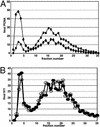
Recruitment of the 9-1-1 complex to DNA by Rad17-RFC. Reaction mixtures (50 μl) containing 32P-labeled 9-1-1 (0.6 pmol), Rad17-RFC (2 pmol), and nicked pBluescript plasmid (0.15 pmol), where indicated, were incubated with or without nucleotide. The mixtures were then loaded onto a 5-ml BioGel A-15M column as described in Materials and Methods. ■, form II (nicked plasmid)/Rad17-RFC/9-1-1/ATPγS-ATP (mixture was incubated with ATPγS for 5 min and then with 8 mM ATP for 5 min); ▴, form II (nicked plasmid)/Rad17-RFC/9-1-1/ATPγS; ●, form II (nicked plasmid)/Rad17-RFC/9-1-1/ATP; ♦, form II (nicked plasmid)/Rad17-RFC/9-1-1; ○, form II (nicked plasmid)/9-1-1/ATP; □, Rad17-RFC/9-1-1/ATPγS.
Figure 6
Figure 6
Sliding assay for PCNA and the 9-1-1 complex. (A) PCNA loading. RFC (2 pmol) and 32P-labeled PCNA (0.6 pmol) were assembled on nicked pBluescript (♦) or BamHI-linearized nicked pBluescript (■) (0.15 pmol) in a 50-μl reaction mixture as described in Materials and Methods. The mixture was incubated in 2 mM ATP for 5 min and then loaded onto a 5-ml Bio-Gel A-15M and eluted as described in Materials and Methods. (B) 9-1-1 loading. Rad17-RFC (2 pmol) and 32P-labeled 9-1-1 (0.6 pmol) were assembled on nicked pBluescript (●) or BamHI-linearized nicked pBluescript (○) (0.15 pmol) in a 50-μl reaction mixture as described in Materials and Methods. The mixture was incubated in 2 mM ATPγS for 5 min and then supplemented with 8 mM ATP and incubated for 5 min. The mixture was loaded onto a 5-ml Bio-Gel A-15M and eluted as described in Materials and Methods.
Figure 7
Figure 7
Electron microscopic evidence for loading of the 9-1-1 complex by Rad17-RFC. (A) 9-1-1 loading on a 6.9-kb nicked plasmid. Large white arrow indicates presumed supercomplex of Rad17-RFC and 9-1-1; small white arrows indicate loaded 9-1-1 protein complexes. (BD) Loaded 9-1-1 complexes (B and C) and Rad17-RFC (D) on a 718-bp circular template with a 40-nt single strand gap. Samples were directly mounted onto thin carbon-coated foils and rotary shadowed with tungsten. Shown in reverse contrast. [Bar = 1,000 bp (A); = 500 bp (B–D).]

References

    1. Abraham R T. Genes Dev. 2001;15:2177–2196. - PubMed
    1. Nyberg K A, Michelson R J, Putnam C W, Weinert T A. Annu Rev Genet. 2002;36:617–656. - PubMed
    1. Osborn A J, Elledge S J, Zou L. Trends Cell Biol. 2002;12:509–516. - PubMed
    1. Rouse J, Jackson S P. Science. 2002;297:547–551. - PubMed
    1. Unsal-Kacmaz K, Makhov A M, Griffith J D, Sancar A. Proc Natl Acad Sci USA. 2002;99:6673–6678. - PMC - PubMed

Publication types

MeSH terms