. 2003 Feb 18;100(4):2106-11.
doi: 10.1073/pnas.0437946100. Epub 2003 Feb 10.

An alpha-syntrophin-dependent pool of AQP4 in astroglial end-feet confers bidirectional water flow between blood and brain

Affiliations

An alpha-syntrophin-dependent pool of AQP4 in astroglial end-feet confers bidirectional water flow between blood and brain

Mahmood Amiry-Moghaddam et al. Proc Natl Acad Sci U S A. .

Abstract

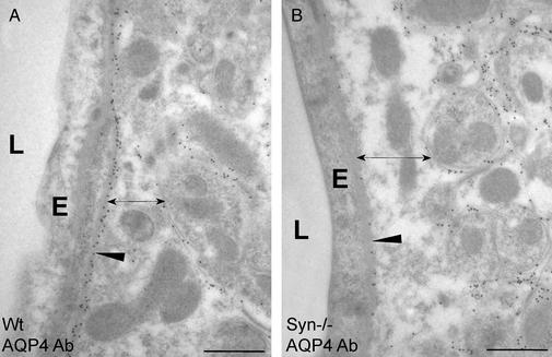
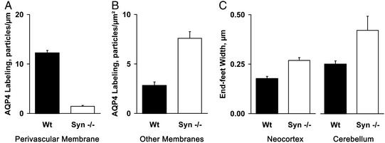
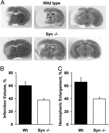
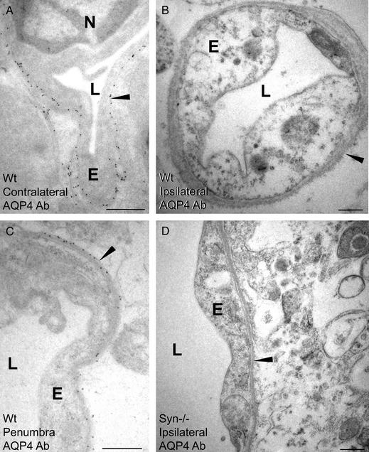
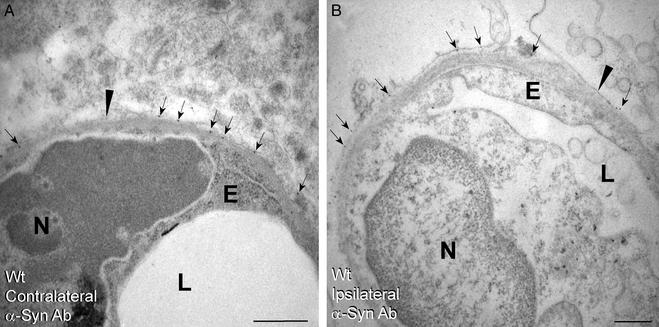
The water channel AQP4 is concentrated in perivascular and subpial membrane domains of brain astrocytes. These membranes form the interface between the neuropil and extracerebral liquid spaces. AQP4 is anchored at these membranes by its carboxyl terminus to alpha-syntrophin, an adapter protein associated with dystrophin. To test functions of the perivascular AQP4 pool, we studied mice homozygous for targeted disruption of the gene encoding alpha-syntrophin (alpha-Syn(-/-)). These animals show a marked loss of AQP4 from perivascular and subpial membranes but no decrease in other membrane domains, as judged by quantitative immunogold electron microscopy. In the basal state, perivascular and subpial astroglial end-feet were swollen in brains of alpha-Syn(-/-) mice compared to WT mice, suggesting reduced clearance of water generated by brain metabolism. When stressed by transient cerebral ischemia, brain edema was attenuated in alpha-Syn(-/-) mice, indicative of reduced water influx. Surprisingly, AQP4 was strongly reduced but alpha-syntrophin was retained in perivascular astroglial end-feet in WT mice examined 23 h after transient cerebral ischemia. Thus alpha-syntrophin-dependent anchoring of AQP4 is sensitive to ischemia, and loss of AQP4 from this site may retard the dissipation of postischemic brain edema. These studies identify a specific, syntrophin-dependent AQP4 pool that is expressed at distinct membrane domains and which mediates bidirectional transport of water across the brain-blood interface. The anchoring of AQP4 to alpha-syntrophin may be a target for treatment of brain edema, but therapeutic manipulations of AQP4 must consider the bidirectional water flux through this molecule.


Figures

Figure 1
Figure 1
Subcellular localization of AQP4 in perivascular astroglial cells in cerebellum under basal conditions. WT (A) and α-Syn−/− (B) mice were killed without other manipulations. Ultrathin sections of brain were incubated with anti-AQP4 and immunogold particles and analyzed by electron microscopy (see Materials and Methods). AQP4 in perivascular end-feet is reduced (arrowhead) and astroglial end-feet width is increased (double-headed arrow) in α-Syn−/−mice. E, capillary endothelial cell; L, lumen. (Bars = 0.5 μm.)
Figure 2
Figure 2
Quantification of AQP4 immunogold labeling and width of perivascular astroglial end-feet under basal conditions. α-Syn−/− (n = 4) and WT (n = 2) mice were killed without other manipulations, and brain sections were prepared for immunogold electron microscopy and quantifications (see Materials and Methods). (A) AQP4 immunogold labeling density of perivascular astroglial end-feet in neocortex. Labeling density represents the number of gold particles divided by the length of the perivascular end-feet membranes (P < 0.01). (B) AQP4 immunogold labeling density of non-end-feet membranes in neocortex. Labeling density represents the number of gold particles divided by the area (P < 0.05). (C) Width of perivascular astroglial end-feet measured in neocortex or cerebellum (P < 0.01). The increase in volume is proportional to the difference in width of end-feet.
Figure 3
Figure 3
Difference in brain injuries sustained by α-Syn−/− and WT mice after ischemia reperfusion. After 90 min of middle cerebral artery occlusion and 23 h of reperfusion, α-Syn−/− (n = 8) and WT (n = 9) mice were killed, and the brains were sectioned. Slices were incubated with triphenyltetrazolium chloride, and infarction volume and hemispheric enlargement were determined (see Materials and Methods). (A) Corresponding serial brain sections reveal smaller infarct zone (unstained) in slices from α-Syn−/− mice compared to WT mice. (B) Quantification of infarction volumes determined by image analysis. (C) Determination of hemispheric enlargement. Brains of α-Syn−/− mice were partially protected (P < 0.05 for both parameters).
Figure 4
Figure 4
Immunogold labeling of AQP4 in brain neocortex after ischemia-reperfusion. WT mice (A–C) and α-Syn−/− mice (D) were subjected to middle cerebral artery occlusion and reperfusion before intracardiac perfusion fixation and immunogold labeling of AQP4 for electron microscopy (see Materials and Methods). Note that AQP4 immunolabeling in WT brain is spared in perivascular membranes (arrowheads) of contralateral hemisphere (A), totally absent from the ipsilateral infarct core (B), and partially reduced in penumbra (C). Ultrastructural derangement including endothelial swelling is much more pronounced in ischemic core from brain of WT mice (B) than in brain of α-Syn−/− mice (D). (Bars = 0.5 μm.) N, nucleus; E, endothelial cell; L, lumen.
Figure 5
Figure 5
Immunogold labeling of α-syntrophin in neocortex from WT mice after ischemia reperfusion. Perivascular membranes (arrowheads) from contralateral hemisphere (A) and ipsilateral hemisphere (B) infarct core showed equivalent α-syntrophin immunolabeling (arrows). (Bars = 0.5 μm.) N, nucleus; E, endothelial cell; L, lumen.

Similar articles

Cited by

References

    1. Jung J S, Bhat R V, Preston G M, Guggino W B, Baraban J M, Agre P. Proc Natl Acad Sci USA. 1994;91:13052–13056. - PMC - PubMed
    1. Nielsen S, Nagelhus E A, Amiry-Moghaddam M, Bourque C, Agre P, Ottersen O P. J Neurosci. 1997;17:171–180. - PMC - PubMed
    1. Rash J E, Yasumura T, Hudson C S, Agre P, Nielsen S. Proc Natl Acad Sci USA. 1998;95:11981–11986. - PMC - PubMed
    1. Neely J D, Amiry-Moghaddam M, Ottersen O P, Froehner S C, Agre P, Adams M E. Proc Natl Acad Sci USA. 2001;98:14108–14113. - PMC - PubMed
    1. Manley G T, Fujimura M, Ma T, Noshita N, Filiz F, Bollen A W, Chan P, Verkman A S. Nat Med. 2000;6:159–163. - PubMed

Publication types

LinkOut - more resources