. 2003 Feb 18;100(4):1920-5.
doi: 10.1073/pnas.0438019100. Epub 2003 Feb 10.

A mechanism converting psychosocial stress into mononuclear cell activation

Affiliations

A mechanism converting psychosocial stress into mononuclear cell activation

Angelika Bierhaus et al. Proc Natl Acad Sci U S A. .

Abstract

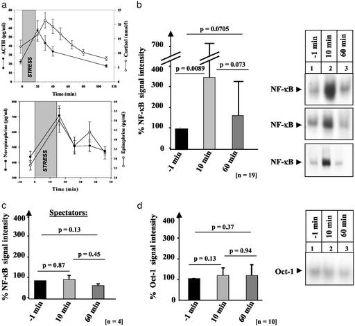
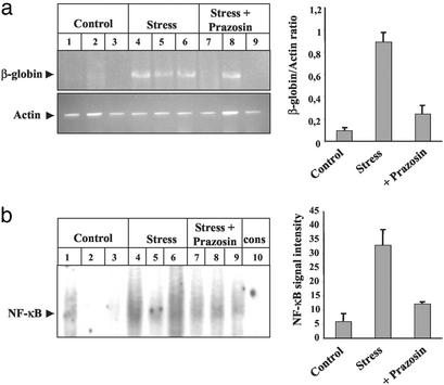
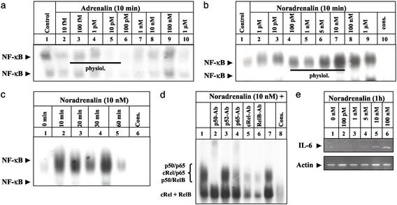
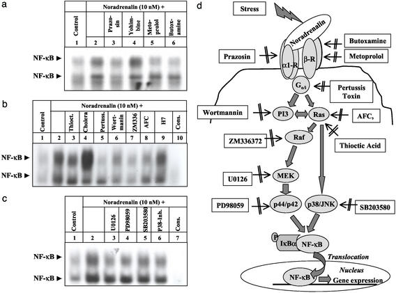
Little is known about the mechanisms converting psychosocial stress into cellular dysfunction. Various genes, up-regulated in atherosclerosis but also by psychosocial stress, are controlled by the transcription factor nuclear factor kappaB (NF-kappaB). Therefore, NF-kappaB is a good candidate to convert psychosocial stress into cellular activation. Volunteers were subjected to a brief laboratory stress test and NF-kappaB activity was determined in peripheral blood mononuclear cells (PBMC), as a window into the body and because PBMC play a role in diseases such as atherosclerosis. In 17 of 19 volunteers, NF-kappaB was rapidly induced during stress exposure, in parallel with elevated levels of catecholamines and cortisol, and returned to basal levels within 60 min. To model this response, mice transgenic for a strictly NF-kappaB-controlled beta-globin transgene were stressed by immobilization. Immobilization resulted in increased beta-globin expression, which could be reduced in the presence of the alpha1-adrenergic inhibitor prazosin. To define the role of adrenergic stimulation in the up-regulation of NF-kappaB, THP-1 cells were induced with physiological amounts of catecholamines for 10 min. Only noradrenaline resulted in a dose- and time-dependent induction of NF-kappaB and NF-kappaB-dependent gene expression, which depended on pertussis-toxin-sensitive G protein-mediated phosphophatidylinositol 3-kinase, Ras/Raf, and mitogen-activated protein kinase activation. Induction was reduced by alpha(1)- and beta-adrenergic inhibitors. Thus, noradrenaline-dependent adrenergic stimulation results in activation of NF-kappaB in vitro and in vivo. Activation of NF-kappaB represents a downstream effector for the neuroendocrine response to stressful psychosocial events and links changes in the activity of the neuroendocrine axis to the cellular response.


Figures

Figure 1
Figure 1
Psychosocial stress induces the transcription factor NF-κB in healthy volunteers undergoing the TSST. (a) ACTH, salivary cortisol (Upper) and AD and NA levels (Lower), expressed as mean ± SEM, of healthy volunteers exposed to the TSST (shaded area). (b and c) NF-κB-binding activity was monitored by EMSA in PBMC before (−1 min), immediately after (10 min), and 60 min after stress induction in 19 volunteers (b) and four spectators of the TSST (c) and evaluated by densitometry. The mean ± SEM is reported. (d) Ten occasionally selected nuclear extracts studied in b were assayed for Oct-1-binding activity and evaluated by densitometry. The mean ± SEM is reported.
Figure 2
Figure 2
Immobilization stress induces NF-κB-dependent gene expression in β-globin transgenic mice. β-globin transgenic mice were left untreated (lanes 1–3) or subjected to immobilization stress for 20 min in the absence (lanes 4–6) or presence (lanes 7–9) of the α1-adrenergic inhibitor prazosin, applied 45 min before immobilization. Three mice were used in each group. (a) Total RNA was prepared from blood and analyzed by RT-PCR for β-globin-transgene (Upper) and β-actin (Lower) transcription. Gel-separated PCR products were quantified by densitometry, and the ratio of β-globin/β-actin was calculated. (b) Nuclear extracts were prepared from the blood investigated above and analyzed for NF-κB-binding activity in EMSA. To confirm NF-κB binding, nuclear extract from an immobilized mouse was competed with a 160-fold molar excess of unlabeled NF-κB consensus oligonucleotides (lane 10). The bar graphs on the right summarize the results obtained in all mice studied. The mean ± SEM is reported.
Figure 3
Figure 3
Physiological concentrations of NA induce functionally significant NF-κB-binding activity in cultured THP-1 monocytic cells. (a and b) THP-1 cells were left untreated (lane 1) or incubated with increasing concentrations of AD (a) and NA (b) for 10 min (lanes 2–9) before NF-κB-binding activity was monitored. NF-κB binding was confirmed by competing with a 160-fold molar excess of unlabeled NF-κB consensus oligonucleotides (b; cons, lane 10). A black line indicates the physiological concentration range of AD and NA. The experiment was repeated two times with identical results and one representative experiment is shown. (c) THP-1 cells were either left untreated (0 h) or stimulated with NA (10 nM) for 10 min to 1 h (lanes 2- 5), and nuclear extracts were assayed for NF-κB-binding activity as above. The experiment was performed twice with identical results and one representative experiment is shown. (d) Characterization of the NF-κB subunits contributing to the NA-induced-binding activity at the NF-κB consensus sequence (lanes 1 and 7) was performed by including 2.5 μg of anti-p50 (lane 2), anti-p52 (lane 3), anti-p65 (lane 4), anti-cRel (lane 5), or anti-relB (lane 6) Abs in the binding reaction. Specificity of NF-κB binding was confirmed as above (lane 10). The position of the different NF-κB complexes formed is indicated at the left. (e) THP-1 cells were either left untreated (0 h) or stimulated with different concentrations of NA (lanes 2–6) for 1 h. Total RNA was prepared and analyzed by RT-PCR with primers specific for human IL-6 and β-actin, respectively.
Figure 4
Figure 4
Biochemical characterization of the signaling pathways involved in NA-dependent NF-κB activation in cultured THP-1 monocytic cells. THP-1 were left untreated (lane 1) or incubated with NA (10 nM) for 10 min in the absence (lane 2) or presence of either the adrenergic inhibitors prazosin (1 nM, lane 3), yohimbine (10 nM, lane 4), metoprolol (100 nM, lane 5), or butoxamine (25 nM, lane 6) (a), or the pathway inhibitors thioctic acid (2 mM, lane 3), cholera toxin (5 μg/ml, lane 4), Ptx (400 ng/ml, lane 5), wortmannin (100 nM, lane 6), ZM336372 (1 μM, lane 7), AFC (50 μM, lane 8), and H7 (100 μM, lane 9) (b), and the MEK and MAPK inhibitors U0126 (50 μM, lane 3), PD98059 (30 μM, lane 4), SB203580 (20 nM, lane 5), and p38 inhibitor (10 μM, lane 6) (c), respectively. Inhibitors were added to the cells 45 min before NA induction. Nuclear extracts were assayed for NF-κB-binding activity, monitored in EMSA. Specificity of NF-κB binding was confirmed as above (cons). The experiments were repeated three (b and c) to five (a) times with identical results and confirmed by NF-κBp65-specific ELISA. One representative experiment is shown. (d) Schematic representation of the proposed mechanism of psychosocial stress-induced NF-κB activation. Psychosocial stress induces NA that binds to α1- and β-adrenergic receptors, which in turn recruit Ptx-sensitive G proteins. G proteins activate directly or indirectly via PI3-kinase Ras interacting with its effector kinase Raf subsequently. Raf phosphorylates MEK-1 and -2, which activates p44/p42-MAPK. In addition, Ras, which is a target of cellular oxidative stress, can directly induce p38-MAPK activation. Activated MAPKs induce as-yet uncharacterized downstream-located signaling pathways that result in phosphorylation and degradation of the NF-κB-specific cytoplasmic inhibitor IκBα and subsequent activation and nuclear translocation of NF-κB. The inhibitors used to identify different steps in the signaling cascades are given in boxes.

References

    1. Rozanski A, Blumenthal J A, Kaplan J. Circulation. 2000;99:2192–2217. - PubMed
    1. Kaprio J, Koskenvuo M, Rita H. Am J Public Health. 1987;77:283–287. - PMC - PubMed
    1. Leor J, Poole W K, Kloner R A. N Engl J Med. 1996;334:413–419. - PubMed
    1. Meisel S R, Kutz I, Dayan K I, Pauzner H, Chetboun I, Arbel Y, David D. Lancet. 1991;338:660–661. - PubMed
    1. Stansfeld S A, Fuhrer R, Shipley M J, Marmot M G. Int J Epidemiol. 2002;31:248–255. - PubMed

Publication types

MeSH terms