. 2003 Feb 18;100(4):2157-62.
doi: 10.1073/pnas.0337195100. Epub 2003 Feb 10.

Multiple routes to memory: distinct medial temporal lobe processes build item and source memories

Affiliations

Multiple routes to memory: distinct medial temporal lobe processes build item and source memories

Lila Davachi et al. Proc Natl Acad Sci U S A. .

Abstract

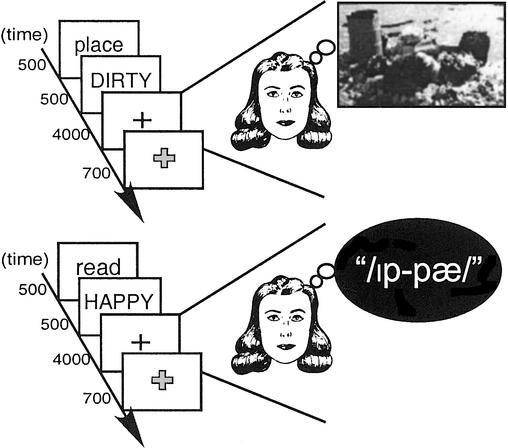
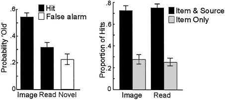
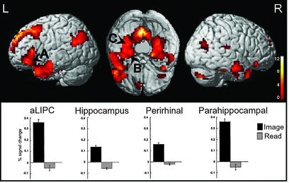
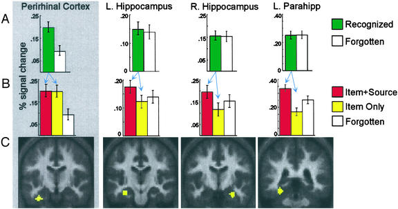
A central function of memory is to permit an organism to distinguish between stimuli that have been previously encountered and those that are novel. Although the medial temporal lobe (which includes the hippocampus and surrounding perirhinal, parahippocampal, and entorhinal cortices) is known to be crucial for recognition memory, controversy remains regarding how the specific subregions within the medial temporal lobe contribute to recognition. We used event-related functional MRI to examine the relation between activation in distinct medial temporal lobe subregions during memory formation and the ability (i) to later recognize an item as previously encountered (item recognition) and (ii) to later recollect specific contextual details about the prior encounter (source recollection). Encoding activation in hippocampus and in posterior parahippocampal cortex predicted later source recollection, but was uncorrelated with item recognition. In contrast, encoding activation in perirhinal cortex predicted later item recognition, but not subsequent source recollection. These outcomes suggest that the subregions within the medial temporal lobe subserve distinct, but complementary, learning mechanisms.


Figures

Figure 1
Figure 1
Encoding conditions performed during fMRI scanning are illustrated.
Figure 2
Figure 2
Subsequent memory is plotted according to overall item recognition and source recollection. (Left) Item recognition was superior after Image than after Read encoding; Read encoding was higher than the false alarm rate. (Right) For recognized items, source recollection after Image and Read trials was comparable.
Figure 3
Figure 3
Statistical maps displayed on a canonical 3D brain reveals regions demonstrating greater activation during Image than during Read trials. Critically, MTL and prefrontal regions demonstrated a greater response during Image compared with Read encoding. Regions included anterior left inferior frontal cortex (A: −54, 24, 3, ≈BA 47/45), left hippocampus (B: −33, −21, −21), left perirhinal cortex (B: −36, −12, −33, ≈BA 35/36), and left parahippocampal cortex (C: −33, −39, −18, ≈BA 37); peak responses did not fall above baseline during Read encoding. Other regions engaged during Image encoding included right MTL and bilateral retrosplenial cortices (complete coordinates are available on request). Note that activations are displayed on left- and right-hemisphere views, as well as on a cerebellumless ventral view (occipital lobe facing top).
Figure 4
Figure 4
Peak signal change during Image encoding revealed qualitatively different activation patterns in left perirhinal cortex (−36, −12, −33), bilateral hippocampus (−33, −21, −21 and 30, −9, −24), and left parahippocampal cortex (−33, −39, −18). (A) Encoding activation, sorted according to whether the item was later recognized (irrespective of source recollection outcome) or forgotten, revealed dissociations between perirhinal cortex and each hippocampal region and between perirhinal and parahippocampal cortices. Perirhinal cortex demonstrated greater activation for items later recognized relative to those later forgotten, whereas neither hippocampus nor parahippocampal cortex differed according to item memory. (B) Time course analyses revealed a main effect of source memory outcome (Item + Source > Item Only) in right and left hippocampus and parahippocampal cortex, but not in perirhinal cortex. As displayed, peak signal change paralleled this pattern. (C) MTL regions of interest rendered on group-averaged structural images; anatomical localization of these ROIs to perirhinal cortex, hippocampus, and parahippocampal cortex, respectively, was confirmed at the individual-subject level.

Similar articles

Cited by

References

    1. Scoville W B, Milner B. J Neurol Neurosurg Psychiatry. 1957;20:11–21. - PMC - PubMed
    1. Squire L R. Psychol Rev. 1992;99:195–231. - PubMed
    1. Schacter D L, Wagner A D, Buckner R L. In: The Oxford Handbook of Memory. Tulving E, Craik F I M, editors. New York: Oxford Univ. Press; 2000. pp. 627–643.
    1. Cohen N J, Eichenbaum H E. Memory, Amnesia, and the Hippocampal System. Cambridge, MA: MIT Press; 1993.
    1. McGaugh J L. Science. 2000;287:248–251. - PubMed

Publication types