. 2003 Feb 18;100(4):2082-7.
doi: 10.1073/pnas.0435872100. Epub 2003 Feb 10.

Visualization of synaptotagmin I oligomers assembled onto lipid monolayers

Affiliations

Visualization of synaptotagmin I oligomers assembled onto lipid monolayers

Yi Wu et al. Proc Natl Acad Sci U S A. .

Abstract

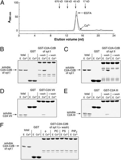
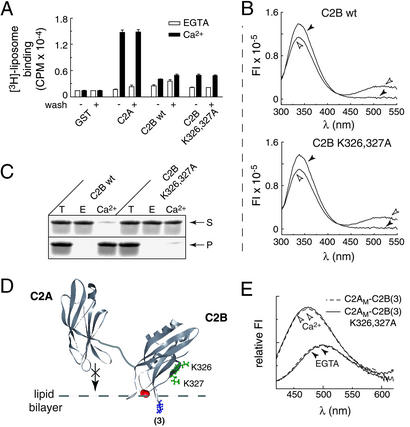
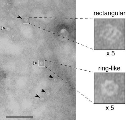
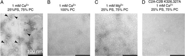
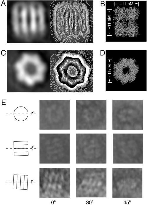
Neuronal exocytosis is mediated by Ca(2+)-triggered rearrangements between proteins and lipids that result in the opening and dilation of fusion pores. Synaptotagmin I (syt I) is a Ca(2+)-sensing protein proposed to regulate fusion pore dynamics via Ca(2+)-promoted binding of its cytoplasmic domain (C2A-C2B) to effector molecules, including anionic phospholipids and other copies of syt. Functional studies indicate that Ca(2+)-triggered oligomerization of syt is a critical step in excitation-secretion coupling; however, this activity has recently been called into question. Here, we show that Ca(2+) does not drive the oligomerization of C2A-C2B in solution. However, analysis of Ca(2+).C2A-C2B bound to lipid monolayers, using electron microscopy, revealed the formation of ring-like heptameric oligomers that are approximately 11 nm long and approximately 11 nm in diameter. In some cases, C2A-C2B also assembled into long filaments. Oligomerization, but not membrane binding, was disrupted by neutralization of two lysine residues (K326,327) within the C2B domain of syt. These data indicate that Ca(2+) first drives C2A-C2B.membrane interactions, resulting in conformational changes that trigger a subsequent C2B-mediated oligomerization step. Ca(2+)-mediated rearrangements between syt subunits may regulate the opening or dilation kinetics of fusion pores or may play a role in endocytosis after fusion.


Figures

Figure 1
Figure 1
Highly purified fragments of syt I–III and VII fail to oligomerize in response to Ca2+. (A) Size-exclusion FPLC was performed using an AKTA Explorer (Amersham Pharmacia Biotech) with a Superdex 200 column (Pharmacia). One-hundred-microliter samples of C2A-C2B from syt I (1.0 μg/μl, after nuclease/salt treatment) plus 1 mM Ca2+ or 1 mM EGTA in Tris-buffered saline were run through the column at a flow rate of 0.5 ml/min. (B) Ten-microgram GST or GST-C2A-C2B from syt I, with (+ wash) or without (−wash) nuclease/salt treatment, was immobilized on glutathione-Sepharose beads. Beads were incubated with 2 μM soluble C2A-C2B syt I for 1.5 h in 150 μl of Hepes buffer plus 0.5% Triton X-100 and either 2 mM EGTA or 1 mM Ca2+. Beads were washed three times with binding buffer and boiled in SDS sample buffer. Seven percent of the total (left two lanes) and 30% of the bound material (remaining lanes) were subjected to SDS/PAGE and visualized by staining with Coomassie blue. (C) Binding of soluble C2A-C2B from syt I (3 μM) to immobilized C2A-C2B from syt II was assayed as described in B. (D) Binding assays were carried out as in B but using C2A-VII. (E) Binding assays were carried out as in B but using C2A-III. (F) Lipid/detergent micelles containing 70 μM PC or PS or PIP2 in 1% Triton X-100 (≈98 μM micelles) were included in the binding assay described in A; rescue of oligomerization was not observed under these conditions.
Figure 2
Figure 2
Isolated C2B does not bind PS/PC membranes with high affinity, but cosediments with liposomes via a secondary mechanism. (A) C2A-C2B, C2A, and C2B domains from syt I were immobilized as GST-fusion proteins. Samples with (+wash) or without (−wash) nuclease/salt treatment (6 μg of protein per data point) to remove bacterial contaminants were assayed for binding 3H-labeled liposomes, (≈0.24 mM 25% PS/75% PC) in 100 μl of Tris-buffered saline buffer as described (38), in either 2 mM EGTA or 0.2 mM Ca2+. Bound liposomes were quantified by liquid scintillation counting. (B) Wild-type and K326,327A mutant C2B bind PS-containing membranes to the same extent as measured by using fluorescence resonance energy transfer. Liposomes composed of 5% 1,2-dioleoyl-sn-glycero-3-phosphoethanolamine-N-(5-dimethylamino-1-naphthalenesulfulfonyl) (dansyl-PE)/25% PS/70% PC were prepared as described (38). Native tryptophan (Trp) residues in C2B (amino acids 390 and 404) served as the energy donors and were excited at 285 nm. The emission spectra of the Trp residues (emission λMAX ≈ 337 nm) and the dansyl-PE acceptor (emission λMAX ≈ 514 nm) were collected from 300 to 550 nm. Proteins (2 μM) were mixed with 11 nM liposomes in either 2 mM EGTA (filled arrowheads) or 0.2 mM Ca2+ (open arrowheads). Fluorescence resonance energy transfer occurred in response to Ca2+ (Upper), indicating some degree of binding (32); however, the Ca2+-binding loops of isolated C2B do not insert into lipid bilayers (31). The K326,327A mutation had no effect on the weak interaction of C2B with membranes (Lower). (C) Cosedimentation of C2B with liposomes is not directly mediated by C2B⋅membrane interactions. Sedimentation assays were carried out as described in Materials and Methods. Thirteen percent of the supernatant (S), pellet (P), and total (T) reaction were subjected to SDS/PAGE and stained with Coomassie blue. Wild-type isolated C2B translocated from the supernatant to the pellet fraction in response to Ca2+; neutralization of two lysine residues (K326,327A) abolished sedimentation without affecting membrane binding. (D) Molecular model depicting the Ca2+-triggered penetration of C2B, in the context of C2AM-C2B, into lipid bilayers. C2B penetrates lipid bilayers when tethered to C2A or C2AM (31). The subscript “M” corresponds to D230,232N substitutions that disrupt C2A⋅membrane penetration (, 58). A 5-[[2-(acetylamino)ethyl]amino] napthalene-1-sulfonic acid (AEDANS) probe was placed in the Ca2+-binding loop 3 of C2B [indicated as (3)] as described (31) to monitor membrane penetration of C2B in the context of C2AM-C2B (31). Lysines 326 and 327 are shown in green; solution structures of C2A (46) and C2B (32) from syt I were rendered in WEBLAB VIEWER LITE (Molecular Simulations)]; the flexible linker that connects them (8) was added by using a drawing program. (E) Liposomes (11 nM; 25% PS/75% PC) and C2AM-C2B (3) (0.5 μM; dashed line) were incubated in Hepes buffer in the presence of 0.1 mM EGTA. AEDANS fluorescence was excited at 336 nm, and the emission spectra were collected from 420 to 600 nm. Ca2+ was then added to a final free concentration of 0.5 mM, and spectra were obtained again. Neutralization of K326,327A (solid line) abolishes oligomerization (Fig. 4D) but does not alter the membrane penetration activity of C2B in the context of C2AM-C2B.
Figure 3
Figure 3
Ca2+-triggered assembly of the cytoplasmic domain of syt I into oligomers on PS/PC monolayers. Rectangular C2A-C2B oligomers are marked with filled arrowheads; ring-like forms are marked with open arrowheads. Examples of individual rectangular and ring-like C2A-C2B oligomers are also shown at ×5 magnification. Lipid monolayers were composed of 25% PS/75% PC; [Ca2+] was 50 μM. (Bar = 100 nm.)
Figure 4
Figure 4
Cation, lipid, and structural requirements for assembly of C2A-C2B-I into oligomers. (A) The image was obtained as described in Fig. 3, except 1 mM Ca2+ was used. (B) Conditions were as in A, except PS was omitted. (C) Conditions were as in A, except 1 mM Ca2+ was replaced with 1 mM Mg2+. (D) Conditions were as in A, except that lysines 326 and 327, which are critical for the oligomerization of C2A-C2B and C2B that harbor contaminants (Fig. 1 A and B) (11, 45), were neutralized by substitution with alanines. This mutation does not affect membrane binding (Fig. 2E) but abolished assembly of oligomeric particles in the presence of 1 mM Ca2+ and 25% PS. Monomeric C2A-C2B K326,327A bound to membranes is below the resolution of our EM assay and was not observed.
Figure 5
Figure 5
Single-particle analysis of syt oligomers. (A) Average maps of the rectangular particles before (Left) and after (Right) contouring. Within a bar, the two electron-dense regions probably correspond to C2A and C2B. At this level of resolution, we cannot discriminate between C2A and C2B, but, because C2B appears to drive oligomerization (Fig. 4D), it is likely that each syt subunit is arranged in a parallel manner. In this case, the membrane anchors of native syt would emerge from the end of the oligomer that contains C2A. (B) Molecular model in which the crystal structure of C2A-C2B from syt III (8) has been packed into a heptameric oligomer; the heptamer is viewed from the side, where four C2A-C2B subunits, vertically oriented, are visible. In this model, the structures of the C2 domains were not altered, but the tandem C2 domains were aligned along their long axes by changing the flexible linker that connects them. (C) Average map of the ring-like particles before (Left) and after (Right) contouring. (D) Top view of the model shown in B; the ends of all seven subunits are visible. (E) Tilt analysis of heptameric particles. In each set of images, a single particle is tilted through 0, 30, and 45° about the axis indicated by the line drawing on the left. (Top) A ring-like particle is analyzed. (Middle and Bottom) Two rectangular particles in the indicated orientations are analyzed.

Similar articles

Cited by

References

    1. Katz B. The Release of Neural Transmitter Substances. Springfield, IL: Thomas; 1969.
    1. Lindau M, Almers W. Curr Opin Cell Biol. 1995;7:509–517. - PubMed
    1. Augustine G J. Curr Opin Neurobiol. 2001;11:320–326. - PubMed
    1. Takei K, Haucke V. Trends Cell Biol. 2001;11:385–391. - PubMed
    1. Chapman E R. Nat Rev Mol Cell Biol. 2002;3:498–508. - PubMed

Publication types