. 2003 Feb 18;100(4):1475-80.
doi: 10.1073/pnas.0437746100. Epub 2003 Feb 10.

Regulatory gene networks and the properties of the developmental process

Collaborators, Affiliations

Regulatory gene networks and the properties of the developmental process

Eric H Davidson et al. Proc Natl Acad Sci U S A. .

Abstract

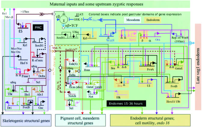
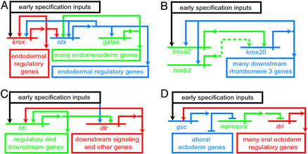
Genomic instructions for development are encoded in arrays of regulatory DNA. These specify large networks of interactions among genes producing transcription factors and signaling components. The architecture of such networks both explains and predicts developmental phenomenology. Although network analysis is yet in its early stages, some fundamental commonalities are already emerging. Two such are the use of multigenic feedback loops to ensure the progressivity of developmental regulatory states and the prevalence of repressive regulatory interactions in spatial control processes. Gene regulatory networks make it possible to explain the process of development in causal terms and eventually will enable the redesign of developmental regulatory circuitry to achieve different outcomes.


Figures

Figure 1
Figure 1
Central portion of the Strongylocentrotus purpuratus embryo endomesoderm GRN, from fertilization to just before gastrulation. The diagram is a recent version of that initially presented in refs. –. Suspected interactions at the cis-regulatory elements represented by the horizontal lines are shown, irrespective of when in the 0- to 30-h period or where in the embryo they are expected to occur [a “view from the genome” GRN (24); for interactions occurring only in given domains and at given periods see ref. and www.its.caltech.edu/∼mirsky/endomes.htm]. Transcriptional regulatory interactions are shown in the indicated spatial domains of the embryo: pmc domain, the skeletogenic micromere lineage; endomes domain, endomesoderm descendant from the sixth cleavage ring of eight “veg2” cells (2, 13, 24). Transcriptional inputs into the cis-regulatory elements of each named gene are indicated by arrows (activation, or permissive of activation) or bars (repression). Outputs from each gene (where known) are indicated by color-coded lines emanating from the bent arrows that symbolize transcription. For evidence see text, refs. –, , , and , and www.its.caltech.edu/∼mirsky/endomes.htm. An arrowhead inserted in an arrow tail indicates an intercellular signaling interaction; small open circles indicate cytoplasmic interactions or specific events off the DNA, e.g., that by which the Soxb1 factor interferes with nuclearization of β-catenin (26). For further details see refs. and and www.its.caltech.edu/∼mirsky/endomes.htm.
Figure 2
Figure 2
Experimental tests of GRN predictions regarding the pmar1 gene. (A) Diagrams of cell transplantation experiments. (Upper) Transplantation of micromere. (Lower) Transplantation of ectoderm precursor cell, or mesomere. Sixteen cell-stage embryos are shown. The red color symbolizes injected constituents, here cadherin mRNA or cadherin mRNA plus pmar1 mRNA, originally introduced into the egg from which the donor fourth-cleavage embryo develops. The micromeres were removed by microsurgery from the recipient embryo and replaced with a micromere or mesomere from the donor embryo, and the embryos were then cultured for 48 h (BD). (B) Replacement with a micromere expressing cadherin mRNA. (C) Replacement with a micromere expressing cadherin plus pmar1 mRNAs. (D) Replacement with a mesomere expressing cadherin plus pmar1 mRNAs as in C. The green stain identifies Msp130, a skeletogenic cell-specific protein, by fluorescence immunocytology. The red stain in D similarly identifies β-catenin to mark the cell junctions. AD are adapted from ref. . (E) Normal localization of sm50 gene expression in skeletogenic pmcs by whole-mount in situ hybridization. (F) Global expression of sm50 in embryo grown from an egg into which pmar1 mRNA had been injected. E and F are adapted from ref. .
Figure 3
Figure 3
Common GRN architectural feature: intergenic reinforcing loops that drive developmental state forward. (A) Relation among krox, otx, and gatae genes from the sea urchin endomesoderm gene network (Fig. 1). (B) Relation between hoxa2 and krox20 in mouse rhombomere specification (from ref. , after refs. –31). (C) Relations between trachealess (trh) and drifter (dfr) regulatory genes (from ref. , after ref. 32). (D) Relation between goosecoid (gsc) gene and deadringer (dri) gene in sea urchin oral ectoderm GRN (G. Amore and E.H.D., unpublished data).

References

    1. Davidson E H. Genomic Regulatory Systems: Development and Evolution. San Diego: Academic; 2001.
    1. Davidson E H, Cameron R A, Ransick A. Development (Cambridge, UK) 1998;125:3269–3290. - PubMed
    1. Angerer L M, Angerer R C. Dev Biol. 2000;218:1–12. - PubMed
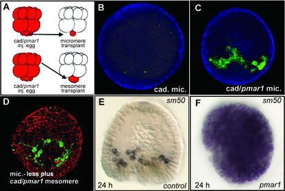
    1. Logan C Y, Miller J R, Ferkowicz M J, McClay D R. Development (Cambridge, UK) 1999;126:345–357. - PubMed
    1. Sherwood D R, McClay D R. Development (Cambridge, UK) 1999;126:1703–1713. - PubMed

Publication types

LinkOut - more resources