. 2003 Feb 18;100(4):1949-54.
doi: 10.1073/pnas.0437901100. Epub 2003 Feb 10.

Hypersusceptibility of cystic fibrosis mice to chronic Pseudomonas aeruginosa oropharyngeal colonization and lung infection

Affiliations

Hypersusceptibility of cystic fibrosis mice to chronic Pseudomonas aeruginosa oropharyngeal colonization and lung infection

Fadie T Coleman et al. Proc Natl Acad Sci U S A. .

Abstract

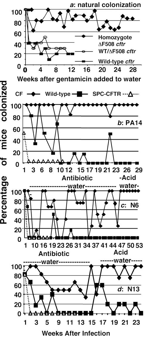
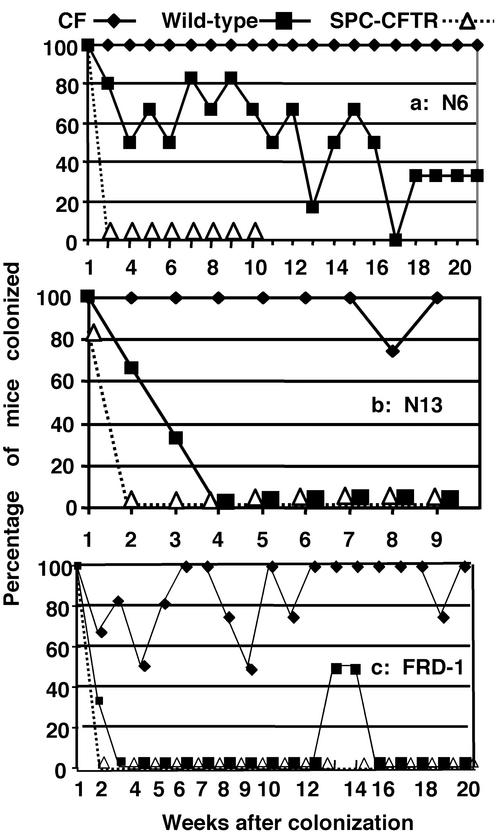
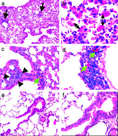
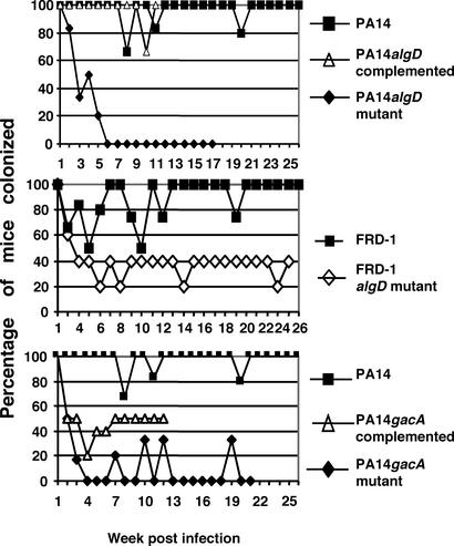
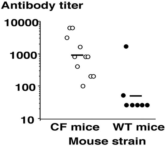
No transgenic cystic fibrosis (CF) mouse model developed to date mimics the major clinical phenotype found in humans with CF, chronic Pseudomonas aeruginosa lung infection. In a transgenic CF transmembrane conductance regulator (cftr) mouse colony, we found WT, heterozygous, and homozygous CF mice housed in the same cage became chronically colonized in the oropharynx with environmental P. aeruginosa when the bacterium was present in drinking water. Elimination of P. aeruginosa from drinking water resulted in clearance in most WT and CF heterozygous, but not homozygous mice. For experimental evaluation, a combination of specific animal husbandry techniques and an oral infection route showed cftr(-/-) mice but not WT mice can be chronically colonized by P. aeruginosa with subsequent lung translocation, yielding a pathologic picture indicative of chronic lung infection. In some instances, mucoid isolates of P. aeruginosa were recovered from lungs, indicating conditions were present for conversion to mucoidy. Overexpression of human CFTR in the lungs of WT mice markedly accelerated the clearance rate of P. aeruginosa, demonstrating that lung levels of CFTR play an important role in defense against infection. P. aeruginosa mutants unable to express the surface polysaccharide alginate or the global regulator GacA were deficient in their ability to colonize the mice. CF mice made potent immune responses to P. aeruginosa outer membrane antigens. Overall, we found that under the proper conditions, transgenic CF mice are hypersusceptible to P. aeruginosa colonization and infection and can be used for evaluations of lung pathophysiology, bacterial virulence, and development of therapies aimed at treating CF lung disease.


Figures

Figure 1
Figure 1
Natural and experimental oropharyngeal colonization of WT or transgenic CF mice by P. aeruginosa. (a) Percentage of mice with the indicated genotype having positive throat cultures of P. aeruginosa initially acquired from contaminated drinking water after the addition of 0.1 mg of gentamicin per ml (arrow) to control bacterial growth in drinking water. (b) Oropharyngeal colonization of P. aeruginosa PA14 in transgenic CF and WT mice, and transgenic SP-C-CFTR mice. (c and d) Colonization of WT, FABP-CFTR, or SP-C-CFTR transgenic mice with P. aeruginosa clinical isolates, N6 and N13, switched from antibiotic water to acid water as indicated. Six to eight mice per group were used initially, although over time some animals died, usually with P. aeruginosa cultured from the lung.
Figure 2
Figure 2
Colonization of WT, FABP-CFTR, or SP-C-CFTR transgenic mice by different strains of P. aeruginosa: N6 (a), N13 (b), and FRD-1 (c). Colonization experiments were done with acidified water to prevent growth of P. aeruginosa.
Figure 3
Figure 3
Histopathologic analysis of lung sections from CF mice with oropharyngeal and/or lung infection with P. aeruginosa. All sections were stained with hematoxylin and eosin. (a) Lung section from an animal with oropharyngeal but not lung infection, showing abundant intraalveolar macrophages and patchy lymphoid aggregates (black arrows) located along lymphatic pathways (original magnification, ×40). (b) Lung section from an animal with oropharyngeal but not lung infection showed increased intraalveolar macrophages (double-headed arrows, original magnification ×100). (c and d) Lung sections from two animals with both oropharyngeal and lung infection with P. aeruginosa. (c) Hyperplastic bronchus-associated lymphoid tissue (BALT) (black arrows); hemosiderin within the BALT stands out as a golden-brown pigment (green arrow, original magnification ×40). (d) Hemosiderin (green arrows) in perilymphatic lymphoid aggregates within the lung (original magnification ×100). (e and f) Lung section from an uninfected CF mouse. No specific pathologic features were noted (original magnifications ×40 and ×100, respectively).
Figure 4
Figure 4
Colonization of FABP-CFTR mice by the strain of P. aeruginosa indicated next to each graph. Colonization experiments were done with antibiotics for strain PA14 and acid water for strain FRD-1. Six to eight mice per group were used initially.
Figure 5
Figure 5
Antibody titers achieved in sera of CF or WT mice after oropharyngeal colonization for >10 weeks with P. aeruginosa. Each symbol represents the titer for one mouse; bar represents geometric mean.

References

    1. Grubb B R, Boucher R C. Physiol Rev. 1999;79:S193–S214. - PubMed
    1. Davidson D J, Dorin J R, Mclachlan G, Ranaldi V, Lamb D, Doherty C, Govan J, Porteous D J. Nat Genet. 1995;9:351–357. - PubMed
    1. Colledge W H, Abella B S, Southern K W, Ratcliff R, Jiang C W, Cheng S H, Macvinish L J, Anderson J R, Cuthbert A W, Evans M J. Nat Genet. 1995;10:445–452. - PubMed
    1. Zeiher B G, Eichwald E, Zabner J, Smith J J, Puga A P, Mccray P B, Capecchi M R, Welsh M J, Thomas K R. J Clin Invest. 1995;96:2051–2064. - PMC - PubMed
    1. Van Doorninck J H, French P J, Verbeek E, Peters R H P C, Morreau H, Bijman J, Scholte B J. EMBO J. 1995;14:4403–4411. - PMC - PubMed

Publication types

MeSH terms