. 2003 Feb;2(1):49-61.
doi: 10.1128/EC.2.1.49-61.2003.

Cell biology of mating in Candida albicans

Affiliations

Cell biology of mating in Candida albicans

Shawn R Lockhart et al. Eukaryot Cell. 2003 Feb.

Abstract

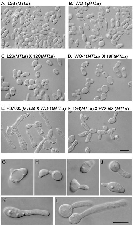
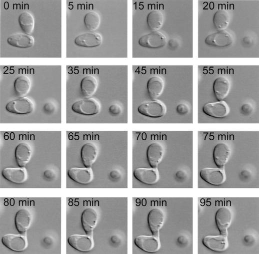
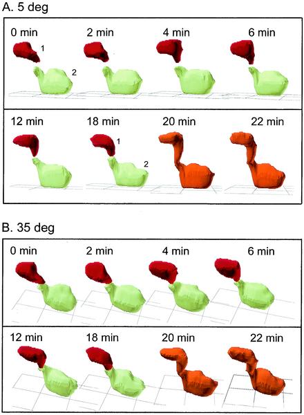
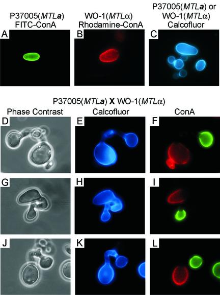
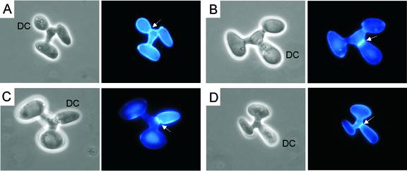
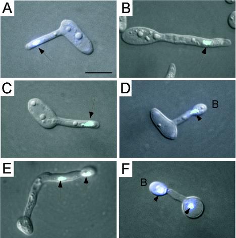
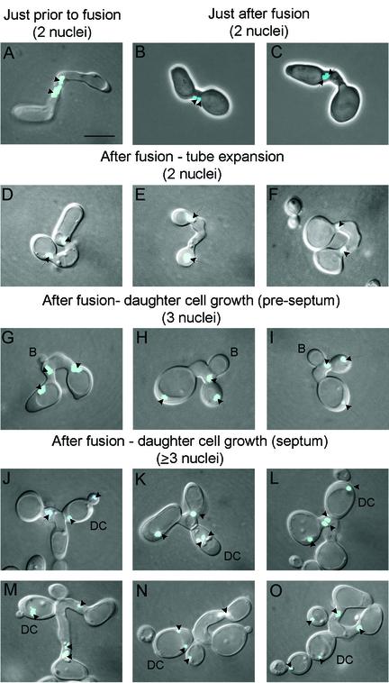
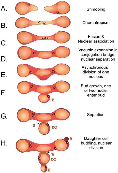
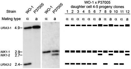
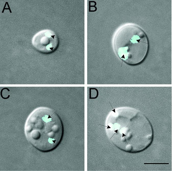
It was recently demonstrated that strains homozygous for either of the mating type-like loci MTLa and MTLalpha of Candida albicans undergo white-opaque switching and that expression of the opaque-phase phenotype greatly enhances mating between strains. Exploiting the latter property to obtain high-frequency mating, we have characterized the cell biology of the mating process of C. albicans. Employing continuous videomicroscopy, computer-assisted three-dimensional reconstruction of living cells, and fluorescence microscopy, we have monitored the mating-associated processes of conjugation, tube formation, fusion, budding, septum formation, and daughter cell development and the spatial and temporal dynamics of nuclear migration and division. From these observations, a model for the stages in C. albicans mating is formulated. The stages include shmooing, chemotropism of conjugation tubes, fusion of tubes and nuclear association, vacuole expansion and nuclear separation in the conjugation bridge, asynchronous nuclear division in the zygote, bud growth, nuclear migration into the daughter cell, septation, and daughter cell budding. Since there was no cytological indication of karyogamy, genetic experiments were performed to assess marker segregation. Recombination was not observed, suggesting that mating takes place in the absence of karyogamy between naturally occurring, homozygous a and alpha strains. This study provides the first description of the cell biology of the mating process of C. albicans.


Figures

FIG. 1.
FIG. 1.
Tube formation occurs exclusively in mixtures of MTLa and MTLα cells. Cells were imaged through DIC optics. (A) Homogeneous culture of strain L26 (MTLa) opaque-phase cells; (B) homogeneous culture of WO-1 (MTLα) opaque-phase cells; (C) mixture of L26 (MTLa) and 12C (MTLa) opaque-phase cells (50:50); (D) mixture of WO-1 (MTLα) and 19F (MTLα) opaque-phase cells (50:50); (E) mixture of P37005 (MTLa) and WO-1 (MTLα) opaque-phase cells (50:50); (F) mixture of L26 (MTLa) and P78048 (MTLα) opaque-phase cells; (G to J) higher-magnification images of incipient tube formation in mixtures of MTLa and MTLα opaque-phase cells; (K and L) tube formation in mixtures of MTLa and MTLα opaque-phase cells. Scale bars, 5 μm.
FIG. 2.
FIG. 2.
Sequence of video images of two cells undergoing fusion in a mixture of MTLa (P37005) and MTLα (WO-1) opaque-phase cells. Cells were imaged through DIC optics. Fusion occurred at 65 min.
FIG. 3.
FIG. 3.
3D-DIAS reconstruction of fusion between two cells in a clump of cells from a mixture of MTLa (P37005) and MTLα (WO-1) cells. The red cell (cell 1) was high up in the clump of cells while the green cell (cell 2) was at the bottom of the clump. Nonfusing cells in the clump were not reconstructed. After fusion occurred, the zygote was color-coded orange. Reconstructed cells are viewed from angles of 5° (A) and 35° (B) relative to the substratum (grid). deg, degree.
FIG. 4.
FIG. 4.
Demonstration by ConA staining that only MTLa and MTLα cells fuse. MTLa cells (strain P37005) were vitally stained with FITC-ConA, MTLα cells were vitally stained with rhodamine-ConA, and the cells were mixed. Fusants were subsequently stained with calcofluor. (A) Unfused MTLa cell stained with FITC-ConA; (B) Unfused MTLα cell stained with rhodamine-ConA; (C) unfused cells stained with calcofluor; (D, G, and J) phase-contrast images of zygotes; (E, H, and K) fluorescent images of calcoflour-stained zygotes; (F, I, and L) fluorescent images of rhodamine-ConA-FITC-ConA-labeled zygotes. Of 100 zygotes, 100% were combinations of green-red, 0% were green-green, and 0% were red-red. Scale bar, 5 μm.
FIG. 5.
FIG. 5.
Daughter cell development and septum formation in C. albicans zygotes. Zygotes formed by MTLa and MTLα crosses were stained with calcofluor to visualize septa. Each of the four examples (A through D) includes a phase-contrast image and a fluorescent image. Arrows point to septa. DC, daughter cell. Scale bar, 5 μm.
FIG. 6.
FIG. 6.
Nuclear distribution in tubes formed by cells in mixed MTLa and MTLα cultures. Nuclei were stained with Hoechst 33342. Fluorescent images of nuclei (blue) are superimposed on phase-contrast images. Arrows point to nuclei. B, terminal bud that forms at the tube apex. Scale bar, 5 μm.
FIG. 7.
FIG. 7.
Nuclear configurations during the different phases of mating. Nuclei were stained with Hoechst 33342. Fluorescent images of nuclei (blue) are superimposed on DIC images. Arrows point to nuclei. B, bud; DC, daughter cell (after septation). Scale bar, 5 μm.
FIG. 8.
FIG. 8.
Model of nuclear dynamics during zygote and daughter cell formation. The consensus sequence of events was interpreted by reviewing a number of mixed cultures of MTLa and MTLα cells. Vacuoles are light brown, and nuclei are dark brown. B, bud; S, septum; DC, daughter cell.
FIG. 9.
FIG. 9.
Examples of Southern blot patterns of WO-1 (MTLα) and P37005 (MTLa) and progeny of a cross probed with both URA3 and NIK1. (A) Southern blots. Note two URA3 bands and one NIK1 band for WO-1 and one URA3 band and two NIK1 bands for P37005. (B) Models of the banding patterns and allelism of the mating type loci of 12 progeny clones of daughter cell 4-5 (Table 3) picked from a cross between WO-1 and P37005. Note that all MTLα progeny exhibited the URA3 and NIK1 banding patterns of the MTLα parent strain and that all MTLa progeny exhibited the banding patterns of the MTLa parent strain. Numbers to the left of the blot are molecular sizes (in kilobases).
FIG. 10.
FIG. 10.
Multinucleated cells from a cross between strain L26 (MTLa) and strain WO-1 (MTLα), of which the mixture was first incubated in modified Lee's medium until fusion occurred (but in which daughter cells did not have a chance to form) and then incubated overnight in sporulation medium. Under these conditions, 20% of the daughter cells were multinucleated. (A to C) Examples of cells with two nuclei; (D) example of a cell with four nuclei. Scale bar, 5 μm.

Similar articles

Cited by

References

    1. Anderson, J. M., and D. R. Soll. 1987. Unique phenotype of opaque cells in the white-opaque transition of Candida albicans. J. Bacteriol. 169:5579-5588. - PMC - PubMed
    1. Bedell, G. W., and D. R. Soll. 1979. Effects of low concentrations of zinc on the growth and dimorphism of Candida albicans: evidence for zinc-resistant and -sensitive pathways for mycelium formation. Infect. Immun. 26:348-354. - PMC - PubMed
    1. Blignaut, E., C. Pujol, S. Lockhart, S. Joly, and D. R. Soll. 2002. Ca3 fingerprinting of Candida albicans isolates from human immunodeficiency virus-positive and healthy individuals reveals a new clade in South Africa. J. Clin. Microbiol. 40:826-836. - PMC - PubMed
    1. Byers, B. 1981. Cytology of the yeast life cycle, p. 59. In J. N. Strethern (ed.), The molecular biology of the yeast Saccharomyces: life cycle and inheritance. Cold Spring Harbor Laboratory Press, Cold Spring Harbor, N.Y.
    1. Byers, B., and L. Goetsch. 1975. Behavior of spindles and spindle plaques in the cell cycle and conjugation in Saccharomyces cerevisiae. J. Bacteriol. 124:511-523. - PMC - PubMed

Publication types