

Nicotine activates immature "silent" connections in the developing hippocampus
- PMID: 12582205
- PMCID: PMC149958
- DOI: 10.1073/pnas.0437947100
Nicotine activates immature "silent" connections in the developing hippocampus
Abstract
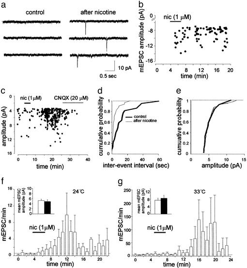
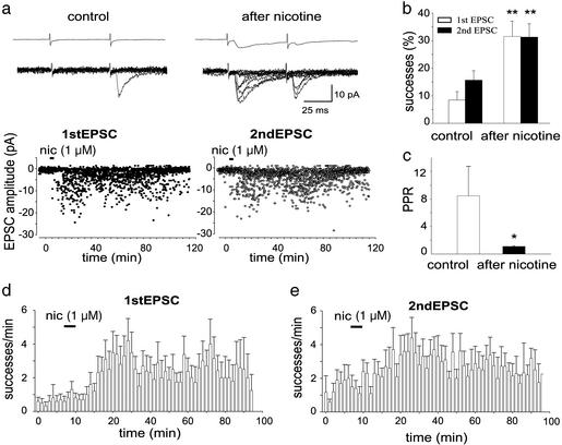
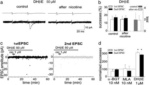
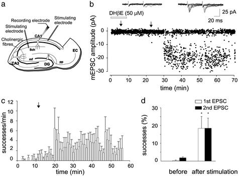
In the hippocampus at birth, most glutamatergic synapses are immature and functionally "silent" either because the neurotransmitter is released in insufficient amount to activate low-affinity alpha-amino-3-hydroxy-5-methyl-4-isoxazole propionate receptors or because the appropriate receptor system is missing or nonfunctional. Here we show that, in the newborn rat, a brief application of nicotine at immature Schaffer collateral-CA1 connections strongly enhances neurotransmitter release and converts presynaptically silent synapses into conductive ones. This effect is persistent and can be mimicked by endogenous acetylcholine released from cholinergic fibers. Thus, during a critical period of postnatal development, activation of nicotinic acetylcholine receptors contributes to the maturation of functional synaptic contacts and the wiring of adult hippocampal circuitry.
Figures
Similar articles
-
Persistent decrease in synaptic efficacy induced by nicotine at Schaffer collateral-CA1 synapses in the immature rat hippocampus.J Physiol. 2004 Sep 15;559(Pt 3):863-74. doi: 10.1113/jphysiol.2004.067041. Epub 2004 Jul 22. J Physiol. 2004. PMID: 15272042 Free PMC article.
-
Correlated network activity enhances synaptic efficacy via BDNF and the ERK pathway at immature CA3 CA1 connections in the hippocampus.Proc Natl Acad Sci U S A. 2007 Aug 7;104(32):13176-81. doi: 10.1073/pnas.0704533104. Epub 2007 Jul 26. Proc Natl Acad Sci U S A. 2007. PMID: 17656555 Free PMC article.
-
The effect of nicotine on spiking activity and Ca2+ dynamics of dendritic spines in rat CA1 pyramidal neurons.Hippocampus. 2008;18(4):376-85. doi: 10.1002/hipo.20401. Hippocampus. 2008. PMID: 18189313
-
Differential Effects of Nicotine Exposure on the Hippocampus Across Lifespan.Curr Neuropharmacol. 2018;16(4):388-402. doi: 10.2174/1570159X15666170714092436. Curr Neuropharmacol. 2018. PMID: 28714396 Free PMC article. Review.
-
Regulation of glutamate synapses by nicotinic acetylcholine receptors in auditory cortex.Neurobiol Learn Mem. 2003 Nov;80(3):285-90. doi: 10.1016/s1074-7427(03)00062-5. Neurobiol Learn Mem. 2003. PMID: 14521870 Review.
Cited by
-
Reversible inhibition of GABAA receptors by alpha7-containing nicotinic receptors on the vertebrate postsynaptic neurons.J Physiol. 2007 Mar 15;579(Pt 3):753-63. doi: 10.1113/jphysiol.2006.124578. Epub 2007 Jan 4. J Physiol. 2007. PMID: 17204496 Free PMC article.
-
Nicotinic modulation of network and synaptic transmission in the immature hippocampus investigated with genetically modified mice.J Physiol. 2006 Oct 15;576(Pt 2):533-46. doi: 10.1113/jphysiol.2006.117572. Epub 2006 Aug 10. J Physiol. 2006. PMID: 16901939 Free PMC article.
-
Regulation of synaptic transmission and plasticity by neuronal nicotinic acetylcholine receptors.Biochem Pharmacol. 2007 Oct 15;74(8):1120-33. doi: 10.1016/j.bcp.2007.07.001. Epub 2007 Jul 7. Biochem Pharmacol. 2007. PMID: 17689497 Free PMC article. Review.
-
Differential regulation of alpha7 nicotinic receptor gene (CHRNA7) expression in schizophrenic smokers.J Mol Neurosci. 2010 Jan;40(1-2):185-95. doi: 10.1007/s12031-009-9233-4. Epub 2009 Aug 13. J Mol Neurosci. 2010. PMID: 19680823 Free PMC article.
-
Chronic neonatal nicotine exposure increases excitation in the young adult rat hippocampus in a sex-dependent manner.Brain Res. 2012 Jan 9;1430:8-17. doi: 10.1016/j.brainres.2011.10.039. Epub 2011 Nov 4. Brain Res. 2012. PMID: 22119395 Free PMC article.
References
Publication types
MeSH terms
Substances
LinkOut - more resources
Full Text Sources
Miscellaneous
