. 2003 Feb 15;31(4):1275-81.
doi: 10.1093/nar/gkg199.

A promoter activity is present in the DNA sequence corresponding to the hepatitis C virus 5' UTR

Affiliations

A promoter activity is present in the DNA sequence corresponding to the hepatitis C virus 5' UTR

Estelle Dumas et al. Nucleic Acids Res. .

Abstract

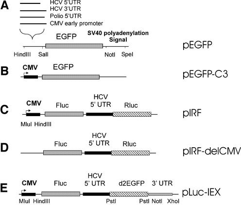
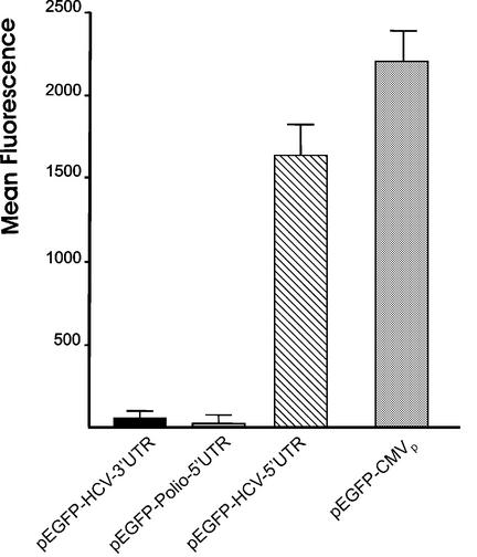
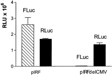
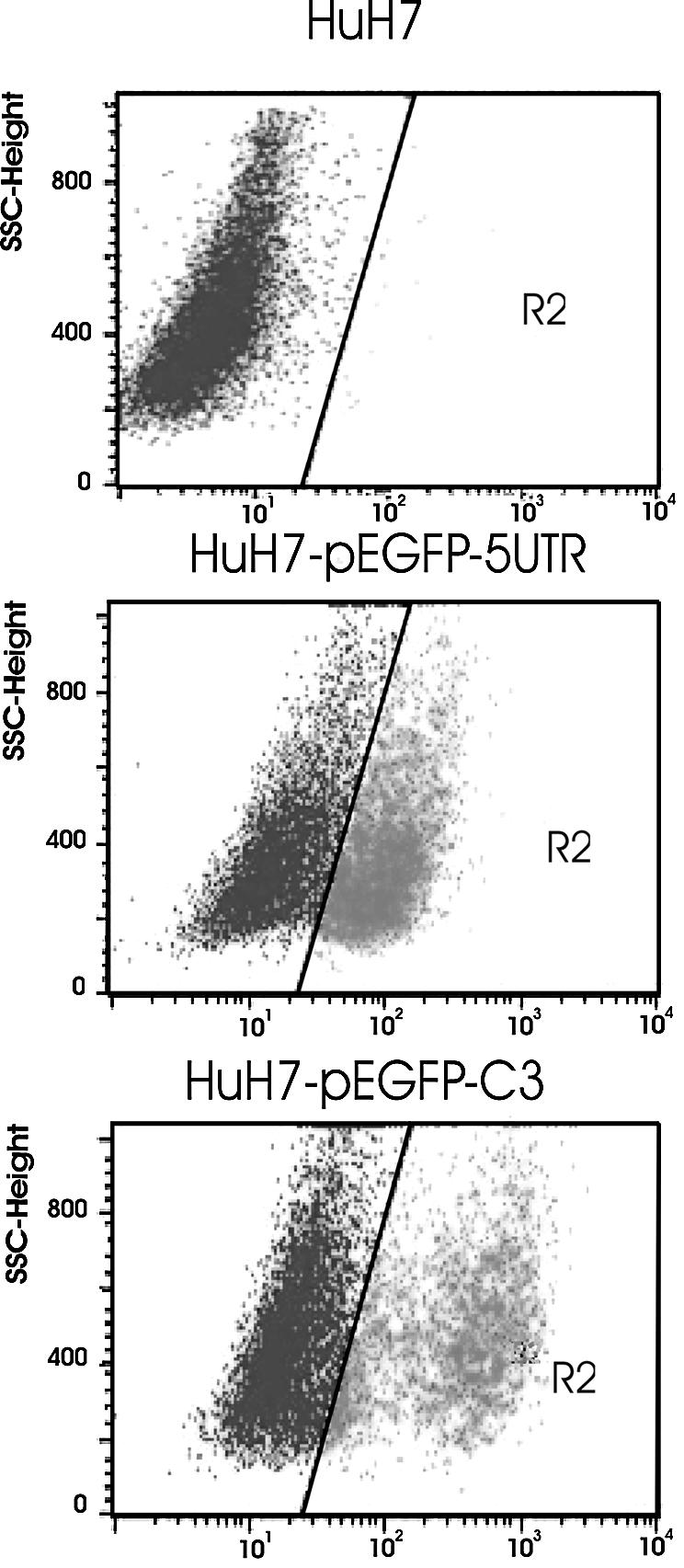
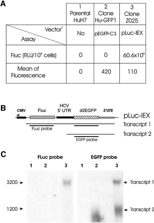
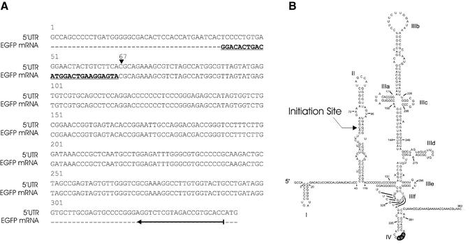
The hepatitis C virus (HCV) 5' untranslated region (UTR) has been extensively studied with regard to its internal ribosomal entry site (IRES) activity. In this work we present results suggesting the existence of a strong promoter activity carried by the DNA sequence corresponding to the HCV 5' UTR. This activity was not detected when the HCV 5' UTR sequence was replaced by HCV 3' UTR or poliovirus 5' UTR sequences. These results were further confirmed by using bicistronic constructions. We demonstrated the presence of an mRNA initiated in this 5' UTR sequence and located the initiation site by the 5' RACE method at nucleotide 67. Furthermore, northern experiments and flow cytometry analysis showed the unambiguous activity of such a promoter sequence in stably transfected cells. Our results strongly suggest that the data obtained using bicistronic DNA constructs carrying the HCV 5' UTR should be analyzed not only at the translational but also at the transcriptional level.


Figures

Figure 1
Figure 1
Schematic representation of plasmid constructions. The various vectors were constructed as described in Materials and Methods. (A and B) monocistronic vectors; (CE) bicistronic vectors.
Figure 2
Figure 2
Analysis of the promoter activity of different DNA sequences. The EGFP expression in transient transfected HuH7 cells was analyzed by flow cytometry. The mean fluorescence was calculated from positive fluorescent cells. The results are the mean of four independent experiments.
Figure 3
Figure 3
Promoter activity associated with the DNA sequence of HCV 5′ UTR. HuH7 cells were transfected with pIRF or pIRF-delCMV vectors and analyzed 48 h later. Luciferase activity (RLU) was measured with a luminometer as described in Materials and Methods. RLuc and FLuc represent Renilla luciferase and firefly luciferase, respectively. The results are the mean of three experiments in duplicate.
Figure 4
Figure 4
Gene expression in stably transfected cells. Flow cytometry analysis of EGFP expression. HuH7 cells were transfected with pEGFP-HCV-5′ UTR (middle) or pEGFP-C3 (bottom). Untransfected cells (top).
Figure 5
Figure 5
Expression from the HCV 5′ UTR sequence in bicistronic vector pLuc-IEX. Cells were transfected with pEGFP-C3 (column and lane 2), pLuc-IEX (column and lane 3) or no construction (column and lane 1), and analyzed for protein expression and transcript synthesis after G418 selection. (A) Luciferase activity (RLU) and d2EGFP expression obtained from flow cytometry experiments; the luciferase activity was expressed for 106 cells; the mean of fluorescence was calculated from positive fluorescent cells. (B) Schematic representation of expected transcripts; transcript 1 is transcribed from the CMV promoter, transcript 2 from the HCV 5′ UTR. (C) Northern blot analysis of transcripts with 10 µg of RNA in the presence of d2EGFP (right) or FLuc (left) 32P-labeled probes, as described in Materials and Methods.
Figure 6
Figure 6
Delineation of the transcription initiation site. (A) The upper sequence (5′ UTR) corresponds to the HCV 5′ UTR sequence. The EGFP mRNA sequence corresponds to that obtained from capped mRNA transcripts derived from HCV 5′ UTR. Bold letters represent the RNA oligonucleotide used in the 5′ RACE method. The vertical arrowhead indicates the position of the initiation site for EGFP transcription. The horizontal arrow indicates the location (nucleotides 321–341) of the primer used for the reverse transcriptase reaction. (B) Position of the initiation site in the IRES structure proposed by Honda et al. (16).

Similar articles

Cited by

References

    1. WHO (2000) Hepatitis C Fact Sheet No. 164, revised October 2000.
    1. Reynolds J.E., Kaminski,A., Carroll,A.R., Clarke,B.E., Rowlands,D.J. and Jackson,R.J. (1996) Internal initiation of translation of hepatitis C virus RNA: the ribosome entry site is at the authentic initiation codon. RNA, 2, 867–878. - PMC - PubMed
    1. Honda M., Ping,L.H., Rijnbrand,R.C., Amphlett,E., Clarke,B., Rowlands,D. and Lemon,S.M. (1996) Structural requirements for initiation of translation by internal ribosome entry within genome-length hepatitis C virus RNA. Virology, 222, 31–42. - PubMed
    1. Odreman-Macchioli F., Baralle,F.E. and Buratti,E. (2001) Mutational analysis of the different bulge regions of the HCV domain II and their influence on IRES translational ability. J. Biol. Chem., 276, 41648–41655. - PubMed
    1. Tanaka T., Kato,N., Cho,M.J. and Shimotohno,K. (1995) A novel sequence found at the 3′ terminus of hepatitis C virus genome. Biochem. Biophys. Res. Commun., 215, 744–749. - PubMed

Publication types

MeSH terms