. 2003 Feb 15;31(4):1292-301.
doi: 10.1093/nar/gkg209.

Characterization of the human beta-globin downstream promoter region

Affiliations

Characterization of the human beta-globin downstream promoter region

Kelly M Leach et al. Nucleic Acids Res. .

Abstract

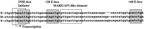
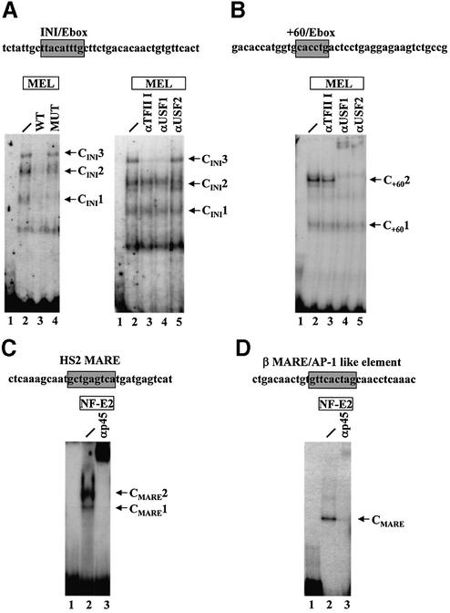
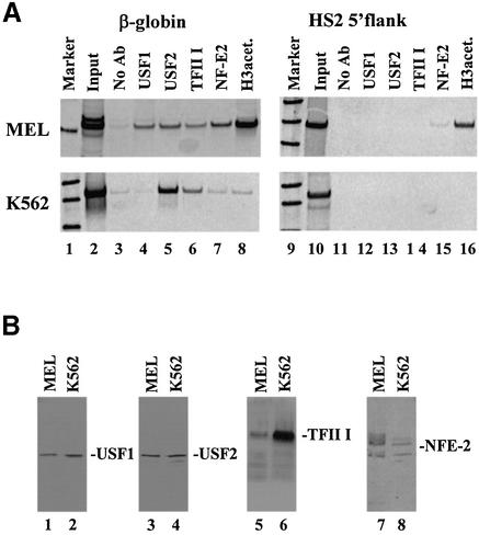
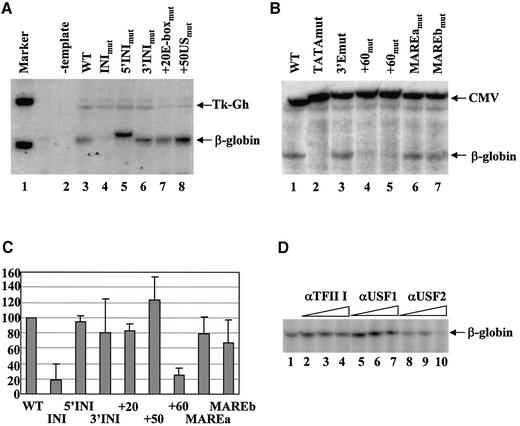
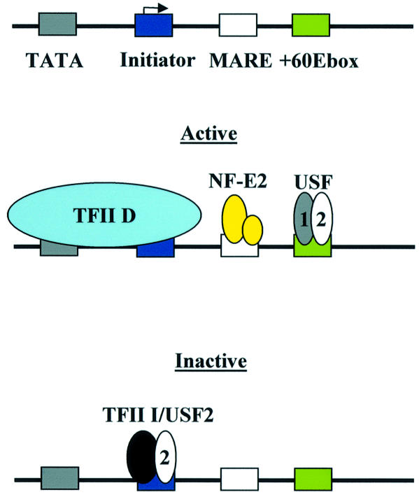
The human beta-globin gene is abundantly expressed specifically in adult erythroid cells. Stage-specific transcription is regulated principally by promoter proximal cis-regulatory elements. The basal promoter contains a non-canonical TATA-like motif as well as an initiator element. These two elements have been shown to interact with the TFII-D complex. Here we show that in addition to the TATA and initiator elements, conserved E-box motifs are located in the beta-globin downstream promoter. One of the E-box motifs overlaps the initiator and this composite element interacts with USF1 and TFII-I in vitro. Another E-box, located 60 bp 3' to the transcription initiation site, interacts with USF1 and USF2. Mutations of either the initiator or the downstream E-box impair transcription of the beta-globin gene in vitro. Mutations of a putative NF-E2-binding site in the downstream promoter region do not affect transcription in vitro. USF1, USF2, TFII-I and p45 can be crosslinked to a beta-globin promoter fragment in MEL cells in vivo, whereas only TFII-I and USF2 crosslink to the beta-globin gene in K562 cells. The summary data demonstrate that in addition to the well-characterized interactions of the TFII-D complex with the basal promoter, E-box motifs contribute to the efficient formation of transcription complexes on the adult beta-globin gene.


Figures

Figure 1
Figure 1
Sequence alignment of the human β-globin downstream promoter region. Shown are three sequences of the adult β-globin downstream promoter region from human (H), mouse (M) and rabbit (R) (26). Shaded boxes highlight the position of E-box motifs (CANNTG). Two of these E-boxes, the one overlapping the initiator and the distal E-box, are conserved in all three species, whereas the E-box located at +20 is only present in the human and rabbit genes. The open box delineates the position of the MARE-like sequence.
Figure 2
Figure 2
Interaction of HLH proteins and NF-E2 with human β-globin downstream promoter sequences in vitro. (A) EMSA was carried out as described in Materials and Methods using a fragment encompassing the initiator/E-box as well as the E-box at +20 (WT). Radiolabeled DNA fragments were incubated with 8 µg MEL protein extracts for 45 min at 30°C (lanes 2–4 and 6–9; lanes 1 and 5, no protein). MEL protein extracts were pre-incubated either without (lane 2) or with a 50-fold excess (compared to the radiolabeled fragment) of unlabeled competitor DNA, either using the wild-type initiator (lane 3) or a mutant fragment in which the initiator/ E-box was mutated (INImut, lane 4). Antibody supershift reactions were post-incubated either without (lane 6) or with specific antibodies raised against TFII-I (αTFII-I, lane 7), USF1 (αUSF1, lane 8) or USF2 (αUSF2, lane 9) for 20 min at 30°C. (B) Characterization of proteins interacting with an E-box motif located 60 bp downstream of the β-globin transcription initiation site. EMSA was carried out with a fragment encompassing the E-box element located 60 bp downstream of the start site of transcription. Radiolabeled fragment was incubated without (lane 1) or with 8 µg MEL protein extract (lanes 2–5). Proteins were post-incubated with specific antibodies raised against TFII-I (αTFII-I, lane 3), USF1 (αUSF1, lane 4) or USF2 (αUSF2, lane 5) for 20 min at 30°C. (C) Interaction of NF-E2 with a consensus MARE sequence from LCR element HS2. A radiolabeled oligonucleotide bearing the NF-E2-binding site from HS2 was incubated with 0.06 µg of a protein fraction enriched for a tethered p45/mafg protein (lane 2; lane 1 depicts the free probe). The binding reaction was pre-incubated with antibodies against p45 (lane 3) for 20 min at 30°C before addition of the radiolabeled fragment. (D) Interaction of NF-E2 with a MARE-sequence from the human β-globin downstream promoter region. A radiolabeled fragment bearing the β-globin downstream MARE like sequence was incubated with 0.2 µg of a protein fraction enriched for the tethered p45/mafg fusion protein (lane 2; lane 1 shows the free probe). Post-incubation with p45 antibodies (lane 3) was done as described in (B).
Figure 3
Figure 3
Characterization of protein–DNA interactions in the human β-globin downstream promoter region in vivo. (A) Chromatin immunoprecipitation (ChIP) experiment analyzing the interaction of proteins with the murine/human β-globin gene or the HS2 5′flanking region in MEL and K562 cells in vivo. Cells were incubated in 1% formaldehyde to crosslink protein–DNA and protein–protein interactions. After sonication, the cells were lysed and the chromatin was precipitated with either no antibody (lanes 3 and 11) or with antibodies specific for USF1 (lanes 4 and 12), USF2 (lanes 5 and 13), TFII-I (lanes 6 and 14), NF-E2 (p45, lanes 7 and 15) or acetylated histone H3 (lanes 8 and 16). DNA was purified from the precipitate and analyzed by PCR for the presence of the murine or human β-globin gene (210 and 321 bp, respectively, lanes 2–8) or the murine or human HS2 5′flank (336 and 565 bp, respectively, lanes 10–16). As positive controls, the input DNA was also analyzed by PCR (lanes 2 and 10). Lanes 1 and 9 show radiolabeled 100 bp markers. (B) Western blot analysis of protein extracts from MEL and K562 cells. Nuclear extracts were prepared from MEL or K562 cells and electrophoresed on denaturing polyacrylamide gels. Proteins were electroblotted to a nitrocellulose membrane, which was then hybridized to antibodies specific for USF1, USF2, TFII-I or NF-E2 (p45) as indicated. Bands were visualized by incubation with horseradish peroxidase-conjugated secondary antibody and autoradiography.
Figure 4
Figure 4
In vivo footprint analysis of a conserved E-box located in the murine adult β-globin downstream promoter region. NIH3T3L1 (lane 2) or MEL (lane 3) cells were treated with DMS. The DNA was purified from these cells and analyzed by linker ligation-mediated PCR as described in Materials and Methods. Lane 1 depicts the G sequencing ladder of the region. The position of the E-box at +60 is indicated on the right. The open circle on the left highlights a G residue that is consistently protected from cleavage by DMS in MEL cells.
Figure 5
Figure 5
Mutations of the initiator and E-boxes in the downstream promoter region impair transcription of the β-globin gene in vitro. In vitro transcription was monitored by primer extension using wild-type and mutant constructs of the β-globin gene and, as an internal control, the human growth hormone gene under control of the thymidine kinase promoter (Tk-Gh) (A) or a CMV construct (CMV) (B) was used. (A) Transcription analysis of mutations in the initiator (INImut), the 5′ region of the initiator (5′INImut), the 3′ region of the initiator (3′INImut), the E-box located 20 bp downstream of the initiation site (+20E-boxmut) and a region 50 bp downstream of the initiation site (+50USmut). Transcription was compared to that of a negative control lacking a DNA template (–template) and to that of the wild-type β-globin gene (WT). The first lane shows a radiolabeled marker (φX174/HinfI, denatured 100 bp marker). β-globin transcription was monitored using the β-glob80 primer. (B) Transcription of the wild-type (WT) and various β-globin gene templates carrying the following mutations: 5′TATAmut, mutation of the β-globin TATA-box; 3′Emut, deletion of the β-globin 3′ enhancer; +60amut, mutation of the E-box at +60; MAREamut and MAREbmut, mutations in the MARE-like element. Transcription of the β-globin gene was monitored using the β-glob90 primer and compared to transcription of the CMV internal control. (C) Quantitative analysis of in vitro transcription of the various promoter mutants shown in (A) and (B). Radiolabeled samples were electrophoresed on sequencing gels and the gels subjected to phosphorimaging. Graphs represent the relative expression levels of each β-globin mutant. RNA levels were normalized using the internal control and WT expression was set as 100%. Results shown are the average over two or three separate experiments. Error bars represent +1 SD from the mean. (D) Analysis of the effect of antibodies against HLH proteins on transcription of the β-globin gene in vitro. In vitro transcription of the wild-type β-globin gene was carried out as described in Figure 2. Protein extracts were pre-incubated with increasing concentrations of antibodies (2, 4 and 8 µl) against TFII-I (lanes 2–4), USF1 (lanes 5–7) and USF2 (lanes 8–10).
Figure 6
Figure 6
Summary and model of protein–DNA interactions at the human β-globin promoter. The human β-globin promoter consists of a TATA-box and an initiator. These sequences are bound by the TFII-D complex in cells expressing the β-globin gene (active) (13,14). Additional sequences required for the formation of active transcription complexes are MARE and E-box elements located in the downstream promoter region. These sequences are bound by NF-E2 (p45 and p18) and USF, respectively. It is proposed that in cells not expressing the β-globin gene (inactive), TFII-D is not bound and the initiator sequence is occupied by protein complexes consisting of TFII-I and USF2.

Similar articles

Cited by

References

    1. Bulger M. and Groudine,M. (1999) Looping versus linking: toward a model for long-distance gene activation. Genes Dev., 13, 2465–2477. - PubMed
    1. Higgs D. (1998) Do LCRs open chromatin domains? Cell, 95, 299–302. - PubMed
    1. Engel J.D. and Tanimoto,K. (2000) Looping, linking and chromatin activity: new insights into β-globin locus regulation. Cell, 100, 499–502. - PubMed
    1. Levings P.P. and Bungert,J. (2002) The human β-globin locus control region: a center of attraction. Eur. J. Biochem., 269, 1589–1599. - PubMed
    1. Reik A., Telling,A., Zitnik,G., Cimbora,D., Epner,E. and Groudine,M. (1998) The locus control region is necessary for gene expression in the human β-globin locus but not for the maintenance of an open chromatin structure in erythroid cells. Mol. Cell. Biol., 18, 5992–6000. - PMC - PubMed

Publication types

MeSH terms