

A Ca2+-induced mitochondrial permeability transition causes complete release of rat liver endonuclease G activity from its exclusive location within the mitochondrial intermembrane space. Identification of a novel endo-exonuclease activity residing within the mitochondrial matrix
- PMID: 12582256
- PMCID: PMC150224
- DOI: 10.1093/nar/gkg205
A Ca2+-induced mitochondrial permeability transition causes complete release of rat liver endonuclease G activity from its exclusive location within the mitochondrial intermembrane space. Identification of a novel endo-exonuclease activity residing within the mitochondrial matrix
Erratum in
- Nucleic Acids Res. 2003 Apr 1;31(7):2024
Abstract
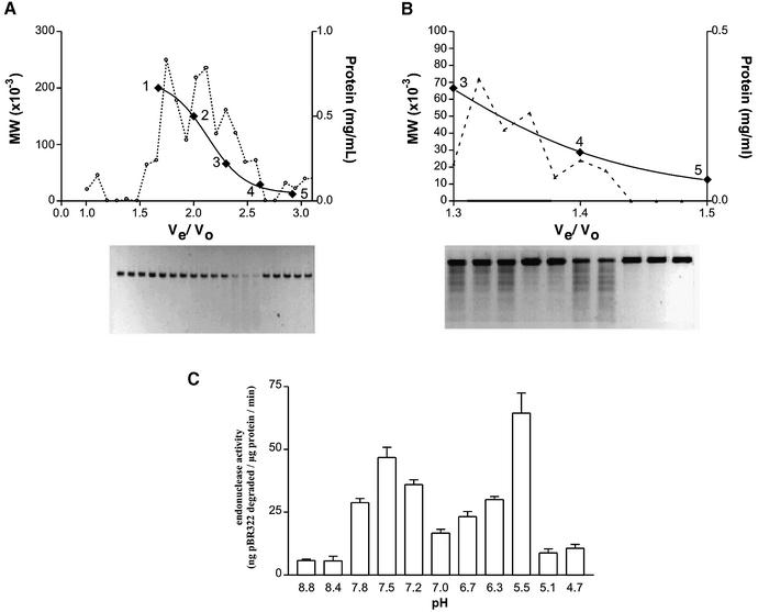
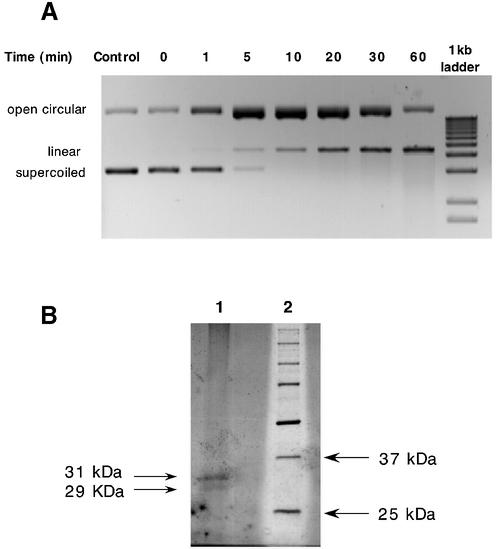
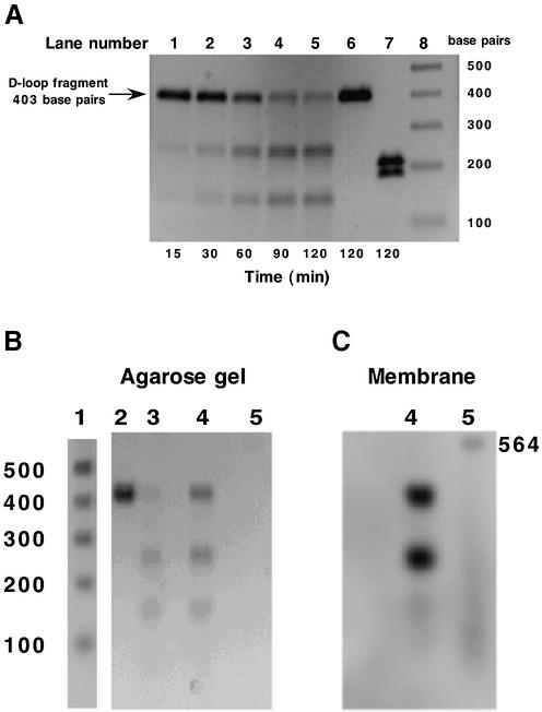
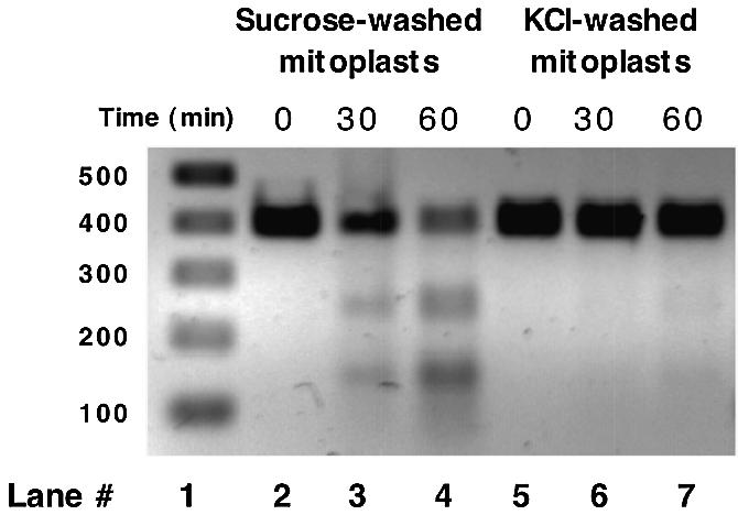
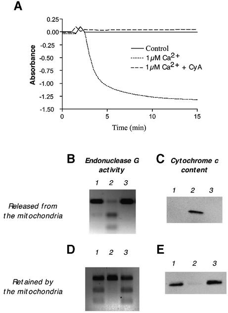
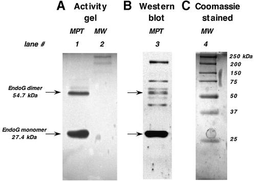
Endonuclease G, a protein historically thought to be involved in mitochondrial DNA (mtDNA) replication, repair, recombination and degradation, has recently been reported to be involved in nuclear DNA degradation during the apoptotic process. As a result, its involvement in mtDNA homeostasis has been called into question and has necessitated detailed analyses of its precise location within the mitochondrion. Data is presented localizing rat liver endonuclease G activity exclusively to the mitochondrial intermembrane space with no activity associated with either the interior face of the inner mitochondrial membrane or with the mitochondrial matrix. Additionally, it is shown that endonuclease G can be selectively released from the mitochondrion via induction of a Ca2+-induced mitochondrial permeability transition and that, upon its release, a further nuclease activity loosely associated with the interior face of the inner mitochondrial membrane and distinct in its properties from that of endonuclease G can be detected.
Figures
Similar articles
-
Tetrandrine-induced apoptosis in rat primary hepatocytes is initiated from mitochondria: caspases and endonuclease G (Endo G) pathway.Toxicology. 2006 Jan 20;218(1):1-12. doi: 10.1016/j.tox.2005.08.024. Epub 2005 Oct 24. Toxicology. 2006. PMID: 16246479
-
Differential permeabilization effects of Ca2+ and valinomycin on the inner and outer mitochondrial membranes as revealed by proteomics analysis of proteins released from mitochondria.Mol Cell Proteomics. 2009 Jun;8(6):1265-77. doi: 10.1074/mcp.M800377-MCP200. Epub 2009 Feb 14. Mol Cell Proteomics. 2009. PMID: 19218587 Free PMC article.
-
Autophagy restricts mitochondrial DNA damage-induced release of ENDOG (endonuclease G) to regulate genome stability.Autophagy. 2021 Nov;17(11):3444-3460. doi: 10.1080/15548627.2021.1874209. Epub 2021 Jan 19. Autophagy. 2021. PMID: 33465003 Free PMC article.
-
Effect of chlorpromazine on transmigration of mitochondrial aspartate aminotransferase in rat liver.Kitasato Arch Exp Med. 1989 Dec;62(4):181-6. Kitasato Arch Exp Med. 1989. PMID: 2640258
-
Discovery, regulation, and action of the major apoptotic nucleases DFF40/CAD and endonuclease G.J Cell Biochem. 2005 Apr 15;94(6):1078-87. doi: 10.1002/jcb.20409. J Cell Biochem. 2005. PMID: 15723341 Review.
Cited by
-
Mitochondrial dynamics and autophagy aid in removal of persistent mitochondrial DNA damage in Caenorhabditis elegans.Nucleic Acids Res. 2012 Sep;40(16):7916-31. doi: 10.1093/nar/gks532. Epub 2012 Jun 20. Nucleic Acids Res. 2012. PMID: 22718972 Free PMC article.
-
Enhanced expression of the DNA damage-inducible gene DIN7 results in increased mutagenesis of mitochondrial DNA in Saccharomyces cerevisiae.Mol Genet Genomics. 2003 Aug;269(5):632-9. doi: 10.1007/s00438-003-0873-8. Epub 2003 Jun 25. Mol Genet Genomics. 2003. PMID: 12827502
-
Point of No Return-What Is the Threshold of Mitochondria With Permeability Transition in Cells to Trigger Cell Death.J Cell Physiol. 2025 Jan;240(1):e31521. doi: 10.1002/jcp.31521. J Cell Physiol. 2025. PMID: 39760157 Free PMC article.
-
EXOG, a novel paralog of Endonuclease G in higher eukaryotes.Nucleic Acids Res. 2008 Mar;36(4):1369-79. doi: 10.1093/nar/gkm1169. Epub 2008 Jan 10. Nucleic Acids Res. 2008. PMID: 18187503 Free PMC article.
-
The flavonoid quercetin induces changes in mitochondrial permeability by inhibiting adenine nucleotide translocase.J Bioenerg Biomembr. 2009 Feb;41(1):41-7. doi: 10.1007/s10863-009-9198-6. Epub 2009 Mar 19. J Bioenerg Biomembr. 2009. PMID: 19296209
References
-
- Gerschenson M., Low,R.L. and Loehr,J. (1994) Levels of the mitochondrial endonuclease during rat cardiac development implicate a role for the enzyme in repair of oxidative damage in mitochondrial DNA. J. Mol. Cell. Cardiol., 26, 31–40. - PubMed
-
- Houmiel K.L., Gerschenson,M. and Low,R.L. (1991) Mitochondrial endonuclease activity in the rat varies markedly among tissues in relation to the rate of tissue metabolism. Biochim. Biophys. Acta, 1079, 197–202. - PubMed
-
- Cote J. and Ruiz-Carrillo,A. (1993) Primers for mitochondrial DNA replication generated by endonuclease G. Science, 261, 765–769. - PubMed
-
- Li L.Y., Luo,X. and Wang,X. (2001) Endonuclease G is an apoptotic DNase when released from mitochondria. Nature, 412, 95–99. - PubMed
Publication types
MeSH terms
Substances
Grants and funding
LinkOut - more resources
Full Text Sources
Molecular Biology Databases
Research Materials
Miscellaneous
