. 2003 Mar;52(3):340-6.
doi: 10.1136/gut.52.3.340.

A rapid decrease in the expression of DMT1 and Dcytb but not Ireg1 or hephaestin explains the mucosal block phenomenon of iron absorption

Affiliations

A rapid decrease in the expression of DMT1 and Dcytb but not Ireg1 or hephaestin explains the mucosal block phenomenon of iron absorption

D M Frazer et al. Gut. 2003 Mar.

Abstract

Background: A large oral dose of iron will reduce the absorption of a subsequent smaller dose of iron in a phenomenon known as mucosal block. Molecular analysis of this process may provide insights into the regulation of intestinal iron absorption.

Aims: To determine the effect of an oral bolus of iron on duodenal expression of molecules associated with intestinal iron transport in rats and to relate this to changes in iron absorption.

Methods: Rats were given an oral dose of iron and duodenal expression of divalent metal transporter 1 (DMT1), Dcytb, Ireg1, and hephaestin (Hp) was determined using the ribonuclease protection assay, western blotting, and immunofluorescence. Iron absorption was measured using radioactive (59)Fe.

Results: A decrease in intestinal iron absorption occurred following an oral dose of iron and this was associated with increased enterocyte iron levels, as assessed by iron regulatory protein activity and immunoblotting for ferritin. Reduced absorption was also accompanied by a rapid decrease in expression of the mRNAs encoding the brush border iron transport molecules Dcytb and the iron responsive element (IRE) containing the splice variant of DMT1. No such change was seen in expression of the non-IRE splice variant of DMT1 or the basolateral iron transport molecules Ireg1 and Hp. Similar changes were observed at the protein level.

Conclusions: These data indicate that brush border, but not basolateral, iron transport components are regulated locally by enterocyte iron levels and support the hypothesis that systemic stimuli exert their primary effect on basolateral transport molecules.


Figures

Figure 1
Figure 1
Duodenal uptake and transfer of 59Fe in rats following a blocking dose of non-radioactive iron. Rats were fasted overnight before being administered 10 mg of oral iron. At various time points thereafter, animals were anaesthetised and uptake (A) and transfer (B) of 59Fe determined as described in the methods section. The number of hours following the initial administration of iron is shown. Data are mean (SEM) of six animals. Statistical significance is shown relative to control animals: *p<0.05, **p<0.01.
Figure 2
Figure 2
Iron regulatory protein (IRP) activity and ferritin expression in rats following orally administered iron. Rats were fasted overnight before being administered 10 mg of oral iron. At various time points thereafter, animals were sacrificed and duodenal enterocytes isolated as described in the methods section. Protein was extracted from enterocytes and analysed by IRP bandshift assay (A) and by western blotting using antibodies specific for ferritin (B). Representative assays from triplicate experiments are shown. The number of hours following iron administration is indicated. βm, β-mercaptoethanol.
Figure 3
Figure 3
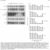
Gene expression following orally administered iron. Rats were fasted overnight before being administered 10 mg of oral iron. At various time points thereafter, animals were sacrificed and duodenal enterocytes isolated as described in the methods section. Total RNA was extracted from enterocytes and gene expression determined by ribonuclease protection assay (RPA) using 5 μg of RNA. Representative RPAs are shown for each gene (A). The number of hours following administration of iron is indicated. Band intensities were quantitated by densitometry, corrected for loading using glyceraldehyde-3-phosphate dehydrogenase (GAPDH) as a control, and graphed as a proportion of GAPDH (B). Data represent mean (SEM) of three animals. Statistical significance is shown relative to the 0 hour time point: *p<0.05, **p<0.01. DMT1, divalent metal transporter 1; DMT1(IRE), IRE splice variant of DMT1; DMT1(non-IRE), non-IRE splice variant of DMT1; Hp, hephaestin; IRE, iron responsive element.
Figure 4
Figure 4
Immunofluorescence for divalent metal transporter 1 (DMT1) and Dcytb protein in the duodenum of rats following oral administration of iron. Rats were fasted overnight before being administered 10 mg of oral iron. At various time points thereafter, animals were sacrificed and duodenal sections analysed by immunofluorescence using antibodies specific for DMT1 or Dcytb, as described in the methods section. Immunohistochemistry was carried out on sections from three animals and representative sections are shown. The number of hours following administration of iron is shown. Original magnification 200×.
Figure 5
Figure 5
Detection of Ireg1 and hephaestin (Hp) protein in the duodenum of rats following oral administration of iron. Rats were fasted overnight before being administered 10 mg of oral iron. At various time points thereafter, animals were sacrificed and duodenal enterocytes isolated, as described in the methods section. Protein was extracted from enterocytes and analysed by western blotting using antibodies specific for Ireg1 (A) or Hp (B). Representative blots are shown for each protein. The number of hours following iron administration is indicated. The sizes (×10−3) of the molecular weight standards are indicated on the right of each blot. Band intensities were quantitated by densitometry, corrected for loading using actin as a control, and graphed as a proportion of actin. Data are mean (SEM) of 5–6 animals. No statistically significant differences were seen between the groups.
Figure 6
Figure 6
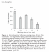
Iron absorption following varying doses of iron. Rats were fasted overnight before being administered an oral dose containing varying amounts of iron. Control animals were given 10 mM HCl only. Intestinal iron absorption was determined using an oral dose of 59Fe administered six hours after the initial dose of iron. Absorption is presented as the percentage of radioactivity retained by the animals five days after dosing. The amount of iron received by each group is indicated (blocking dose of iron). Data are mean (SEM) of three animals. Statistical significance is shown relative to control animals: *p<0.05.
Figure 7
Figure 7
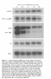
Gene expression following varying doses of oral iron. Rats were fasted overnight before being administered an oral dose containing varying amounts of iron. Control animals were given 10 mM HCl only. Six hours after this dose, animals were sacrificed and duodenal enterocytes isolated, as described in the methods section. Total RNA was extracted from enterocytes and gene expression determined by ribonuclease protection assay using 5 μg of RNA. Representative assays from triplicate experiments are shown. The amount of iron received by each group is indicated (iron blocking dose). DMT1, divalent metal transporter 1; DMT1(IRE), IRE splice variant of DMT1; DMT1(non-IRE), non-IRE splice variant of DMT1; Hp, hephaestin; IRE, iron responsive element.

References

    1. Conrad ME, Barton JC. Factors affecting iron balance. Am J Hematol 1981;10:199–225. - PubMed
    1. Crichton RR, Charloteaux-Wauters M. Iron storage and transport. Eur J Biochem 1987;164:485–506. - PubMed
    1. Lombard M, Chua E, O’Toole P. Regulation of intestinal non-haem iron absorption. Gut 1997;40:435–9. - PMC - PubMed
    1. McCance RA, Widdowson EM. Absorption and excretion of iron. Lancet 1937;ii:680–4. - PMC - PubMed
    1. Bothwell TH, Pirzio-Biroli G, Finch CA. Iron absorption I. Factors influencing absorption. J Lab Clin Med 1958;51:24–36. - PubMed

Publication types

MeSH terms