. 2003 Mar;77(5):2807-18.
doi: 10.1128/jvi.77.5.2807-2818.2003.

Simian virus 40 small tumor antigen induces deregulation of the actin cytoskeleton and tight junctions in kidney epithelial cells

Affiliations

Simian virus 40 small tumor antigen induces deregulation of the actin cytoskeleton and tight junctions in kidney epithelial cells

Viyada Nunbhakdi-Craig et al. J Virol. 2003 Mar.

Abstract

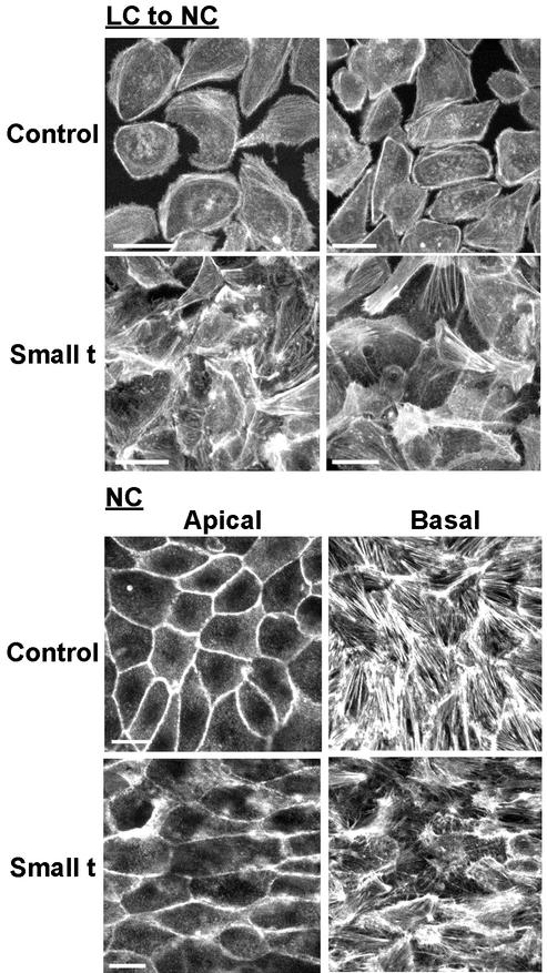
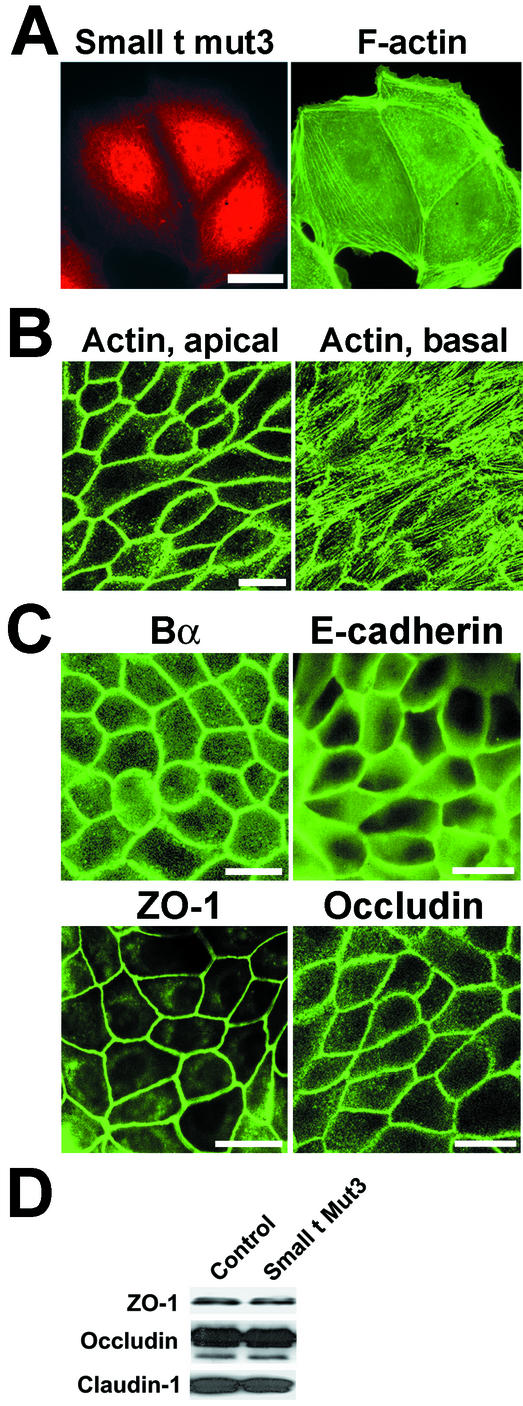
There is increasing evidence that the transforming DNA tumor virus simian virus 40 (SV40) is associated with human malignancies. SV40 small tumor antigen (small t) interacts with endogenous serine/threonine protein phosphatase 2A (PP2A) and is required for the transforming activity of SV40 in epithelial cells of the lung and kidney. Here, we show that expression of SV40 small t in epithelial MDCK cells induces acute morphological changes and multilayering. Significantly, it also causes severe defects in the biogenesis and barrier properties of tight junctions (TJs) but does not prevent formation of adherens junctions. Small t-induced TJ defects are associated with a loss of PP2A from areas of cell-cell contact; altered distribution and reduced amounts of the TJ proteins ZO-1, occludin, and claudin-1; and marked disorganization of the actin cytoskeleton. Small t-mediated F-actin rearrangements encompass increased Rac-induced membrane ruffling and lamellipodia, Cdc42-initiated filopodia, and loss of Rho-dependent stress fibers. Indeed, these F-actin changes coincide with elevated levels of Rac1 and Cdc42 and decreased amounts of RhoA in small t-expressing cells. Notably, these cellular effects of small t are dependent on its interaction with endogenous PP2A. Thus, our findings provide the first evidence that, in polarized epithelial cells, expression of small t alone is sufficient to induce deregulation of Rho GTPases, F-actin, and intercellular adhesion, through interaction with endogenous PP2A. Because defects in the actin cytoskeleton and TJ disruption have been linked to loss of cell polarity and tumor invasiveness, their deregulation by PP2A and small t likely contributes to the role of SV40 in epithelial cell transformation.


Figures

FIG. 1.
FIG. 1.
Analysis of stable MDCK cell populations by phase-contrast microscopy. Control MDCK cells stably transfected with the empty vector alone and two separate populations of MDCK-small t cells stably expressing SV40 small t were grown in NC medium and analyzed by phase-contrast microscopy. (A) Cycling cells. (B) Confluent cells. (C) Postconfluent cells grown for 5 days. Representative images are shown; similar results were obtained with at least seven distinct clones and with pooled stable cell populations of transfectants. The morphology of control cells was similar to that of nontransfected, parental MDCK cells (not shown). Bar, 40 μm.
FIG. 2.
FIG. 2.
Analysis of SV40 small t distribution by confocal microscopy in stably transfected MDCK cells. MDCK-small t cells were grown on Transwell filters in NC medium and analyzed by confocal microscopy with monoclonal anti-small t antibodies. Bars, 60 μm. (A) Representative images of two separate small t populations are shown in the left and middle panels. A representative multinucleated cell is enlarged in the right panel; the arrow indicates the absence of small t from the plasma membrane. (B) Thirty-two x-y sections were performed across the cells; successive sections 11, 16, 22, and 30 are shown. (C) Representative transversal x-z view of the cells. The double arrow indicates the thickness of the normal MDCK monolayer.
FIG. 3.
FIG. 3.
Analysis of PP2A subunit distribution in MDCK-small t cells. (A) Control and MDCK-small t cells were grown on Transwell filters in NC medium and analyzed by confocal microscopy with anti-A, anti-Bα, and anti-C subunit antibodies. Bars, 10 μm. Note that under our experimental conditions, the monoclonal anti-C antibody used here failed to recognize nuclear PP2A, in contrast to the anti-A antiserum. (B) Total cell lysates were prepared from control MDCK or MDCK-small t cells cultured in NC medium, immunoprecipitated with anti-small t antibodies, and analyzed by immunoblotting for the presence of small t and the A and C subunits of PP2A.
FIG. 4.
FIG. 4.
Effects of SV40 small t on the distribution of junctional proteins during TJ biogenesis. Confluent control and MDCK-small t cells were Ca2+ starved overnight in LC medium to induce complete TJ disruption and then switched for the indicated times to NC medium to induce TJ assembly. Cells were analyzed by confocal microscopy for the distribution of ZO-1, occludin, claudin-1, and E-cadherin (E-cad.). Bars, 10 μm.
FIG. 5.
FIG. 5.
Expression of SV40 small t inhibits TJ assembly. (A and B) Confluent control and MDCK-small t cells were Ca2+ starved overnight in LC medium and then switched to NC medium. (A) Equivalent amounts of proteins (∼40 μg) from detergent-soluble (lanes S) and -insoluble (lanes I) fractions were prepared from the cells 5 or 24 h after the Ca2+ switch and analyzed by Western blotting for the presence of junctional proteins. (B) Equivalent amounts of proteins (∼50 μg) from cytosolic (lanes Cy) and membrane (lanes M) fractions were prepared from the cells 24 h after the Ca2+ switch and analyzed by Western blotting for the presence of ZO-1, small t, and PP2A subunits. (C) Confluent MDCK cells stably expressing low basal levels of small t were grown for 3 days on Transwell filters in NC medium. Identical subsets of cells were preincubated without (noninduced) or with (induced) sodium butyrate to enhance expression of small t. The cells were incubated for 90 min in LC medium containing 1 mM EGTA to induce complete TJ opening (t = 0; TER < 30 Ω · cm2) and then switched to NC medium for the indicated times to induce TJ resealing. Results are expressed as the percentage of the relative initial resistance (TER = 1,300 ± 100 Ω · cm2) measured prior to the Ca2+ switch in noninduced cells cultured in NC medium. Values are the means ± standard deviations of duplicate determinations performed in four separate experiments with four distinct populations of transfectants. The representative immunoblot in the upper panel shows the total expression levels of small t in the cells before (lane −) and after (lane +) treatment with sodium butyrate.
FIG. 6.
FIG. 6.
Expression of SV40 small t induces TJ defects in MDCK cells cultured in NC medium. Cells were grown for 3 days on Transwell filters in NC medium. (A) Control cells and two separate populations of small t-expressing MDCK cells were analyzed by confocal microscopy for the distribution of junctional proteins. Bars, 10 μm. (B) Equivalent amounts of proteins (∼30 μg) from total lysates prepared from control (lane C) and small t-expressing (lane St) cells were analyzed by Western blotting for the presence of junctional proteins. Note the decreased levels of TJ proteins in small t-expressing cells. In particular, the upper band corresponding to slow-migrating, phosphorylated occludin species present in control cell lysates is nearly undetectable in extracts prepared from MDCK-small t cells. (C) Paracellular diffusion of [3H]mannitol and [3H]inulin was measured before (noninduced) and after (induced) preincubation of subsets of MDCK-small t cells with sodium butyrate, as described for Fig. 5C. Results are expressed as the percentage of tracer flux measured in noninduced cells and are the means ± standard deviations of triplicate determinations performed in four distinct experiments with four separate cell populations.
FIG. 7.
FIG. 7.
Expression of SV40 small t in MDCK cells induces the reorganization of the actin cytoskeleton. (A) Confluent control and MDCK-small t cells were Ca2+ starved overnight in LC medium and then switched for 2 h from LC to NC medium. (B) Confluent control and MDCK-small t cells were grown in NC medium. For both panels, cells were fixed and analyzed by confocal microscopy for the distribution of F-actin by using FITC-labeled phalloidin. Representative apical and basal x-y sections are shown for cells cultured in NC medium (B). Bars, 10 μm.
FIG.8.
FIG.8.
Analysis of MDCK cells expressing small t mutant 3. (A to C) MDCK-small tmutant 3 cells expressing a PP2A binding-defective small t mutant were grown in NC medium and analyzed by confocal microscopy. Bars, 10 μm. (A) Cells were Ca2+ starved overnight in LC medium, switched for 2 h from LC to NC medium, and double labeled with mouse anti-small t and FITC-phalloidin. (B) F-actin distribution at the apical and basal sections of the monolayer. (C) Polarized cells were analyzed for the distribution of Bα, E-cadherin, occludin, and ZO-1. The staining pattern of claudin-1 and of PP2A A and C subunitsin MDCK-small tmutant 3 cells was similar to that in control cells (not shown). (D) Equivalent amounts of proteins (∼30 μg) from total lysates prepared from control and MDCK-small tmutant 3 cells were analyzed by immunoblotting for the presence of TJ proteins.
FIG. 9.
FIG. 9.
Analysis of the role of expressed wild-type and mutant small t proteins in the regulation of Rho GTPases and F-actin rearrangements in MDCK cells. Control, MDCK-small t (Wt Small t), and MDCK-small tmutant 3 (Small t mut3) cells were cultured at low densities in NC medium and serum starved overnight by incubation in DMEM containing 0.2% dialyzed FBS (HyClone). (A to C) Serum-starved cells were stimulated for 10 min with either EGF (50 ng/ml; Upstate Biotechnology) (A), bradykinin (1 μM; Sigma) (B), or LPA (10 μM; Sigma) (C). Cells were then labeled with FITC-phalloidin and analyzed by immunofluorescence microscopy for the distribution of F-actin. Bars, 10 μm (A) and 5 μm (B and C). Note that F-actin patterns in MDCK-small tmutant 3 and control cells (not shown) were indistinguishable. (D) Equivalent amounts of proteins (∼100 μg) from total cell lysates were simultaneously analyzed by Western blotting for the expression levels of RhoA, Rac1, and Cdc42. A representative blot is shown; similar results were found in two other experiments.

Similar articles

Cited by

References

    1. Cereijido, M., L. Shoshani, and R. G. Contreras. 2000. Molecular physiology and pathophysiology of tight junctions. I. Biogenesis of tight junctions and epithelial polarity. Am. J. Physiol. Gastrointest. Liver Physiol. 279:G477-G482. - PubMed
    1. Choi, Y., I. Lee, and S. R. Ross. 1988. Requirement for the simian virus 40 small tumor antigen in tumorigenesis in transgenic mice. Mol. Cell. Biol. 8:3382-3390. - PMC - PubMed
    1. Denker, B. M., and S. K. Nigam. 1998. Molecular structure and assembly of the tight junction. Am. J. Physiol. 274:F1-F9. - PubMed
    1. Ellman, M., I. Bikel, J. Figge, T. Roberts, R. Schlossman, and D. M. Livingston. 1984. Localization of the simian virus 40 small t antigen in the nucleus and cytoplasm of monkey and mouse cells. J. Virol. 50:623-628. - PMC - PubMed
    1. Evers, E. E., G. C. Zondag, A. Malliri, L. S. Price, J. P. ten Klooster, R. A. van der Kammen, and J. G. Collard. 2000. Rho family proteins in cell adhesion and cell migration. Eur. J. Cancer 36:1269-1274. - PubMed

Publication types