. 2003 Mar;77(5):2832-42.
doi: 10.1128/jvi.77.5.2832-2842.2003.

Quantitative role of the human papillomavirus type 16 E5 gene during the productive stage of the viral life cycle

Affiliations

Quantitative role of the human papillomavirus type 16 E5 gene during the productive stage of the viral life cycle

Sybil M Genther et al. J Virol. 2003 Mar.

Abstract

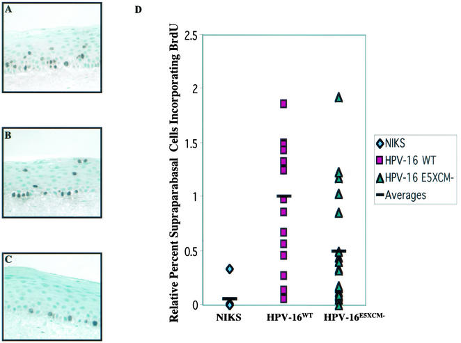
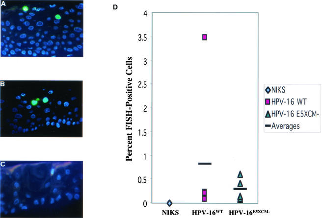
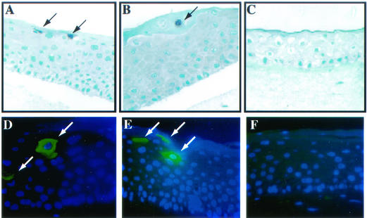
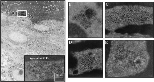
Human papillomaviruses (HPVs) are small circular DNA viruses that cause warts. Infection with high-risk anogenital HPVs, such as HPV type 16 (HPV16), is associated with human cancers, specifically cervical cancer. The life cycle of HPVs is intimately tied to the differentiation status of the host epithelium and has two distinct stages: the nonproductive stage and the productive stage. In the nonproductive stage, which arises in the poorly differentiated basal epithelial compartment of a wart, the virus maintains itself as a low-copy-number nuclear plasmid. In the productive stage, which arises as the host cell undergoes terminal differentiation, viral DNA is amplified; the capsid genes, L1 and L2, are expressed; and progeny virions are produced. This stage of the viral life cycle relies on the ability of the virus to reprogram the differentiated cells to support DNA synthesis. Papillomaviruses encode multiple oncoproteins, E5, E6, and E7. In the present study, we analyze the role of one of these viral oncogenes, E5, in the viral life cycle. To assess the role of E5 in the HPV16 life cycle, we introduced wild-type (WT) or E5 mutant HPV16 genomes into NIKS, a keratinocyte cell line that supports the papillomavirus life cycle. By culturing these cells under conditions that allow them to remain undifferentiated, a state similar to that of basal epithelial cells, we determined that E5 does not play an essential role in the nonproductive stage of the HPV16 life cycle. To determine if E5 plays a role in the productive stage of the viral life cycle, we cultured keratinocyte populations in organotypic raft cultures, which promote the differentiation and stratification of epithelial cells. We found that cells harboring E5 mutant genomes displayed a quantitative reduction in the percentage of suprabasal cells undergoing DNA synthesis, compared to cells containing WT HPV16 DNA. This reduction in DNA synthesis, however, did not prevent amplification of viral DNA in the differentiated cellular compartment. Likewise, late viral gene expression and the perturbation of normal keratinocyte differentiation were retained in cells harboring E5 mutant genomes. These data demonstrate that E5 plays a subtle role during the productive stage of the HPV16 life cycle.


Figures

FIG. 1.
FIG. 1.
Schematic displaying the DNA sequence of the first 28 amino acids of HPV16 E5WT ORF (A) and the corresponding 28-amino-acid E5XCM− mutant (B). To construct the E5XCM− HPV16 mutant, a single nucleotide, adenine, was deleted in codon 10 of the E5 ORF, disrupting the XcmI restriction site (underlined) and leading to a frameshift with a stop codon (TAA) encountered at codon 28. Only the first 10 amino acids of E5XCM− are derived from the E5 ORF.
FIG. 2.
FIG. 2.
Southern analysis of low-molecular-weight (Hirt) DNA extracted from three populations each of stably transfected NIKS containing the HPV16WT (A) or HPV16E5XCM− (B) viral DNA. Hirt DNA from passage 2 populations of NIKS cells harboring HPV16WT or HPV16E5XCM− genomes (lanes 1 to 3 and 5 to 7), untransfected NIKS cells and W12E cells grown on feeders were left uncut (lanes 1 to 4) or were cut with BamHI (to linearize viral genomes) (lanes 5 to 8) and subjected to Southern analysis using an HPV16-specific probe. Arrows indicate the migration of open circular (oc), linear (lin), and supercoiled (sc) HPV16 genomes as well as a bacterially synthesized spike plasmid DNA that was added to the cells at the time of lysis to assess efficiency and reproducibility of recovery of low molecular weight DNAs using the Hirt extraction protocol.
FIG. 3.
FIG. 3.
Presence and phosphorylated state of EGFR in NIKS cells. (A) Levels of P-EFGR (as detected by immunoprecipitation and Western analysis; see Materials and Methods) in NIKS cells either grown on fibroblast feeders in normal F medium containing 5% FBS and EGF (10 ng/ml) (lane 1), serum-starved for 24 h in F medium containing 0.1% FBS (lane 2), or serum-starved in F medium containing 0.1% FBS for 24 h followed by the addition of EGF (10 ng/ml) for 15 min (lane 3). Note the induction of P-EGFR in serum-starved, EGF stimulated cells. Indicated by the arrows at left is the migration position of P-EGFR and immunoglobulin (IgG) from the immunoprecipitation using anti-EGFR antibody (Ab-15; Labvision). The blot was probed with an antiphosphotyrosine antibody (PY20; Santa Cruz). (B) Levels of total EGFR protein (as detected by direct Western analysis using anti-EGFR antibody) (Ab-15; Labvision) in untransfected NIKS (N); three independently derived, transfected populations of NIKS cells harboring the WT HPV16 genome (W1, W2, and W3); three independently derived, transfected populations of NIKS cells harboring the E5XCM− mutant HPV16 genome (M1, M2, and M3); and W12E cells (W12) grown in the absence (−) or presence (+) of EGF (10 ng/ml). Note the similar range in levels of EGFR in different populations of NIKS cells harboring the WT or E5XCM− mutant HPV16 genomes and the similar modest reduction in levels of EGFR in cells grown in the presence of EGF at 10 ng/ml.
FIG. 4.
FIG. 4.
Immunohistochemical analyses of the differentiation program in organotypic raft cultures. Shown are organotypic raft cultures of HPV16WT-harboring NIKS cells (A, D, and G), HPV16E5XCM−-harboring NIKS cells (B, E, and H), and untransfected NIKS cells (C, F, and I) that were maintained on a dermal equivalent of collagen embedded with fibroblasts. The cultures were lifted to the liquid-air interface after 4 days in culture and were harvested 11 days postlift. The rafts were fixed in 4% formalin, embedded in paraffin, and cut into 4-μm-thick serial sections. Cross sections from each sample stained with hematoxylin and eosin (A to C) reveal normal stratification of the keratinocyte cultures, with no gross morphological differences between the HPV16WT (A)- and HPV16E5XCM− (B)-harboring rafts. Immunohistochemical staining for terminal differentiation markers of the epithelium reveals that there is also no difference in the differentiation program between HPV16WT- and HPV16E5XCM−-harboring rafts. K10 was detected by immunohistochemical staining using an anti-K10 antibody (clone Ck 8.60). (D to F) Positive cells, staining brown, were localized to the suprabasal compartment of the epithelium, as expected. (G to I) Filaggrin was detected with an antifilaggrin antibody. Positive cells localized to the granular layer of the rafts, as expected.
FIG. 5.
FIG. 5.
Analysis of DNA synthesis in organotypic raft cultures. HPV16 reprograms terminally differentiating cells to undergo unscheduled DNA synthesis. Shown are organotypic raft cultures of HPV16WT-harboring NIKS cells (A), HPV16 E5XCM−-harboring NIKS cells (B), and untransfected NIKS cells (C). BrdU was added to culture media 8 h prior to harvest. BrdU incorporation was detected by immunohistochemical staining using an antibody to BrdU. Shown is BrdU-specific immunohistochemical staining (brown nuclei) with hematoxylin counterstain (blue nuclei). BrdU was detected only in the basal compartment of untransfected NIKS cells (C). In contrast, BrdU was detected in both the basal and suprabasal compartment of HPV16WT- and HPV16E5XCM−-harboring NIKS (A and B). However, the graph (D) demonstrates that there is a quantitative reduction (approximately twofold) in the percentage of BrdU-positive cells in the supraparabasal compartment of HPV16E5XCM− mutant-harboring rafts compared with that of HPV16WT rafts (D). To obtain the data graphed, BrdU-positive cells from 7 populations of untransfected NIKS cells, 8 populations of independently derived populations of NIKS cells harboring HPV16WT genomes, and 10 independently derived populations of NIKS cells harboring HPV16 E5XCM− genomes were counted from four independent experiments (10 fields per slide at a magnification of ×40) as described in Materials and Methods.
FIG. 6.
FIG. 6.
FISH analysis on sections of organotypic raft cultures of NIKS cells harboring HPV 16 WT (A) and E5 mutant (B) genomes as well as untransfected (C) NIKS cells, using an HPV16-specific probe. The sections were counterstained with DAPI (blue nuclei). FISH-positive nuclei (green), indicating amplification of viral DNA, were found in the terminally differentiating compartment of rafts generated with NIKS cells harboring either HPV16WT (A) or HPV16E5XCM− genomes (B), but not in rafts generated with untransfected NIKS (C). (D) Histogram in which the frequency of cells harboring amplified copies of HPV16 were quantified in raft cultures subjected to HPV16-specific FISH analysis, as described in Materials and Methods. No significant difference in the frequency of cells with amplified copies of HPV16 genomes was evident between the cell populations harboring WT or E5 mutant viral genomes (P = 0.8551).
FIG. 7.
FIG. 7.
Immunohistochemical and immunofluorescent staining for late viral gene products in organotypic raft cultures. Shown are histological sections of organotypic raft cultures generated with WT and E5 mutant-harboring NIKS cells, or untransfected NIKS cells. For L1 immunohistochemistry, cross sections from each sample were incubated with an anti-L1 antibody (CamVir 1) and detected using the Vectastain ABC Kit. L1-positive cells, staining brown, are indicated by arrows. The sections were counterstained with hematoxylin, which stains nuclei blue. L1 was detected in both the WT (A) and E5 mutant (B) HPV16-positive raft cultures, but not in the untransfected NIKS raft cultures (C). For E1∧E4 immunofluorescence, cross sections from each sample were incubated with an anti-E1∧E4 antibody (TVG-402) and detected using a fluorescent-conjugated secondary antibody (AlexaFluor 488; Molecular Probes). E1∧E4-positive cells are indicated by arrows. The sections were counterstained with DAPI (blue nuclei). The E1∧E4 protein also was detected in both the WT (D) and E5 mutant (E) HPV16-positive NIKS raft cultures but not in untransfected NIKS raft cultures (F).
FIG. 8.
FIG. 8.
Ultrastructural analysis of HPV16 WT and E5 mutant HPV16 rafts. Shown are electron micrographs of thin sections taken from rafts generated with NIKS cells harboring WT (A) or E5 mutant (B to E) HPV16 genomes. (A) Low magnification (×2,700) of an entire cross section of a raft generated with NIKS harboring WT HPV16 genomes. The white box in panel A indicates the region shown at high magnification (×54,000) in the inset. An aggregate of VLPs measuring ∼55 nm were observed. A 1-cm ruler representing 185 nm is shown. Particles similar in size to these were also observed in multiple nuclei in E5 mutant HPV16-harboring rafts. (B to E) High magnification of examples of aggregates of VLPs measuring ∼55 nm from different rafts of two independent populations of E5 mutant HPV16 harboring NIKS cells (magnification, ×54,000[B] or ×55,000 [C to E]).

Similar articles

Cited by

References

    1. Adam, J. L., M. W. Briggs, and D. J. McCance. 2000. A mutagenic analysis of the E5 protein of human papillomavirus type 16 reveals that E5 binding to the vacuolar H+-ATPase is not sufficient for biological activity, using mammalian and yeast expression systems. Virology 272:315-325. - PubMed
    1. Allen-Hoffman, B. L., S. L. Schlosser, C. A. Ivarie, L. F. Meisner, and O. C. S. L. 2000. Normal growth and differentiation in a spontaneously immortalized near-diploid human keratinocyte cell line, NIKS. J. Investig. Dermatol. 114:444-455. - PubMed
    1. Baker, C. C., W. C. Phelps, V. Lindgren, M. J. Braun, M. A. Gonda, and P. M. Howley. 1987. Structural and transcriptional analysis of human papillomavirus type 16 sequences in cervical carcinoma cell lines. J. Virol. 61:962-971. - PMC - PubMed
    1. Banks, L., C. Edmonds, and K. H. Vousden. 1990. Ability of the HPV16 E7 protein to bind RB and induce DNA synthesis is not sufficient for efficient transforming activity in NIH3T3 cells. Oncogene 5:1383-1389. - PubMed
    1. Bouvard, V., G. Matlashewski, Z. M. Gu, A. Storey, and L. Banks. 1994. The human papillomavirus type 16 E5 gene cooperates with the E7 gene to stimulate proliferation of primary cells and increases viral gene expression. Virology 203:73-80. - PubMed

Publication types

MeSH terms

Substances

LinkOut - more resources