

Nucleocapsid-independent specific viral RNA packaging via viral envelope protein and viral RNA signal
- PMID: 12584316
- PMCID: PMC149775
- DOI: 10.1128/jvi.77.5.2922-2927.2003
Nucleocapsid-independent specific viral RNA packaging via viral envelope protein and viral RNA signal
Abstract
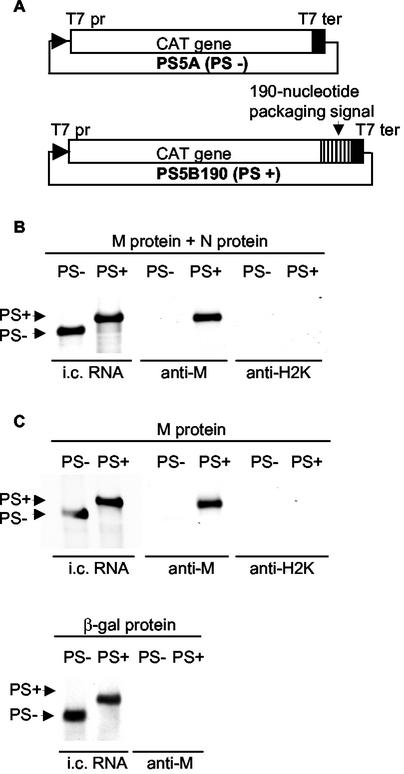
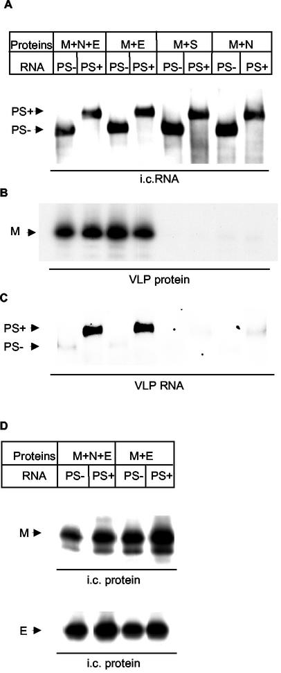
For any of the enveloped RNA viruses studied to date, recognition of a specific RNA packaging signal by the virus's nucleocapsid (N) protein is the first step described in the process of viral RNA packaging. In the murine coronavirus a selective interaction between the viral transmembrane envelope protein M and the viral ribonucleoprotein complex, composed of N protein and viral RNA containing a short cis-acting RNA element, the packaging signal, determines the selective RNA packaging into virus particles. In this report we show that expressed coronavirus envelope protein M specifically interacted with coexpressed noncoronavirus RNA transcripts containing the short viral packaging signal in the absence of coronavirus N protein. Furthermore, this M protein-packaging signal interaction led to specific packaging of the packaging signal-containing RNA transcripts into coronavirus-like particles in the absence of N protein. These findings not only highlight a novel RNA packaging mechanism for an enveloped virus, where the specific RNA packaging can occur without the core or N protein, but also point to a new, biologically important general model of precise and selective interaction between transmembrane proteins and specific RNA elements.
Figures
Similar articles
-
Characterization of the coronavirus M protein and nucleocapsid interaction in infected cells.J Virol. 2000 Sep;74(17):8127-34. doi: 10.1128/jvi.74.17.8127-8134.2000. J Virol. 2000. PMID: 10933723 Free PMC article.
-
Characterization of nucleocapsid-M protein interaction in murine coronavirus.Adv Exp Med Biol. 2001;494:577-82. doi: 10.1007/978-1-4615-1325-4_85. Adv Exp Med Biol. 2001. PMID: 11774528 No abstract available.
-
Cooperation of an RNA packaging signal and a viral envelope protein in coronavirus RNA packaging.J Virol. 2001 Oct;75(19):9059-67. doi: 10.1128/JVI.75.19.9059-9067.2001. J Virol. 2001. PMID: 11533169 Free PMC article.
-
Coronavirus genomic RNA packaging.Virology. 2019 Nov;537:198-207. doi: 10.1016/j.virol.2019.08.031. Epub 2019 Aug 30. Virology. 2019. PMID: 31505321 Free PMC article. Review.
-
The nucleocapsid of vesicular stomatitis virus.Sci China Life Sci. 2012 Apr;55(4):291-300. doi: 10.1007/s11427-012-4307-x. Epub 2012 May 9. Sci China Life Sci. 2012. PMID: 22566085 Review.
Cited by
-
The human pandemic coronaviruses on the show: The spike glycoprotein as the main actor in the coronaviruses play.Int J Biol Macromol. 2021 May 15;179:1-19. doi: 10.1016/j.ijbiomac.2021.02.203. Epub 2021 Mar 2. Int J Biol Macromol. 2021. PMID: 33667553 Free PMC article. Review.
-
Evaluation of kinetics and thermodynamics of interaction between immobilized SARS-CoV-2 nucleoprotein and specific antibodies by total internal reflection ellipsometry.J Colloid Interface Sci. 2021 Jul 15;594:195-203. doi: 10.1016/j.jcis.2021.02.100. Epub 2021 Mar 10. J Colloid Interface Sci. 2021. PMID: 33761394 Free PMC article.
-
Genetic and molecular biological analysis of protein-protein interactions in coronavirus assembly.Adv Exp Med Biol. 2006;581:163-73. doi: 10.1007/978-0-387-33012-9_29. Adv Exp Med Biol. 2006. PMID: 17037525 Free PMC article. Review. No abstract available.
-
Search for potential target site of nucleocapsid gene for the design of an epitope-based SARS DNA vaccine.Immunol Lett. 2008 Jun 15;118(1):65-71. doi: 10.1016/j.imlet.2008.03.003. Epub 2008 Apr 8. Immunol Lett. 2008. PMID: 18440652 Free PMC article.
-
Interaction of the coronavirus infectious bronchitis virus membrane protein with beta-actin and its implication in virion assembly and budding.PLoS One. 2009;4(3):e4908. doi: 10.1371/journal.pone.0004908. Epub 2009 Mar 16. PLoS One. 2009. PMID: 19287488 Free PMC article.
References
Publication types
MeSH terms
Substances
Grants and funding
LinkOut - more resources
Full Text Sources
