. 2003 Mar;77(5):3031-40.
doi: 10.1128/jvi.77.5.3031-3040.2003.

An umbraviral protein, involved in long-distance RNA movement, binds viral RNA and forms unique, protective ribonucleoprotein complexes

Affiliations

An umbraviral protein, involved in long-distance RNA movement, binds viral RNA and forms unique, protective ribonucleoprotein complexes

Michael Taliansky et al. J Virol. 2003 Mar.

Abstract

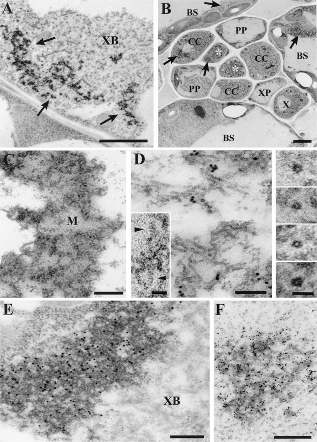
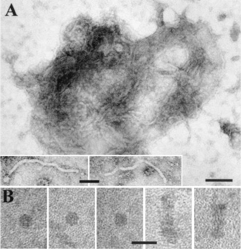
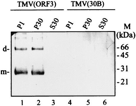
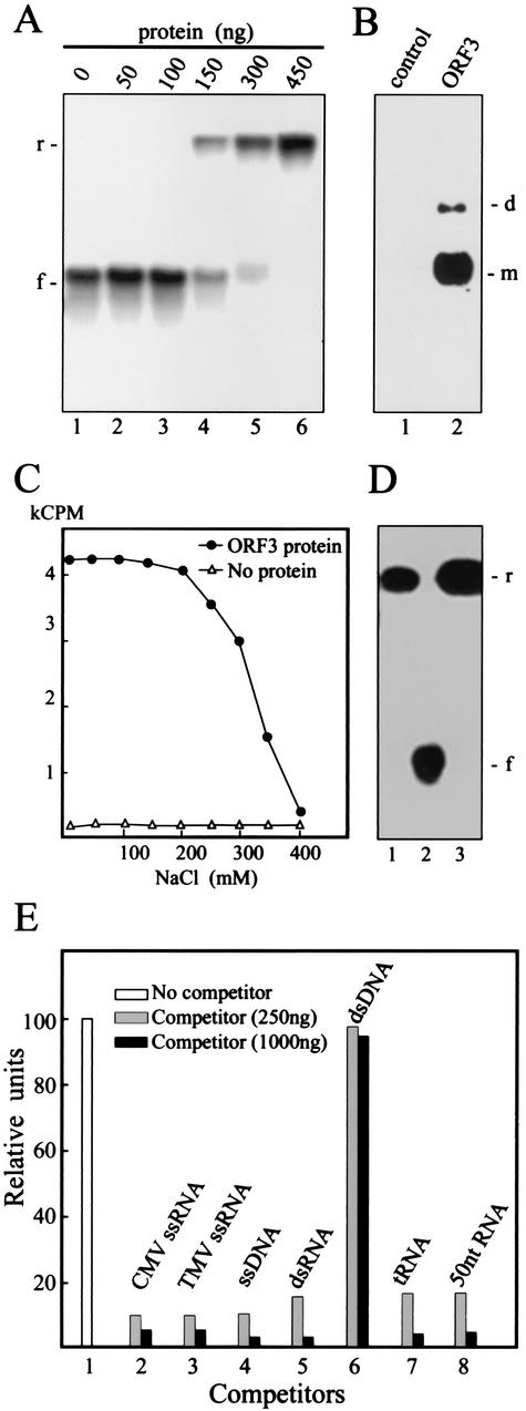
Umbraviruses are different from most other viruses in that they do not encode a conventional capsid protein (CP); therefore, no recognizable virus particles are formed in infected plants. Their lack of a CP is compensated for by the ORF3 protein, which fulfils functions that are provided by the CPs of other viruses, such as protection and long-distance movement of viral RNA. When the Groundnut rosette virus (GRV) ORF3 protein was expressed from Tobacco mosaic virus (TMV) in place of the TMV CP [TMV(ORF3)], in infected cells it interacted with the TMV RNA to form filamentous ribonucleoprotein (RNP) particles that had elements of helical structure but were not as uniform as classical virions. These RNP particles were observed in amorphous inclusions in the cytoplasm, where they were embedded within an electron-dense matrix material. The inclusions were detected in all types of cells and were abundant in phloem-associated cells, in particular companion cells and immature sieve elements. RNP-containing complexes similar in appearance to the inclusions were isolated from plants infected with TMV(ORF3) or with GRV itself. In vitro, the ORF3 protein formed oligomers and bound RNA in a manner consistent with its role in the formation of RNP complexes. It is suggested that the cytoplasmic RNP complexes formed by the ORF3 protein serve to protect viral RNA and may be the form in which it moves through the phloem. Thus, the RNP particles detected here represent a novel structure which may be used by umbraviruses as an alternative to classical virions.


Figures

FIG.1.
FIG.1.
Electron micrographs showing the localization in TMV(ORF3)-infected N. benthamiana cells of RNP particles containing the ORF3 protein. (A) Section of an infected companion cell showing typical ORF3 protein-related cytoplasmic inclusions (indicated by arrows), associated with an X body (XB). Bar, 2 μm. (B) Typical section of a class V vein showing the presence of the ORF3 protein-related inclusions (indicated by arrows) in bundle sheath (BS) cells, companion cells (CC), and sieve elements (indicated by asterisks). In other sections, inclusions were also detected in all other types of cells, including phloem parenchyma (PP) and xylem parenchyma (XP) cells. X, xylem vessel. Bar, 2 μm. (C) Section showing an inclusion composed of a complex of filamentous structures embedded in an electron-dense matrix (M). Bar, 200 nm. (D) Section showing filamentous particles at higher magnification. The section was labeled by in situ hybridization with an RNA probe specific for TMV(ORF3) positive-strand RNA. Bar, 100 nm. Inserts show selected transverse sections of the filamentous particles, showing the electron-lucent central hole and parts of two particles which show helical structure (arrowheads). Bars in inserts, 50 nm. (E) Immunogold labeling of a section containing an ORF3 protein-related inclusion, using rabbit antibody against the ORF3 protein. XB, X body. Bar, 200 nm. (F) In situ hybridization, as in panel D, of a section containing an ORF3 protein-specific inclusion. Bar, 500 nm.
FIG. 2.
FIG. 2.
Electron micrographs of cytoplasmic RNP complexes isolated from N. benthamiana leaves systemically infected with TMV(ORF3). (A) Typical RNP complexes with buoyant densities in Cs2SO4 of 1.34 to 1.45 g/ml. Similar complexes were isolated from N. benthamiana plants infected with GRV. Bar, 100 nm. Inserts show individual filamentous particles within complexes. Insert bar, 50 nm. (B) Small rods and disks with buoyant densities of 1.22 to 1.29 g/ml isolated from TMV(ORF3)-infected plants. Bar, 25 nm.
FIG. 3.
FIG. 3.
Western blot analysis of fractionated extracts from N. benthamiana plants infected with TMV(ORF3) or TMV(30B). Fractions were enriched for nuclei and chloroplasts (P1), membranes and mitochondria (P30), or soluble proteins (S30), and 20 μl of each fraction was loaded. The blot was probed with antibodies against a synthetic oligopeptide corresponding to part of the deduced sequence of the GRV ORF3 protein (see Materials and Methods). The positions of the molecular mass markers (M) and of the dimers (d) and monomers (m) of the ORF3 protein are indicated to the right and left of the blot, respectively.
FIG. 4.
FIG. 4.
Fractionation of the cytoplasmic RNP complexes isolated from plants infected with TMV(ORF3) by Cs2SO4 density gradient centrifugation. (A) Diagram showing density (ρ) gradient in the fractions obtained after centrifugation. The positions of the rods and disks (rods) and cytoplasmic RNP complexes (RNP-C) determined by electron microscopy and of fractions possessing infectivity are indicated. (B) Western blot analysis of the fractions, using antibody against the GRV ORF3 protein. The positions of the dimers (d) and monomers (m) of the ORF3 protein are indicated. Fraction b (bottom) is a sample of the resuspended pellet from the gradient. (C) Electrophoretic protein analysis (SDS-PAGE) of a partially purified preparation of the cytoplasmic RNP complexes (pooled fractions 11 to 14). Gel was stained with Coomassie blue (lane 1) or analyzed by Western blotting as in panel B (lane 2). The positions of the molecular mass markers (M) and of the dimers (d) and monomers (m) of the ORF3 protein are indicated to the right and left of the gel, respectively. (D) Dot blot hybridization analysis of TMV(ORF3) RNA contained in combined fractions 3 to 6, 7 to 10, and 11 to 14. Hybridization was with a probe specific to the ORF3 nucleotide sequence.
FIG. 5.
FIG. 5.
Oligomerization of the ORF3 protein in vitro. (A) Western blot analysis, using antibody against the GRV ORF3 protein, of the recombinant ORF3-His protein isolated from TMV(ORF3-His)-infected plants and fractionated by SDS-PAGE (lane 1). Control sample isolated from TMV(30B)-infected plants was loaded in lane 2. The positions of the molecular mass markers (M) and of the dimers (d) and monomers (m) of the ORF3 protein are indicated to the right and left of the blot, respectively. (B) Sedimentation analysis of the ORF3-His protein oligomers in a 10 to 30% sucrose density gradient. Fractions obtained after centrifugation were subjected to Western blot analysis as in panel A. The positions in the gradient of the molecular mass markers (M) (carbonic anhydrase [29 kDa], BSA [66 kDa], alcohol dehydrogenase [150 kDa], β-amylase [200 kDa], and apoferritin [443 kDa]) are shown above the blot. The positions of dimers (d) and monomers (m) of the ORF3-His protein are indicated to the right of the blot.
FIG. 6.
FIG. 6.
Analysis of nucleic acid binding by the ORF3-His protein. (A) Gel retardation electrophoresis assay for RNA binding by the ORF3 protein. Increasing amounts of the ORF3 protein were incubated in 15 μl of binding buffer A (see Materials and Methods) with 2ng of 32P-labeled GRV RNA transcript, and the mixtures were electrophoresed in 1% nondenaturing agarose gel. The amount of the ORF3 protein used in the assay is indicated above the lanes. The positions of free (f) and retarded (r) RNAs are shown to the left of the gel. (B) North-Western blot analysis of RNA binding by the ORF3 protein. After SDS-PAGE, the ORF3 protein was transferred onto a nitrocellulose membrane and renatured. The membrane was treated with 32P-labeled GRV RNA transcript. The ORF3 protein was substituted by BSA mixed with an equivalent protein preparation from plants infected with the empty TMV(30B) vector as a control. The positions of dimers (d) and monomers (m) are indicated to the right of the blot. (C) Salt stability of RNA-ORF3 protein complex. The ORF3 protein (450 ng) was incubated with labeled GRV RNA (4 ng) in the presence of the indicated concentrations of NaCl. After incubation, the mixtures were analyzed by nitrocellulose membrane filter-binding assay. RNA binding was quantified by determining the radioactivity of the membrane by liquid scintillation counting. kCPM, 1,000 cpm. (D) Gel retardation electrophoresis assay for RNA binding by the ORF3 protein (400 ng) as in panel A but in the presence of 350 mM (lane 1) or 200 mM (lane 3) NaCl. Lane 2 contained RNA without the ORF3 protein, preincubated in 350 mM NaCl. The positions of free (f) and retarded (r) RNAs are shown to the right of the gel. (E) Competition binding assay of the ORF3 protein to GRV ssRNA. The protein (450 ng in 15 μl) was incubated with labeled GRV RNA (4 ng) either in the absence (column 1) or in the presence of the following unlabeled competitors: CMV ssRNA (column 2), TMV ssRNA (column 3), bacteriophage M13 ssDNA (column 4), simian rotavirus SA11 dsRNA (column 5), plasmid dsDNA fragment (SmaI-digested pUC18 DNA) (column 6), tRNA (column 7), and rRNA hydrolyzed by NaOH (about 50 nucleotides in length) (column 8). The amounts of the competitors are indicated. After incubation, the mixtures were analyzed by the nitrocellulose membrane filter-binding assay. RNA binding was quantified by densitometry of autoradiographic images in relative units (100 units corresponds to RNA binding by the ORF3 protein without competitor).

Similar articles

Cited by

References

    1. Bentley, M., M. J. Ladu, C. Rajan, G. S. Getz, and C. A. Reardon. 2002. Apolipoprotein E structural requirements for the formation of SDS-stable complexes with beta-amiloid-(1-40): the role of salt bridges. Biochem. J. 366:273-279. - PMC - PubMed
    1. Canto, T., D. A. M. Prior, K.-H. Hellwald, K. J. Oparka, and P. Palukaitis. 1997. Characterization of cucumber mosaic virus. IV. Movement protein and coat protein are both essential for cell-to-cell movement of cucumber mosaic virus. Virology 237:237-248. - PubMed
    1. Carrington, J. C. 2000. RNA silencing: moving targets. Nature 408:150-151. - PubMed
    1. Carrington, J. C., K. D. Kasschau, S. K. Mahajan, and M. C. Schaad. 1996. Cell-to-cell and long-distance transport of viruses in plants. Plant Cell 8:1669-1681. - PMC - PubMed
    1. Demler, S. A., D. G. Rucker, and G. A. de Zoeten. 1993. The chimeric nature of the genome of pea enation mosaic virus: the independent replication of RNA-2. J. Gen. Virol. 74:1-14. - PubMed

Publication types

MeSH terms

LinkOut - more resources