. 2003 Mar;77(5):3041-9.
doi: 10.1128/jvi.77.5.3041-3049.2003.

Nef-mediated disruption of HLA-A2 transport to the cell surface in T cells

Affiliations

Nef-mediated disruption of HLA-A2 transport to the cell surface in T cells

Matthew R Kasper et al. J Virol. 2003 Mar.

Abstract

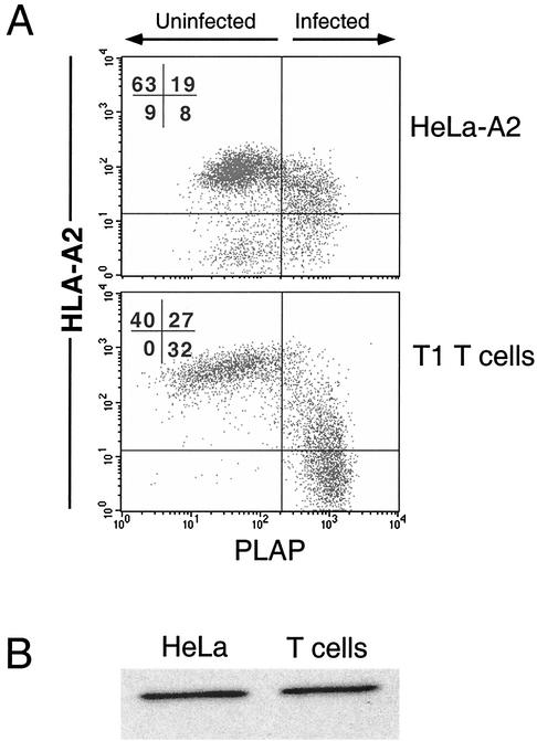
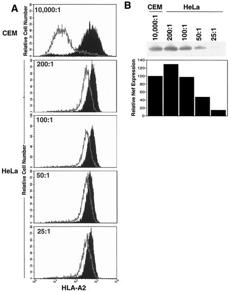
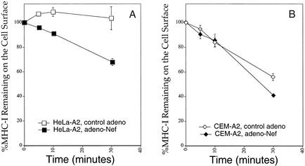
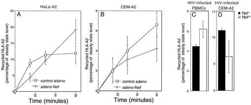
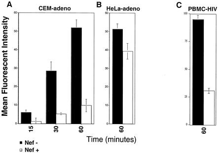
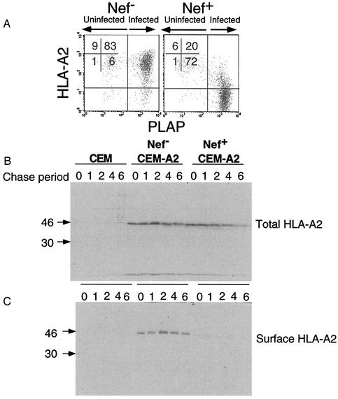
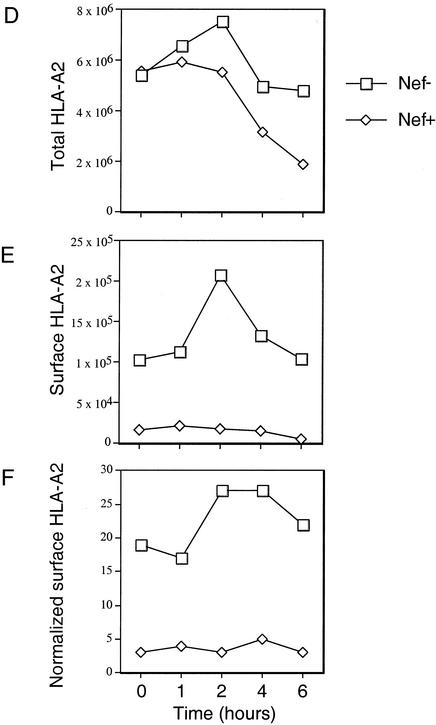
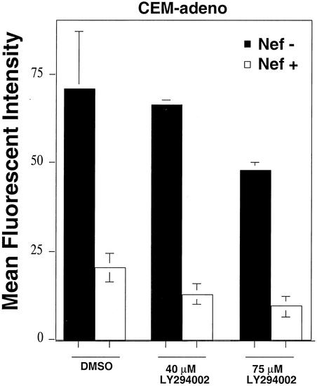
Human immunodeficiency virus type 1 (HIV-1) Nef is a key pathogenic factor necessary for the development of AIDS. One important function of Nef is to reduce cell surface levels of major histocompatibility complex class I (MHC-I) molecules, thereby protecting HIV-infected cells from recognition by cytotoxic T lymphocytes. The mechanism of MHC-I downmodulation by Nef has not been clearly elucidated, and its reported effect on MHC-I steady-state levels ranges widely, from 2-fold in HeLa cells to 200-fold in HIV-infected primary T cells. Here, we directly compared downmodulation of HLA-A2 in HIV-infected HeLa cells to that in T cells. We found that similar amounts of Nef protein resulted in a much more dramatic downmodulation of HLA-A2 in T cells than in HeLa cells. A comparison of Nef's effects on HLA-A2 endocytosis, recycling, and transport rates indicated that the most prominent effect of Nef on HLA-A2 in T cells was to inhibit transport to the cell surface. The phosphatidylinositol 3-kinase inhibitor, LY294002, previously reported to inhibit Nef-mediated MHC-I downmodulation in astrocytic cells, did not directly affect Nef's ability to block transport of MHC-I to the cell surface in T cells.


Figures

FIG. 1.
FIG. 1.
HIV-infected T cells downmodulate MHC-I more efficiently than HIV-infected HeLa cells. (A) HeLa-A2 cells or T1 T cells were transduced with an HIV (NL-PIenv) pseudotyped with VSV-G protein as described in Materials and Methods. At 48 h, the cells were harvested and stained for HLA-A2 and PLAP. Control uninfected cells fell to the left of the vertical line, and control HLA-A2-negative cells fell below the horizontal line. (B) Western blot analysis of Nef expression in HeLa and T1 cells was performed on the indicated cell lines normalized for total protein and infection rate. Results shown are typical of at least three separate experiments.
FIG. 2.
FIG. 2.
Nef expressed in an adenoviral vector downmodulates MHC-I more efficiently in T cells than in HeLa cells. HeLa-A2 and CEM-A2 cells were transduced with the indicated amount of adeno-Nef or control-adeno as described in Materials and Methods. (A) After 48 h, the cells were stained with the HLA-A2-specific monoclonal antibody BB7.2 to assess HLA-A2 expression levels by using flow cytometry. Nef reduced the expression of HLA-A2 by 11-fold in T cells and 1.2- to 2.7-fold in HeLa cells. (B) Western blot analysis assessing Nef expression levels revealed that HeLa cells treated with adeno-Nef at an estimated MOI of 100:1 (as determined in 293 cells) had Nef expression similar to that of CEM T cells treated at an MOI of 10,000:1. Results shown are typical of at least five separate experiments.
FIG. 3.
FIG. 3.
Nef has a small effect on endocytosis rates in HeLa cells and a lesser effect on endocytosis rates in T cells. The endocytosis rates of MHC-I in CEM-A2 (A) or HeLa-A2 (B) cells 24 h after treatment with adeno-Nef or control-adeno were measured as described in Materials and Methods. Shown are the means ± standard deviations for one experiment performed in triplicate. Results shown are typical of at least two separate experiments. At the MOIs of adenovirus used in these experiments (100:1 for HeLa and 10,000:1 for CEM T cells), Nef was expressed at similar levels (Fig. 2).
FIG. 4.
FIG. 4.
Nef does not significantly inhibit recycling of MHC-I to the cell surface in HeLa cells or T cells. The rates of MHC-I recycling in HeLa-A2 (A) or CEM-A2 (B) cells 24 h after treatment with adeno-Nef or control-adeno were measured as described in Materials and Methods. Shown are the means ± standard deviations for three separate experiments. At the MOIs of adenovirus used in these experiments (50:1 for HeLa and 10,000:1 for CEM T cells), there was about half as much Nef expressed in HeLa cell lysates as in T-cell lysates normalized for total protein (Fig. 2B). Additional experiments did not detect a significant Nef-dependent inhibition of HLA-A2 recycling in HIV-infected primary T cells (C) or HIV-infected CEM T cells (D) after a 60-min incubation.
FIG. 5.
FIG. 5.
Nef blocks the transport of newly synthesized MHC-I to the cell surface in T cells. (A) Transport of HLA-A2 to the cell surface in T cells transduced with a Nef-expressing or control adenovirus at an MOI of 10,000:1. Shown here is a summary graph of the MFI at each time point corrected for recycling and background expression as described in Materials and Methods. The means ± standard deviations for two separate experiments are shown. (B) Transport of HLA-A2 to the cell surface in HeLa cells was measured as described for panel A, except that only the 60-min time point was measured. At the MOI of adenovirus used in these experiments (100:1), Nef expression was comparable to that in CEM T cells transduced at an MOI of 10,000:1 (Fig. 2). These results, from assays performed in duplicate, are typical of at least three experiments. (C) Transport of HLA-A2 to the cell surface in primary T cells infected with HXB-EP with or without Nef expression (5). Shown here is the average MFI ± standard deviation of newly synthesized HLA-A2 in PLAP+ (HIV-infected) cells after 60 min of incubation with or without Nef expression.
FIG. 6.
FIG. 6.
Nef blocks the transport of newly synthesized MHC-I to the cell surface in T cells. (A) CEM and CEM-A2 T cells were infected with NL-PIenv with or without nef expression, harvested after 48 h, and stained for HLA-A2 and PLAP. As shown, almost all the cells are infected and Nef-expressing cells had approximately 50-fold less HLA-A2 expression. (B and C) HIV-infected CEM and CEM-A2 T cells were pulse labeled with [35S]methionine and cysteine and were then surface labeled with biotin as described in Materials and Methods. Lysates were made, and HLA-A2 was immunoprecipitated with monoclonal antibody BB7.2. (B) One-third of the immunoprecipitate was analyzed by SDS-PAGE to assess total HLA-A2 expression. (C) Two-thirds of the immunoprecipitate was reprecipitated with avidin beads to isolate cell surface HLA-A2 prior to analysis by SDS-PAGE. (D to F) The amount of total HLA-A2 (panel D) or surface HLA-A2 (panel E) or the percentage of recovered surface HLA-A2 relative to total HLA-A2 (panel F) was determined from the gels shown in panels B and C by using a phosphorimager. Results obtained were similar in three independent experiments.
FIG. 6.
FIG. 6.
Nef blocks the transport of newly synthesized MHC-I to the cell surface in T cells. (A) CEM and CEM-A2 T cells were infected with NL-PIenv with or without nef expression, harvested after 48 h, and stained for HLA-A2 and PLAP. As shown, almost all the cells are infected and Nef-expressing cells had approximately 50-fold less HLA-A2 expression. (B and C) HIV-infected CEM and CEM-A2 T cells were pulse labeled with [35S]methionine and cysteine and were then surface labeled with biotin as described in Materials and Methods. Lysates were made, and HLA-A2 was immunoprecipitated with monoclonal antibody BB7.2. (B) One-third of the immunoprecipitate was analyzed by SDS-PAGE to assess total HLA-A2 expression. (C) Two-thirds of the immunoprecipitate was reprecipitated with avidin beads to isolate cell surface HLA-A2 prior to analysis by SDS-PAGE. (D to F) The amount of total HLA-A2 (panel D) or surface HLA-A2 (panel E) or the percentage of recovered surface HLA-A2 relative to total HLA-A2 (panel F) was determined from the gels shown in panels B and C by using a phosphorimager. Results obtained were similar in three independent experiments.
FIG. 7.
FIG. 7.
LY294002 does not inhibit Nef's ability to disrupt transport of HLA-A2 to the cell surface in CEM-A2 T cells. T cells were transduced with a Nef-expressing or control adenovirus at an MOI of 10,000:1. Transport of newly synthesized HLA-A2 to the cell surface was measured flow cytometrically, with or without the addition of 40 or 75 μM LY294002 as described in Materials and Methods. Shown here is a summary graph of the MFI at each time point corrected for recycling and background expression as described in Materials and Methods. The means ± standard deviations are shown. Results were similar in two independent experiments. DMSO, dimethyl sulfoxide.

Similar articles

Cited by

References

    1. Altschuler, Y., S. Liu, L. Katz, K. Tang, S. Hardy, F. Brodsky, G. Apodaca, and K. Mostov. 1999. ADP-ribosylation factor 6 and endocytosis at the apical surface of Madin-Darby canine kidney cells. J. Cell Biol. 147:7-12. - PMC - PubMed
    1. Benichou, S., M. Bomsel, M. Bodeus, H. Durand, M. Doute, F. Letourneur, J. Camonis, and R. Benarous. 1994. Physical interaction of the HIV-1 Nef protein with β-COP, a component of non-clathrin-coated vesicles essential for membrane traffic. J. Biol. Chem. 269:30073-30076. - PubMed
    1. Bresnahan, P., W. Yonemoto, S. Ferrell, D. Williams-Herman, R. Geleziunas, and W. Greene. 1998. A dileucine motif in HIV-1 Nef acts as an internalization signal for CD4 downregulation and binds the AP-1 clathrin adaptor. Cur. Biol. 8:1235-1238. - PubMed
    1. Caplan, S., N. Naslavsky, L. M. Hartnell, R. Lodge, R. S. Polishchuk, J. G. Donaldson, and J. S. Bonifacino. 2002. A tubular EHD1-containing compartment involved in the recycling of major histocompatibility complex class I molecules to the plasma membrane. EMBO J. 21:2557-2567. - PMC - PubMed
    1. Chen, B., R. Gandhi, and D. Baltimore. 1996. CD4 down-modulation during infection of human T cells with human immunodeficiency virus type 1 involves independent activities of vpu, env, and nef. J. Virol. 70:6044-6053. - PMC - PubMed

Publication types

MeSH terms

Substances