. 2003 Mar;77(5):3181-90.
doi: 10.1128/jvi.77.5.3181-3190.2003.

Efficient replication of hepatitis C virus genotype 1a RNAs in cell culture

Affiliations

Efficient replication of hepatitis C virus genotype 1a RNAs in cell culture

Keril J Blight et al. J Virol. 2003 Mar.

Abstract

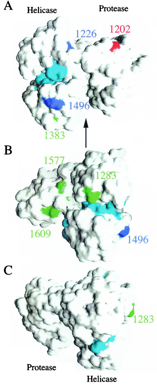
Hepatitis C virus (HCV) genotype 1 (subtypes 1a and 1b) is responsible for the majority of treatment-resistant liver disease worldwide. Thus far, efficient HCV RNA replication has been observed only for subgenomic and full-length RNAs derived from genotype 1b isolates. Here, we report the establishment of efficient RNA replication systems for genotype 1a strain H77. Replication of subgenomic and full-length H77 1a RNAs required the highly permissive Huh-7.5 hepatoma subline and adaptive amino acid substitutions in both NS3 and NS5A. Replication could be detected by RNA quantification, fluorescence-activated cell sorting, and metabolic labeling of HCV-specific proteins. Replication efficiencies were similar for subgenomic and full-length RNAs and were most efficient for HCV RNAs lacking heterologous RNA elements. Interestingly, both subtype 1a and 1b NS3 adaptive mutations are surface exposed and present on only one face of the NS3 structure. The cell culture-adapted subtype 1a replicons should be useful for basic replication studies and for antiviral development. These results are also encouraging for the development of adapted replicons for the remaining HCV genotypes.


Figures

FIG. 1.
FIG. 1.
Schematic representation of HCV RNAs used in this study. The 5′ and 3′ NTR structures are shown, and open reading frames are depicted as open boxes with the polyprotein cleavage products indicated. The first 21 amino acids of the core coding region (solid box), the neo gene (Neo; shaded box), and the EMCV IRES (EMCV; solid line) are illustrated. The nomenclature adopted for each construct is displayed on the right, and throughout this report, the HCV RNAs are prefaced by either H or Con1 to indicate H77- or Con1-derived sequences, respectively.
FIG. 2.
FIG. 2.
Detection of HCV proteins in cell clones supporting H77 subgenomic replication. Two days postseeding, monolayers of the cell clones H-1, H-2, and H-3 and parental Huh-7.5 cells (Mock) were incubated for 10 h in the presence of [35S]methionine and [35S]cysteine. The labeled cells were lysed and immunopreciptated with HCV-positive human serum (JHF, anti-NS3, NS4B, and NS5A), and the labeled proteins were separated by SDS- 10% PAGE. The mobilities of molecular-mass standards are indicated on the left, and the migrations of NS3, NS4B, and NS5A are shown on the right.
FIG. 3.
FIG. 3.
Colony-forming abilities of H77 subgenomic RNAs containing mutations in NS3. (A) Huh-7.5 cells were electroporated with 0.5 μg (each) of the subgenomic replicons H/SG-Neo (L+I), H/SG-Neo (D+I), Con1/SG-Neo (I), and H/SG-Neo (pol). Forty-eight hours later, the cells were subjected to G418 selection, and the resulting colonies were fixed and stained with crystal violet. Representative dishes after 2.5 × 10⁴ cells were plated are illustrated. The percentage below each dish refers to the calculated G418 transduction efficiency of the replicon that was determined by serially titrating transfected cells from 2 × 10⁵ to 1 × 10³ cells per 100-mm-diameter dish, together with feeder cells electroporated with the pol replicon. The resulting G418-resistant foci were counted for at least three cell densities, and the relative G418 transduction efficiency was expressed as a percentage after dividing the number of colonies by the number of electroporated Huh-7.5 cells initially plated. (B) One microgram (each) of the subgenomic RNAs H/SG-Neo (L+I), H/SG-Neo (L), H/SG-Neo (D), and H/SG-Neo (pol) was transfected into Huh-7.5 cells, and after 3 weeks of G418 selection, the transduction efficiency was determined as described for panel A. Dishes seeded with 2 × 10⁵ electroporated cells are depicted, with the relative transduction efficiencies shown below.
FIG. 4.
FIG. 4.
Detection of HCV proteins and RNA in Huh-7.5 cells transiently transfected with subgenomic and full-length HCV RNAs. (Top) Ninety-six hours after RNA transfection of Huh-7.5 cells, the monolayers were labeled with 35S protein-labeling mixture and lysed, and NS3, NS4B, and NS5A were analyzed by immunoprecipitation, SDS- 10% PAGE, and autoradiography. The positions of the molecular-mass standards are given on the left, and HCV-specific proteins are indicated on the right. (Middle) Total cellular RNA was extracted 96 h posttransfection, and HCV RNA levels were quantified as described in Materials and Methods. The ratio of HCV RNA to the pol negative control is shown (HCV RNA/pol). (Bottom) Ninety-six hours after transfection, cells were fixed with 4% paraformaldehyde, permeabilized with 0.1% saponin, stained for HCV NS3, and analyzed by FACS. The percentages of cells expressing NS3 relative to an isotype-matched irrelevant IgG are displayed. Values of <1% were considered negative (−). ND, not determined.
FIG. 5.
FIG. 5.
Replication of HCV RNAs with and without heterologous elements. (A) Huh-7.5 cells were transfected with 1 μg (each) of full-length and subgenomic RNAs, and 2 × 10⁵ cells were plated in 35-mm-diameter wells. Ninety-six hours posttransfection, the Huh-7.5 cells were labeled with [35S]methionine and [35S]cysteine for 10 h. The cells were lysed, and HCV proteins were isolated by immunoprecipitation using a patient serum specific for NS3, NS4B, and NS5A. HCV proteins and the positions of protein molecular-mass standards (in kilodaltons) are shown. In lane 1, half the amount of immunoprecipitated sample was loaded (0.5x). The ratio of HCV RNA relative to the pol negative control (HCV RNA/pol) is shown below each lane. (B) Transfected Huh-7.5 cells were fixed with 4% paraformaldehyde and permeabilized with 0.1% saponin, and the frequency of cells expressing NS3 antigen was quantified by FACS. The percentage of cells expressing NS3 relative to pol-transfected cells and an isotype-matched IgG was determined and is shown in the upper left corner of each plot. The median fluorescence intensity of the gated positive cells is shown in the upper right corner of each plot. FSC-H, forward scatter; FL1-H, fluorescence. (C) One microgram (each) of H/FL-Neo (L+I) and H/SG-Neo (pol) RNA was electroporated into Huh-7.5 cells, and 2 × 10⁶ cells were plated on 100-mm-diameter dishes. G418 selection was applied 48 h posttransfection, and after 3 weeks in culture, G418-resistant foci were fixed and stained with crystal violet. The G418 transduction efficiency, displayed below each dish, was determined as described in the legend to Fig. 3.
FIG. 5.
FIG. 5.
Replication of HCV RNAs with and without heterologous elements. (A) Huh-7.5 cells were transfected with 1 μg (each) of full-length and subgenomic RNAs, and 2 × 10⁵ cells were plated in 35-mm-diameter wells. Ninety-six hours posttransfection, the Huh-7.5 cells were labeled with [35S]methionine and [35S]cysteine for 10 h. The cells were lysed, and HCV proteins were isolated by immunoprecipitation using a patient serum specific for NS3, NS4B, and NS5A. HCV proteins and the positions of protein molecular-mass standards (in kilodaltons) are shown. In lane 1, half the amount of immunoprecipitated sample was loaded (0.5x). The ratio of HCV RNA relative to the pol negative control (HCV RNA/pol) is shown below each lane. (B) Transfected Huh-7.5 cells were fixed with 4% paraformaldehyde and permeabilized with 0.1% saponin, and the frequency of cells expressing NS3 antigen was quantified by FACS. The percentage of cells expressing NS3 relative to pol-transfected cells and an isotype-matched IgG was determined and is shown in the upper left corner of each plot. The median fluorescence intensity of the gated positive cells is shown in the upper right corner of each plot. FSC-H, forward scatter; FL1-H, fluorescence. (C) One microgram (each) of H/FL-Neo (L+I) and H/SG-Neo (pol) RNA was electroporated into Huh-7.5 cells, and 2 × 10⁶ cells were plated on 100-mm-diameter dishes. G418 selection was applied 48 h posttransfection, and after 3 weeks in culture, G418-resistant foci were fixed and stained with crystal violet. The G418 transduction efficiency, displayed below each dish, was determined as described in the legend to Fig. 3.
FIG. 6.
FIG. 6.
Locations of NS3 adaptive mutations. Solvent-accessible surface of the NS3/4A crystal structure (23) highlighting the locations of several adaptive mutations. Adaptive mutations described in this paper are colored blue, while published mutations from references and are colored red and green, respectively. The seven conservedmotifs of the RNA helicase are colored cyan. The numbering corresponds to the genotype 1 sequences. (A) The NS4A peptide and protease domain are on the right, with the helicase domain on the left. (B and C) Rotations (90 and 180°, respectively) about a vertical axis (represented by the arrow in panel A).

References

    1. Blight, K. J., A. Grakoui, H. L. Hanson, and C. M. Rice. 2002. The molecular biology of hepatitis C virus, p. 81-108. In J.-H. J. Ou (ed.), Hepatitis viruses. Kluwer Academic Publishers, Boston, Mass.
    1. Blight, K. J., A. A. Kolykhalov, and C. M. Rice. 2000. Efficient initiation of HCV RNA replication in cell culture. Science 290:1972-1974. - PubMed
    1. Blight, K. J., J. A. McKeating, and C. M. Rice. 2002. Highly permissive cell lines for hepatitis C virus genomic and subgenomic RNA replication. J. Virol. 76:13001-13014. - PMC - PubMed
    1. Cornberg, M., H. Wedemeyer, and M. P. Manns. 2001. Hepatitis C: therapeutic perspectives. Forum 11:154-162. - PubMed
    1. Cornberg, M., H. Wedemeyer, and M. P. Manns. 2002. Treatment of chronic hepatitis C with PEGylated interferon and ribavirin. Curr. Gastroenterol. Rep. 4:23-30. - PubMed

Publication types

Substances

LinkOut - more resources