. 2003 Mar;77(5):3204-16.
doi: 10.1128/jvi.77.5.3204-3216.2003.

Subcellular localization of herpes simplex virus type 1 UL51 protein and role of palmitoylation in Golgi apparatus targeting

Affiliations

Subcellular localization of herpes simplex virus type 1 UL51 protein and role of palmitoylation in Golgi apparatus targeting

Naoki Nozawa et al. J Virol. 2003 Mar.

Abstract

The herpes simplex virus type 1 (HSV-1) UL51 gene products are virion-associated phosphoproteins with apparent molecular masses of 27, 29, and 30 kDa in HSV-1-infected cells. In this study, we have investigated the intracellular localization and distribution of UL51 protein both in infected cells and in transfected cells expressing only UL51. We found that this protein colocalized closely with Golgi marker proteins such as the Golgi-58K protein and GM130 in transfected cells expressing only UL51. However, in infected cells, the UL51 protein localized to the juxtanuclear region but only partially colocalized with the Golgi maker proteins. Mutant protein analysis revealed that the N-terminal 15 amino acid residues of the UL51 protein sufficed for this Golgi localization property. The UL51 protein redistributed on addition of brefeldin A. This was prevented by pretreatment with 2-deoxyglucose and sodium azide, which results in ATP depletion, but not by pretreatment with NaF and AlCl(3), which activates heterotrimeric G proteins. Moreover, we found that palmitoylation of the UL51 protein through the N-terminal cysteine at position 9 was necessary for its Golgi localization. Protease digestion analysis suggested that the UL51 protein localized on the cytoplasmic face of the membrane in UL51-transfected cells, while in infected cells it localized mainly to the inside of cytoplasmic vesicles and/or the viral envelope. Transmission immunoelectron microscopy revealed an association of UL51 protein-specific labeling with cytoplasmic virions and also with some membranous structure. We infer from these observations that internalization of UL51 protein into the cytoplasmic vesicle and/or virion may occur in association with viral envelopment in HSV-infected cells.


Figures
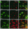
FIG. 1.
FIG. 1.
Intracellular localization of the UL51 protein in ST51 cells. ST51 cells were fixed; double stained with the anti-UL51 rabbit polyclonal antibody (a, d, and g) and mouse monoclonal antibodies against Golgi-58K protein (b), GM130 (e), or β-COP (h); and then reacted with anti-rabbit IgG-conjugated FITC or anti-mouse IgG-conjugated TRITC. The merged images are shown in panels c, f, and i. The insets show high magnification of the juxtanuclear region.
FIG. 2.
FIG. 2.
Intracellular localization of the UL51 protein in HSV-1-infected U251 cells. Mock-infected (a) and HSV-1-infected (b to j) U251 cells were fixed at 6 (b and e to j), 12 (c), and 24 (d) h p.i. as described in Materials and Methods. The samples were double stained with the anti-UL51 rabbit polyclonal antibody (a to e and h) and anti-Golgi-58K protein mouse monoclonal antibody (f) or anti-GM130 mouse monoclonal antibody (i) and then reacted with anti-rabbit IgG-conjugated FITC or anti-mouse IgG-conjugated TRITC. The merged images are shown in panels g and j. Infected cells were pretreated with normal goat serum to block nonspecific antibody reaction. The insets show high magnification of the juxtanuclear region.
FIG. 3.
FIG. 3.
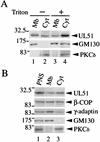
Subcellular distribution of the UL51 protein. (A) Lysates of U251 cells transiently expressing the UL51w/t protein were separated into the membranous pellet (Mb) (lanes 1 and 3) and the cytosolic supernatant (Cyt) (lanes 2 and 4) by ultracentrifugation (120,000 × g, 1 h) in the absence (lanes 1 and 2) or presence (lanes 3 and 4) of 1% Triton X-100 as described in Materials and Methods. The samples were subjected to SDS-PAGE and analyzed by Western blotting with the anti-UL51 rabbit polyclonal antibody (upper panel) and mouse monoclonal antibodies against GM130 (middle panel) or PKCδ (lower panel). (B) ST51 cells were homogenized, the resulting PNS (lane 1) was ultracentrifuged (120,000 × g, 1 h), and the membranous pellets (lane 2) and the cytosolic supernatants (lane 3) were collected. The samples were separated by SDS-PAGE and analyzed by Western blotting with the anti-UL51 rabbit polyclonal antibody and mouse monoclonal antibodies against β-COP, γ-adaptin, GM130, or PKCδ.
FIG. 4.
FIG. 4.
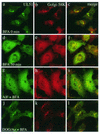
Effect of BFA on the distribution of UL51 protein and Golgi-58K protein. ST51 cells were pretreated with either AlF (g to i) or DOG-Az (j to l) for 10 min at 37°C and mock treated (a to c) or treated with BFA (5 μg/ml) (d to l) for 30 min at 37°C. Cells were then fixed with 4% paraformaldehyde and permeabilized with 0.1% Triton X-100. The cells were double stained with anti-UL51 rabbit polyclonal antibody and anti-Golgi-58K protein mouse monoclonal antibody and then reacted with anti-rabbit IgG-conjugated FITC and anti-mouse IgG-conjugated TRITC. Fluorescence images were obtained with the Bio-Rad MRC 1024 confocal imaging system.
FIG. 5.
FIG. 5.
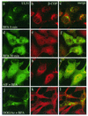
Effect of BFA on the distribution of UL51 protein and β-COP. ST51 cells were pretreated with either AlF (g to i) or DOG-Az (j to l) for 10 min at 37°C and mock treated (a to c) or treated with BFA (5 μg/ml) (d to l) for 30 min at 37°C. Cells were then fixed with 4% paraformaldehyde and permeabilized with 0.1% Triton X-100. The cells were double stained and fluorescence images were obtained as described in the legend to Fig. 4.
FIG. 6.
FIG. 6.
Effect of N- and C-terminal deletions on the Golgi targeting of UL51 protein. (A) Construction of the UL51 deletion mutant proteins. All of the constructs were inserted into pcDNA3.1(+) as described in Materials and Methods. Highly conserved amino acid sequences in alphaherpesviruses are shown by vertical lines in green. Proline-rich region are shown in red. (B) Expression of the UL51 deletion mutant proteins. U251 cells were transfected with pcDNA-UL51w/t or each plasmid and labeled with [35S]methionine as described in Materials and Methods. Cell lysates were subjected to immunoprecipitation with anti-UL51 rabbit polyclonal antibody, and the labeled proteins were separated by SDS-PAGE and detected by autoradiography with the Fujix Bioimaging Analyser BAS 2000 system. The main bands of each deletion protein are shown by black dots. Molecular mass markers (in kilodaltons) are shown on the left. (C) Intracellular localization of the UL51 deletion mutant proteins. U251 cells were transfected with each plasmid. After 24 h, cells were fixed with 4% paraformaldehyde and permeabilized with 0.1% Triton X-100. The samples were stained with the anti-UL51 rabbit polyclonal antibody and then reacted with anti-rabbit IgG-conjugated FITC. Fluorescence images were obtained with the Bio-Rad MRC 1024 confocal imaging system.
FIG. 7.
FIG. 7.
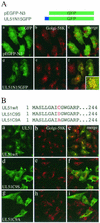
Mutational analysis of the first 15 amino acid residues of the UL51 protein. (A) The first 15 amino acids of UL51w/t were fused to the N terminus of GFP in frame (UL51N15GFP). U251 cells were transfected with pEGFP-N3 (panels a to c) or UL51N15GFP (d to f). At 24 h posttransfection, the cells were fixed and permeabilized as described in Materials and Methods. The samples were reacted with anti-Golgi-58K protein mouse monoclonal antibody (panels b and e). The inset shows high magnification of the juxtanuclear region. (B) The cysteine at position 9 of UL51w/t was replaced by serine (UL51C9S) or alanine (UL51C9A). U251 cells were transfected with UL51w/t (panels a to c), UL51C9S (panels d to f), or UL51C9A (panels g to i). The samples were double stained with anti-UL51 rabbit polyclonal antibody (panels a, d, and g) and anti-Golgi-58K protein mouse monoclonal antibody (panels b, e, and h). Fluorescence images were obtained with the Bio-Rad MRC 1024 confocal imaging system.
FIG. 8.
FIG. 8.
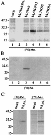
Palmitoylation of UL51 protein. (A and B) Transfected U251 cells were labeled with [35S]methionine (A) or [3H]palmitic acid (B) as described in Materials and Methods. Cell lysates were immunoprecipitated with anti-UL51 rabbit polyclonal antibody (lanes 1 and 3 to 6) or preimmune rabbit serum (lane 2), and the labeled proteins were mixed with the sample buffer (without β-mercaptoethanol). The samples were separated by SDS-PAGE and detected by autoradiography. (C) HSV-1-infected U251 cells were labeled with [35S]methionine or [3H]palmitic acid. At 9 h p.i., cells were harvested and the lysates were immunoprecipitated and analyzed as described in Materials and Methods.
FIG. 9.
FIG. 9.
Membrane binding of nonpalmitoylated UL51 mutant proteins. Transfected U251 cells were harvested at 24 h posttransfection. The cells were homogenized, the resulting PNS was ultracentrifuged (120,000 × g, 1 h), and then the membranous pellets (Mb) and the cytosolic supernatants (Cyt) were collected as described in Materials and Methods. The samples were separated by SDS-PAGE and analyzed by Western blotting with the anti-UL51 rabbit polyclonal antibody and mouse monoclonal antibodies against GM130, β-COP, or PKCδ.
FIG. 10.
FIG. 10.
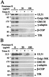
Sensitivity of UL51 protein to proteinase K. U251 cells expressing only the UL51w/t protein (A) and HSV-1-infected U251 cells (B) were treated with proteinase K in the absence (−) or presence (+) of 1% Triton X-100. All samples were then separated by SDS-PAGE and analyzed by Western blotting with the anti-UL51 rabbit polyclonal antibody and with mouse monoclonal antibodies against the Golgi-58K protein, GM130, γ-adaptin, β-COP, or TfR. Positions of molecular mass markers (in kilodaltons) are indicated on the left.
FIG. 11.
FIG. 11.
TIEM of HSV-1-infected U251 cells. Thin sections were prepared as described in Materials and Methods and incubated with preimmune rabbit serum (A) or the anti-UL51 rabbit polyclonal antibody (B) after treatment with 20% normal goat serum to block nonspecific antibody reactions. Samples were then incubated with anti-rabbit IgG-conjugated 10-nm-diameter gold particles. After extensive rinsing, sections were stained with uranyl acetate and lead citrate and examined with a Hitachi H7100 transmission electron microscope at 100 kV. Some of the enveloped capsids (arrows) were labeled. Labeling was also found in L-particle-like structures (large arrowheads) and along with membranous structures (small arrowheads). The inset shows an enveloped intracellular capsid labeled for UL51 proteins. Bars, 200 nm.

Similar articles

Cited by

References

    1. Albrecht, J. C., J. Nicholas, D. Biller, K. R. Cameron, B. Biesinger, C. Newman, S. Wittmann, M. A. Craxton, H. Coleman, B. Fleckenstein, and R. W. Honess. 1992. Primary structure of the herpesvirus saimiri genome. J. Virol. 66:5047-5058. - PMC - PubMed
    1. Avitabile, E., S. D. Gaeta, M. R. Torrisi, P. L. Ward, B. Roizman, and G. Campadelli-Fiume. 1995. Redistribution of microtubules and Golgi apparatus in herpes simplex virus-infected cells and their role in viral exocytosis. J. Virol. 69:7472-7482. - PMC - PubMed
    1. Baer, R. J., A. T. Bankier, M. D. Biggin, P. L. Deininger, P. J. Farrell, T. J. Gibson, G. Hatfull, G. S. Hudson, S. C. Satchwell, C. Seguin, P. Tuffnell, and B. G. Barrell. 1984. DNA sequence and expression of the B95-8 Epstein-Barr virus genome. Nature (London) 310:207-211. - PubMed
    1. Baines, J. D., R. J. Jacob, L. Simmerman, and B. Roizman. 1995. The herpes simplex virus 1 UL11 proteins are associated with cytoplasmic and nuclear membranes and with nuclear bodies of infected cells. J. Virol. 69:825-833. - PMC - PubMed
    1. Baines, J. D., and B. Roizman. 1992. The UL11 gene of herpes simplex virus 1 encodes a function that facilitates nucleocapsid envelopment and egress from cells. J. Virol. 66:5168-5174. - PMC - PubMed

Publication types

MeSH terms

LinkOut - more resources