. 2003 Mar;77(5):3247-56.
doi: 10.1128/jvi.77.5.3247-3256.2003.

Effects of point mutations in the major capsid protein of beet western yellows virus on capsid formation, virus accumulation, and aphid transmission

Affiliations

Effects of point mutations in the major capsid protein of beet western yellows virus on capsid formation, virus accumulation, and aphid transmission

V Brault et al. J Virol. 2003 Mar.

Abstract

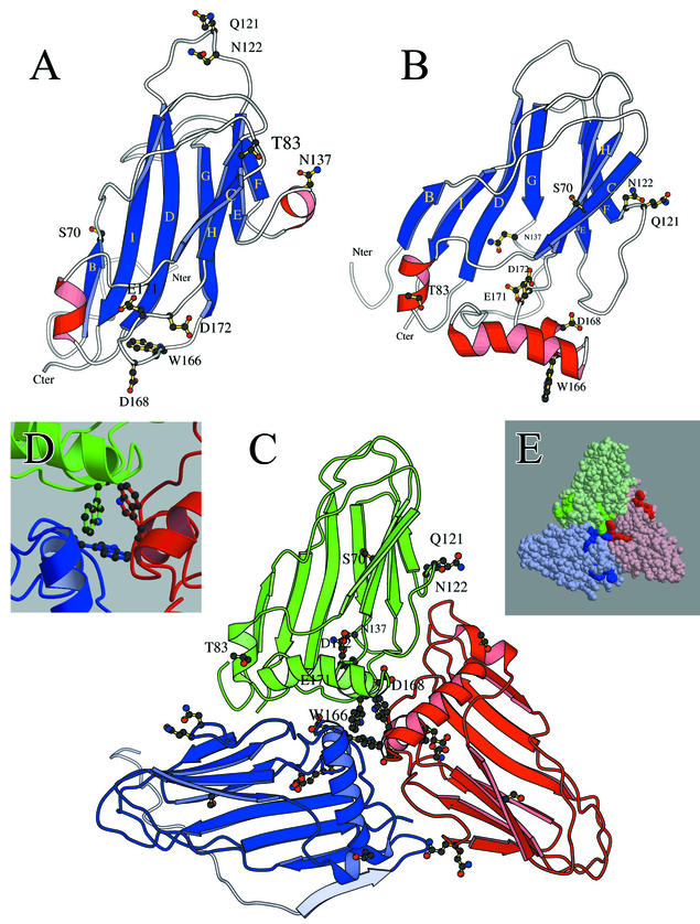
Point mutations were introduced into the major capsid protein (P3) of cloned infectious cDNA of the polerovirus beet western yellows virus (BWYV) by manipulation of cloned infectious cDNA. Seven mutations targeted sites on the S domain predicted to lie on the capsid surface. An eighth mutation eliminated two arginine residues in the R domain, which is thought to extend into the capsid interior. The effects of the mutations on virus capsid formation, virus accumulation in protoplasts and plants, and aphid transmission were tested. All of the mutants replicated in protoplasts. The S-domain mutant W166R failed to protect viral RNA from RNase attack, suggesting that this particular mutation interfered with stable capsid formation. The R-domain mutant R7A/R8A protected approximately 90% of the viral RNA strand from RNase, suggesting that lower positive-charge density in the mutant capsid interior interfered with stable packaging of the complete strand into virions. Neither of these mutants systemically infected plants. The six remaining mutants properly packaged viral RNA and could invade Nicotiana clevelandii systemically following agroinfection. Mutant Q121E/N122D was poorly transmitted by aphids, implicating one or both targeted residues in virus-vector interactions. Successful transmission of mutant D172N was accompanied either by reversion to the wild type or by appearance of a second-site mutation, N137D. This finding indicates that D172 is also important for transmission but that the D172N transmission defect can be compensated for by a "reverse" substitution at another site. The results have been used to evaluate possible structural models for the BWYV capsid.


Figures

FIG. 1.
FIG. 1.
(A) Genetic organization of BWYV RNA. Important ORFs are represented by numbered rectangles, and untranslated sequences are represented by horizontal lines. The major coat protein (P3) gene and the P3 R and S domains are shown below. The thick arrows indicate the positions of primers used to analyze progeny virus RNA sequences as described in the text. (B) Alignment of the S-domain sequences of BWYV and PLRV and positions of the BWYV S-domain mutations. The alignment was produced by SEQLAB. The vertical lines indicate identical amino acids, and the semicolons indicate similar amino acids (S and T, R and K, and F and Y). The positions of epitopes 5 and 10 (20) in the PLRV capsid are indicated below the PLRV sequence. The positions of point mutations introduced into the BWYV S domain are shown above the sequences.
FIG. 2.
FIG. 2.
Amplification in protoplasts of BWYV RNA carrying mutations in the P3 cistron. (A) Northern blot analysis of total RNA extracted from 200,000 protoplasts inoculated with transcripts of BW0 (lanes 2 and 8), S70A (lane 3), T83A (lane 4), E171Q (lane 5), D172N (lane 6), R7A/R8A (lane 9), Q121E/N122D (lane 10), W166R (lane 11), and D168N (lane 12). RNA extracted from mock-inoculated protoplasts was loaded in lanes 1 and 7. The positions of genomic (g) and subgenomic (sg) RNAs are indicated on the right. (B) Detection of encapsidated viral RNA from infected protoplasts. Total RNA was extracted under conditions in which only virion-packaged RNA remains intact. The lane numbering is as in panel A. In panels A and B, the blots on the left were hybridized with a digoxigenin-labeled riboprobe complementary to the 3′-terminal 196 residues of BWYV RNA. In the blots on the right, the same riboprobe was 32P labeled. (C) Immunodetection of BWYV P3 and RT proteins in protein extracts from protoplasts infected with the above-mentioned mutants. The upper part of the blot was probed with an RT domain-specific antiserum (15), and the lower part was probed with a P3-specific antiserum. The lines on the left indicate the positions of 82-, 49-, 33.5-, and 28.5-kDa molecular-mass markers. Minor species detected by the RT domain-specific antiserum in the upper blot are assumed to be RT protein degradation products.
FIG. 3.
FIG. 3.
Distributions of nucleotide mutations detected in viral progeny RNA following agroinfection of N. clevelandii with wild-type or mutant BWYV cDNA constructs (A) or after successful aphid transmission of virus from agroinfected leaves or purified virus solutions to M. perfoliata (B). The results obtained with the two different types of inoculum did not differ significantly and have been combined. For each construct, the column to the immediate right indicates the total number of reverse transcription-PCR clones sequenced and the number of plants (pl) that were analyzed. The column on the far right indicates the number of nucleotide substitutions observed per 1,000 nt sequenced in the progeny. The position of the primary mutation in each mutant clone is indicated by a triangle. An open triangle indicates that the primary mutation was conserved in the progeny, and a solid triangle indicates that the primary mutation had undergone reversion to the wild-type sequence (observed only for some D172N progeny). The circles symbolize the different second-site nucleotide mutations which were detected in the progeny reverse transcription-PCR clones; an open circle represents a silent mutation, and a solid circle represents a mutation which resulted in an amino acid change in P3 (the wild-type amino acid at each such position is shown above the line, and the observed amino acid substitution is shown below). Stacks of symbols indicate that the mutation in question was detected multiple times. The ladder at the top of each panel refers to the amino acid (aa) position in P3.
FIG. 4.
FIG. 4.
BWYV P3 S-domain residues predicted to be in β-sheets (horizontal lines) and α-helices (loops) in the Terradot model (symbols above the sequence) and in the alternative model proposed in this paper (symbols below the sequence). The positions of the BWYV counterparts of PLRV epitopes 5 and 10 are indicated by thick lines.
FIG.5.
FIG.5.
(A and B) Structures of the BWYV S domain (A conformation) predicted in the Terradot model (19) (A) and in this paper (B). The views look down on the outer surface, and amino acid residues targeted for mutation in the present work are identified. The blue ribbons correspond to β-sheets, and the red coils correspond to α-helices (Fig. 4). The R domain, which is believed to extend into the capsid interior from the N terminus of the S domain (Nter), is not shown. (C) Asymmetric trimer of subunits in the A (green), B (red), and C (blue) conformations derived from our BWYV P3 S-domain structure (panel B). Amino acids targeted for mutation are identified in the green subunit. For the blue subunit, the R domain is shown in light blue. The β-sheet (light blue ribbon) near the C terminus of the R domain was predicted by computer modeling (unpublished observations), but the rest of the R domain sequence is depicted as an arbitrarily drawn disordered structure. (D) Positions of W166 residues on the subunits at the center of the trimer shown in panel C. Chains in different subunits are colored as in panel C. (E) Solid-surface representation of the structure shown in panel C. The darker colors on each subunit indicate the positions of residues corresponding to PLRV epitope 5 (the elongated structures near the sides of the triangle) and epitope 10 (the trilobate structures at the center).

References

    1. Brault, V., J. F. J. M. van den Heuvel, M. Verbeek, V. Ziegler-Graff, A. Reutenauer, E. Herrbach, J. C. Garaud, H. Guilley, K. Richards, and G. Jonard. 1995. Aphid transmission of beet western yellows luteovirus requires the minor capsid readthrough protein P74. EMBO J. 14:650-659. - PMC - PubMed
    1. Brault, V., J. Mutterer, D. Scheidecker, M. T. Simonis, E. Herrbach, K. Richards, and V. Ziegler-Graff. 2000. Effects of point mutations in the readthrough domain of beet western yellows virus minor capsid protein on virus accumulation in planta and on transmission by aphids. J. Virol. 74:1140-1148. - PMC - PubMed
    1. Bruyère, A., V. Brault, V. Ziegler-Graff, M. T. Simonis, J. F. J. M. van den Heuvel, K. Richards, H. Guilley, G. Jonard, and E. Herrbach. 1997. Effects of mutations in the beet western yellows virus readthrough protein on its expression and packaging, and on virus accumulation, symptoms and aphid transmission. Virology 230:323-334. - PubMed
    1. Cheng, S. L., L. L. Domier, and C. J. D'Arcy. 1994. Detection of the readthrough protein of barley yellow dwarf virus. Virology 202:1003-1006. - PubMed
    1. Dolja, V. V., and E. V. Koonin. 1991. Phylogeny of capsid proteins of small icosahedral RNA plant viruses. J. Gen. Virol. 72:1481-1486. - PubMed

Publication types

LinkOut - more resources