. 2003 Mar 4;100(5):2700-5.
doi: 10.1073/pnas.0438035100. Epub 2003 Feb 13.

Development of murine ischemic cardiomyopathy is associated with a transient inflammatory reaction and depends on reactive oxygen species

Affiliations

Development of murine ischemic cardiomyopathy is associated with a transient inflammatory reaction and depends on reactive oxygen species

Oliver Dewald et al. Proc Natl Acad Sci U S A. .

Abstract

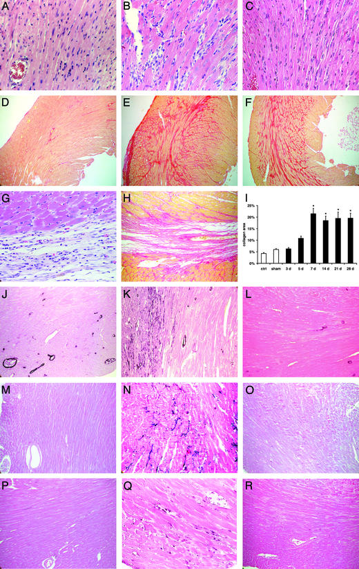
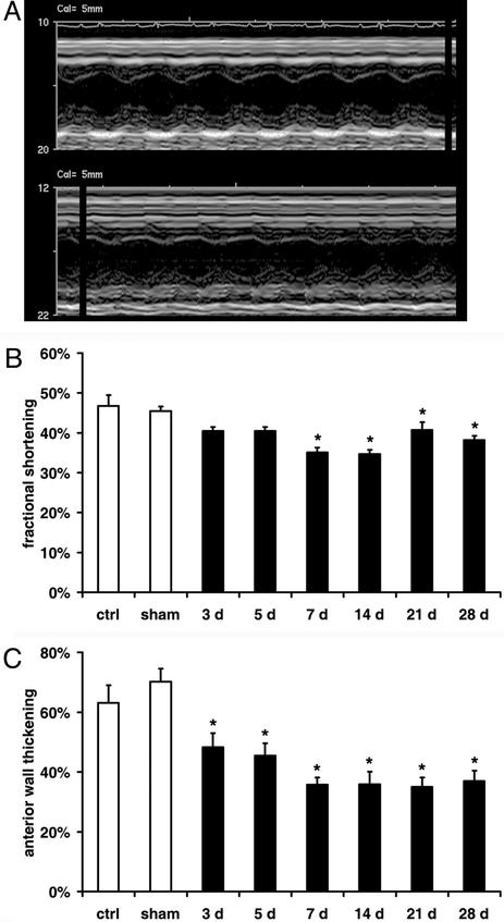
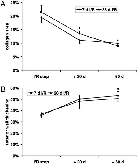
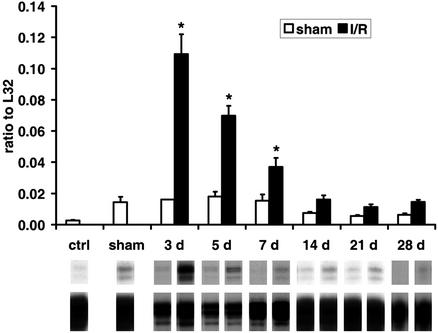
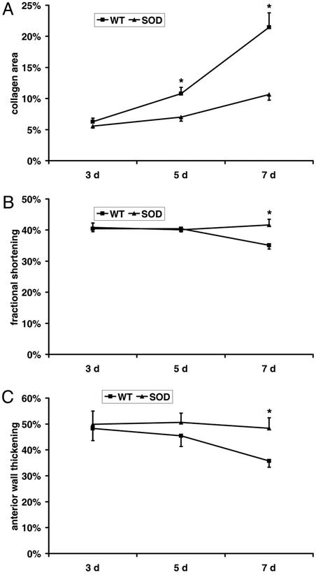
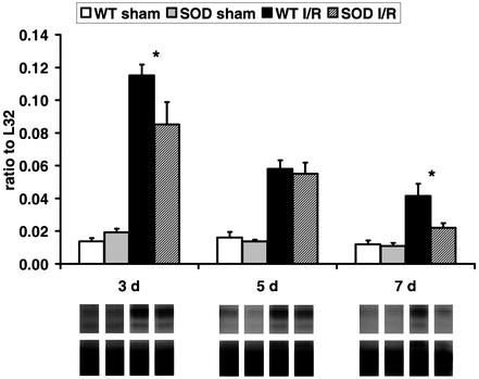
We examined the effects of daily repetitive brief (15 min) myocardial ischemia and reperfusion (I/R) in WT C57BL6 and extracellular superoxide dismutase (EC-SOD)-overexpressing mice. In the absence of myocardial necrosis, I/R resulted in persistent fibrosis in ischemic areas of C57/BL6 mice associated with persistent global and segmental anterior wall dysfunction. The I/R protocol induced chemokines (peak 3 days) followed sequentially by infiltration of macrophages and myofibroblasts (5 days). Fibrosis peaked at 7 days and was stable at 28 days despite regression of the chemokine and cellular response. Discontinuation of I/R at 7 or 28 days led to regression of fibrosis and ventricular dysfunction. In contrast, the EC-SOD mice developed markedly less chemokine induction, cell response, and fibrosis, with no ventricular dysfunction. Reversible fibrosis and ventricular dysfunction are features of human hibernating myocardium. The reduction of the cellular and functional response in EC-SOD mice suggests a role for reactive O(2) in the pathogenesis of ischemic cardiomyopathy.


Figures

Figure 1
Figure 1
Histopathological findings in C57/BL6 mice undergoing repetitive I/R protocol (daily 15-min I/R). Hematoxylin and eosin staining of the left ventricular AW in sham (A), 5-day I/R (B), and 28-day I/R (C) mouse (×400) demonstrates marked cellular infiltration after 5 days of brief repetitive I/R. Collagen deposition stained with picrosirius red of the same area in sham (D), 7-day I/R (E), and 28-day I/R (F) mouse (×100). Note that fibrotic changes are interstitial and there is no myocyte loss. In contrast, (G) myocardial infarction (1 h of ischemia and 7 days of reperfusion) is associated with replacement fibrosis (H) (×400 and 200). (I) Quantitative analysis of the collagen deposition in the left ventricular AW revealed significant increase in fibrosis after 7 days of brief repetitive I/R (*, P < 0.05 vs. respective shams). (J) Staining for α-smooth muscle actin shows only positive arteriolar wall in sham animals, but (K) after 5 days of brief repetitive I/R identifies phenotypically modulated myofibroblasts infiltrating the interstitial space (×200). (L) Myofibroblasts diminish after 28 days of I/R. (M) Tenascin expression was not found in sham mice (×200). Tenascin staining reached a maximum after 5 days (N) and diminished thereafter until 28 days (O) of I/R. (P) Sham hearts contain few macrophages stained with F4/80, in contrast to (Q) extensive macrophage infiltration after 5 days of I/R (×200). (R) Macrophages diminish thereafter until 28 days.
Figure 2
Figure 2
Ventricular function during the repetitive I/R protocol. (A) Short axis M-mode echocardiography picture of sham animal (Upper) shows normokinetic left ventricular AW in contrast to 7 days of I/R mouse with hypokinetic AW (Lower). (B) Fractional shortening indicates normal global function in sham mice and significant dysfunction in I/R animals. (C) Quantitation of AW thickening identifies significant persistent regional ventricular dysfunction. *, P < 0.05 vs. respective shams.
Figure 3
Figure 3
Regression of fibrosis and ventricular dysfunction after discontinuation of repetitive I/R protocol. (A) Fibrosis regression in the left ventricular AW after discontinuation of the 7- and 28-day I/R protocol followed by 30- and 60-day recovery period. (B) AW thickening during the regression process. *, P < 0.05 vs. respective I/R groups.
Figure 4
Figure 4
mRNA expression of chemokines in C57/BL6 mice undergoing repetitive I/R. (Upper) MCP-1 is highly induced after 3 days of I/R and normalizes after 14 days when compared with shams. (Lower) A representative transcript band with a corresponding housekeeping gene band. *, P < 0.05 vs. respective shams.
Figure 5
Figure 5
Fibrosis and ventricular function in EC-SOD-overexpressing mice. (A) Collagen area of C57/BL6 mice compared with the EC-SOD mice revealed significantly less fibrosis in EC-SOD hearts after the repetitive brief I/R protocol. (B) WT mice exhibit decreased fractional shortening after 7 days of I/R, suggesting global dysfunction, whereas EC-SOD-overexpressing mice show normal function. (C) In addition, WT mice show diminished AW thickening, suggesting regional dysfunction, in contrast to EC-SOD mice, which demonstrate normal regional function. *, P < 0.05 in C57/BL6 vs. EC-SOD I/R groups.
Figure 6
Figure 6
Comparison of chemokine induction in C57/BL6 and EC-SOD mice. (Upper) MCP-1 evaluation shows no difference between C57/BL6 and EC-SOD shams. The I/R protocol induced significantly less MCP-1 in EC-SOD than in C57/BL6 mice. MCP-1 expression in EC-SOD mice after 7 days of I/R is not significantly different from the corresponding shams. (Lower) A representative transcript band with corresponding housekeeping gene band. *, P < 0.05 in C57/BL6 vs. EC-SOD I/R groups.

References

    1. Rahimtoola S H. Circulation. 1985;72:V123–V135. - PubMed
    1. Kloner R A, Bolli R, Marban E, Reinlib L, Braunwald E. Circulation. 1998;97:1848–1867. - PubMed
    1. Vanoverschelde J L, Wijns W, Borgers M, Heyndrickx G, Depre C, Flameng W, Melin J A. Circulation. 1997;95:1961–1971. - PubMed
    1. Elsasser A, Schlepper M, Klovekorn W P, Cai W J, Zimmermann R, Muller K D, Strasser R, Kostin S, Gagel C, Munkel B, et al. Circulation. 1997;96:2920–2931. - PubMed
    1. Frangogiannis N G, Shimoni S, Chang S M, Ren G, Dewald O, Gersch C, Shan K, Aggeli C, Reardon M, Letsou G V, et al. J Am Coll Cardiol. 2002;39:1468–1474. - PubMed

Publication types

MeSH terms