. 2003 Feb;131(2):824-37.
doi: 10.1104/pp.010538.

In plants, 3-o-methylglucose is phosphorylated by hexokinase but not perceived as a sugar

Affiliations

In plants, 3-o-methylglucose is phosphorylated by hexokinase but not perceived as a sugar

Sandra Cortès et al. Plant Physiol. 2003 Feb.

Abstract

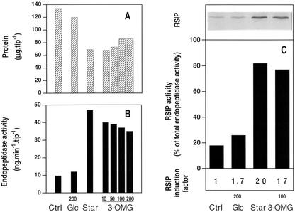
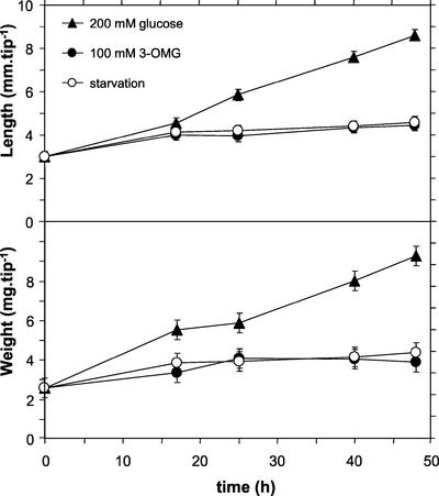
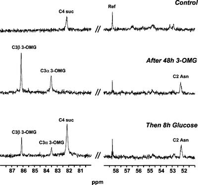
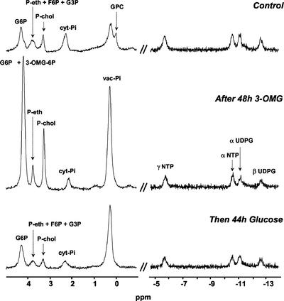
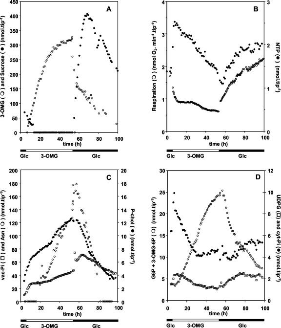
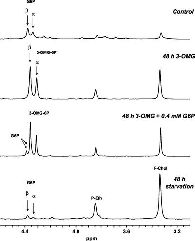
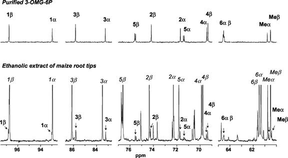
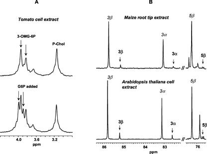
In plants, sugars are the main respiratory substrates and important signaling molecules in the regulation of carbon metabolism. Sugar signaling studies suggested that sugar sensing involves several key components, among them hexokinase (HXK). Although the sensing mechanism of HXK is unknown, several experiments support the hypothesis that hexose phosphorylation is a determining factor. Glucose (Glc) analogs transported into cells but not phosphorylated are frequently used to test this hypothesis, among them 3-O-methyl-Glc (3-OMG). The aim of the present work was to investigate the effects and fate of 3-OMG in heterotrophic plant cells. Measurements of respiration rates, protein and metabolite contents, and protease activities and amounts showed that 3-OMG is not a respiratory substrate and does not contribute to biosynthesis. Proteolysis and lipolysis are induced in 3-OMG-fed maize (Zea mays L. cv DEA) roots in the same way as in sugar-starved organs. However, contrary to the generally accepted idea, phosphorous and carbon nuclear magnetic resonance experiments and enzymatic assays prove that 3-OMG is phosphorylated to 3-OMG-6-phosphate, which accumulates in the cells. Insofar as plant HXK is involved in sugar sensing, these findings are discussed on the basis of the kinetic properties because the catalytic efficiency of HXK isolated from maize root tips is five orders of magnitude lower for 3-OMG than for Glc and Man.


Figures

Figure 1
Figure 1
Effects of 3-OMG on protein content (A), proteolytic activity (B), and RSIP expression (C) in maize root tips. Incubation of freshly excised root tips during 4 h in 200 mm Glc, defined as control (Ctrl). Otherwise, excised root tips were incubated for 48 h without carbon substrate (Star), with 200 mm Glc (Glc), or in the presence of 10, 50, 100, or 200 mm 3-OMG, and analyzed for proteins (A), endopeptidase activity (B), and western-blot RSIP detection (C) as described in “Materials and Methods.” C, Total proteins from one root tip equivalent (between 134 and 65 μg protein according to A) were loaded in each lane for western-blot RSIP analysis. The percentage of endopeptidase activity due to RSIP in control (Ctrl), 200 mm Glc-fed (Glc), Glc-starved (Star), and 100 mm 3-OMG-fed root tips was measured after immunoprecipitation experiments with anti-RSIP antibodies. Data are the mean of five (protein), three (endopeptidase activities), and two (immunoprecipitation) independent experiments. sds are less than 15%.
Figure 2
Figure 2
Growth of excised maize root tips in different nutritive media. Three sets of maize root tips were incubated either with or without the hexose indicated by symbols. Each point was obtained from a sample of 20 root tips. Data are the mean ± sd of two independent experiments.
Figure 3
Figure 3
13C NMR spectra of excised maize root tips in different nutritive media: regions used for quantification of sugars and amino acids. Excised maize root tips were first perifused with a medium containing 200 mm Glc during 4 h (Control, top). Then, 100 mm 3-OMG was substituted for Glc for 48 h (middle). Finally, root tips were perifused with 200 mm Glc (bottom). Ref, 13CH2 resonance of ethanol contained in a capillary. Spectra were acquired at 100.61 MHz with a free induction decay (FID) resolution of 1.4 Hz, a 45° radiofrequency (RF) pulse (45 μs), and 3,000 transients repeated every 1 s.
Figure 4
Figure 4
31P NMR spectra of excised maize root tips in different nutritive media: regions used for quantification of phosphoesters and nucleoside phosphates. Excised maize root tips were first perifused with a medium containing 200 mm Glc during 4 h (Control, top). Then, 100 mm 3-OMG was substituted for Glc during 48 h (middle). Finally, the sample was perifused with 200 mm Glc (bottom). GPC, glycerylphosphorylcholine. Spectra were acquired at 161.98 MHz with an FID resolution of 1.7 Hz, a 45° RF pulse (32.5 μs), and 1,000 transients repeated every 0.6 s.
Figure 5
Figure 5
Time courses of respiration and metabolite contents in excised maize root tips. Metabolite concentrations, deduced from spectral intensities as described in “Materials and Methods,” are plotted as a function of the incubation time. The composition of the external medium is indicated at the bottom. Data are from one representative experiment of three. A, Time courses of intracellular hexose concentrations. B, Time courses of respiration rate and NTP concentration. C, Time courses of concentrations of starvation markers: Asn, P-chol, and vac-Pi. D, Time courses of concentrations of cyt-Pi, UDPG, and C6 hexose phosphates.
Figure 6
Figure 6
31P NMR spectra of acid extracts of maize root tips. Samples of 2,000 excised root tips were filtered and frozen in liquid N2 after 4-h incubation with 200 mm Glc (Control, top spectrum), or 48-h incubation with 100 mm 3-OMG (second spectrum from top), or 48-h incubation without substrate (bottom spectrum). The third spectrum from top was recorded after an addition of 0.4 mm G6P into the extract of 3-OMG-treated root tips. Acid extracts were prepared as described in “Materials and Methods” and pH was adjusted at 7.5. Spectra were acquired at 161.98 MHz with an FID resolution of 0.6 Hz, a 60° RF pulse (15 μs), and 2,048 transients repeated every 3.5 s. The exponential apodization was 0.5 Hz.
Figure 7
Figure 7
13C NMR identification of 3-OMG-6P. Synthesis of 3-OMG-6P from 3-OMG and ATP using yeast HXK, and isolation using exclusion-diffusion chromatography as described in “Materials and Methods.” Top, 13C NMR spectrum of purified 3-OMG-6P. Bottom, 13C NMR spectrum of an ethanolic extract of 1,000 maize root tips incubated with 100 mm 3-OMG for 48 h. Arrows and bold labels indicate the resonances of 3-OMG-6P, whereas italic labels identify 3-OMG resonances. Spectra were acquired at 100.61 MHz with an FID resolution of 0.7 Hz, a 60° RF pulse (10 μs), and 2,048 transients repeated every 1.75 s. The exponential apodization was 0.5 Hz.
Figure 8
Figure 8
NMR identification of 3-OMG-6P in extracts of different species. A, 31P NMR spectra of tomato cell extracts. Cell extracts were prepared from 4-d-old cells incubated during 24 h with 100 mm 3-OMG. The figure shows the phosphomonoester resonance interval of the 31P NMR spectra of one acid extract before (top) and after (bottom) addition of 2 mm G6P in the sample. It contained 80 mm trans-1,2-diaminocyclohexane-N,N,N′,N′-tetraacetic acid (CDTA) and pH was adjusted to 6.9 to improve resolution in this spectral interval. These spectra are the sum of 1,024 transients obtained with the acquisition parameters reported in Figure 6. B, 13C NMR spectra of maize root and Arabidopsis cell extracts. Comparison of two intervals of the 13C NMR spectra of acid extracts of maize root tips (top) and Arabidopsis cells (bottom) proving the presence of 3-OMG-6P in these cells incubated for 24 h with 100 mm 3-OMG. The selected 3-OMG-6P resonances (bold labels) are characteristic of the 13C NMR spectrum of this molecule (Fig. 7). The intense 3-OMG resonances (italic labels) reveal the accumulation of the precursor in the cells. No CDTA was added to this sample, and pH was 7.0. Spectra were obtained by adding 6,144 transients recorded with the acquisition parameters reported in Figure 7.

Similar articles

Cited by

References

    1. ap Rees T. Hexose phosphate metabolism by non-photosynthetic tissues of higher plants. In: Preiss J, editor. The Biochemistry of Plants. Vol. 14. New York: Academic Press; 1988. pp. 1–33.
    1. Axelos M, Curie C, Mazzolini L, Bardet C, Lescure B. A protocol for transient gene expression in Arabidopsis thaliana protoplasts isolated from cell suspension cultures. Plant Physiol Biochem. 1992;30:123–128.
    1. Bock K, Pedersen C. Carbon-13 nuclear magnetic resonance spectroscopy of monosaccharides. In: Tipson RS, Horton D, editors. Advances in Carbohydrates Chemistry and Biochemistry. New York: Academic Press; 1983. pp. 27–66.
    1. Bradford MM. A rapid and sensitive method for the quantification of microgram quantities of proteins utilizing the principle of protein-dye binding. Anal Biochem. 1976;72:248–254. - PubMed
    1. Brouquisse R, Evrard A, Rolin D, Raymond P, Roby C. Regulation of protein degradation and protease expression by mannose in maize root tips. Pi sequestration by mannose may hinder the study of its signaling properties. Plant Physiol. 2001;125:1485–1498. - PMC - PubMed

Publication types

MeSH terms

LinkOut - more resources