

Congenital talipes equinovarus (clubfoot): a disorder of the foot but not the hand
- PMID: 12587918
- PMCID: PMC1571059
- DOI: 10.1046/j.1469-7580.2003.00147.x
Congenital talipes equinovarus (clubfoot): a disorder of the foot but not the hand
Abstract
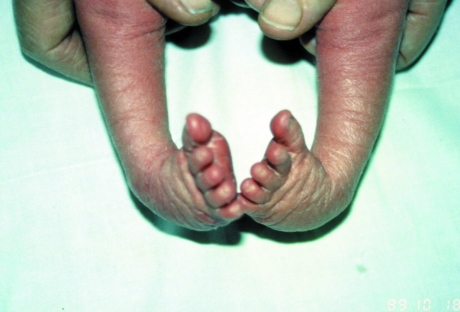
Idiopathic (non-syndromic) congenital talipes equinovarus, or clubfoot, is a poorly understood but common developmental disorder of the lower limb, which affects at least 2 per 1000 Scottish births (ISD data). It is defined as a fixation of the foot in a hand-like orientation--in adduction, supination and varus--with concomitant soft tissue abnormalities. Despite advances in treatment, disability often persists. The aetiology of the condition has been little studied and is poorly understood. Neurological, muscular, bony, connective tissue and vascular mechanisms have been proposed, but the only firm evidence is that the mildest cases appear to be associated with intra-uterine posture. There is evidence for a genetic contribution to congenital talipes equinovarus aetiology. Its incidence varies with ethnic group, and we found that a family history is present in 24-50% of cases, depending on the population studied. Complex segregation analysis suggests that the most likely inheritance pattern is a single gene of major effect operating against a polygenic background. Possible mechanisms for congenital talipes equinovarus are discussed.
Figures
References
-
- Atlas S, Menacho LCS, Ures S. Some new aspects in the pathology of clubfoot. Clin. Orthop. 1980;149:224–228. - PubMed
-
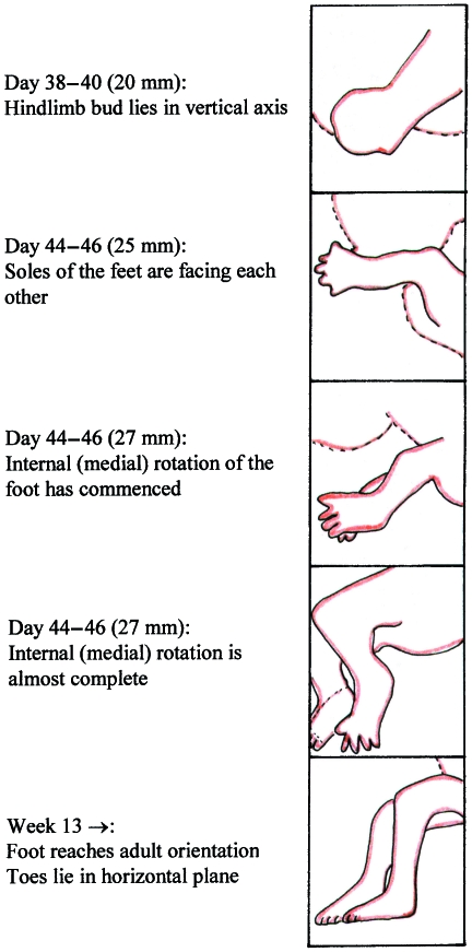
- Bareiter D. Prenatal development of the foot and ankle. J. Am. Podiatric Med. Assocn. 1995;85:753–764. - PubMed
-
- Barker S, Chesney D, Sharp L, et al. J. Med. Genet. 2001;38:S34.
-
- Barker S, MacNicol M. Seasonal distribution of idiopathic congenital talipes equinovarus in Scotland. J. Pediatr. Orthop. 2001;10:1–5. - PubMed
-
- Böhm M. The embryologic origin of club-foot. JBJS. 1929;XI:229–259.
Publication types
MeSH terms
LinkOut - more resources
Full Text Sources
