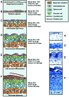
Covering the limb--formation of the integument
- PMID: 12587926
- PMCID: PMC1571060
- DOI: 10.1046/j.1469-7580.2003.00142.x
Covering the limb--formation of the integument
Abstract
An organism's outermost covering, the integument, has evolved to fulfil a diverse range of functions. Skin provides a physical barrier, an environment for immunological surveillance, and also performs a range of sensory, thermoregulatory and biosynthetic functions. Examination of the skin of limb digits reveals a range of skin types including the thickened hairless epidermis of the toe pads (palmar or plantar epidermis) and thinner epidermis between the hair follicles (interfollicular epidermis) of hairy skin. An important developmental function of skin is to give rise to a diverse group of appendages including hair follicles, with associated sebaceous glands (or feathers and scales in chick), eccrine sweat glands and the nail. A key question is how does this morphological variety arise from the single-layered epithelium covering embryonic limb buds? This review will attempt to address this question by linking the extensive morphological/anatomical data on maturation of epidermis and its appendages with (1) current research into the range, plasticity and location of the putative epidermal stems cells; (2) molecular/microenvironmental regulation of epidermal stem cell lineages and lineage choice; and (3) regulation of the differentiation pathways, focusing on differentiation of the interfollicular epidermis.
Figures


Similar articles
-
Designer skin: lineage commitment in postnatal epidermis.Trends Cell Biol. 2002 Apr;12(4):185-92. doi: 10.1016/s0962-8924(02)02263-8. Trends Cell Biol. 2002. PMID: 11978538 Review.
-
Lgr6 marks stem cells in the hair follicle that generate all cell lineages of the skin.Science. 2010 Mar 12;327(5971):1385-9. doi: 10.1126/science.1184733. Science. 2010. PMID: 20223988
-
Bmp7 mediates early signaling events during induction of chick epidermal organs.Dev Dyn. 2004 Sep;231(1):22-32. doi: 10.1002/dvdy.20096. Dev Dyn. 2004. PMID: 15305284
-
Hair Follicle and Sebaceous Gland De Novo Regeneration With Cultured Epidermal Stem Cells and Skin-Derived Precursors.Stem Cells Transl Med. 2016 Dec;5(12):1695-1706. doi: 10.5966/sctm.2015-0397. Epub 2016 Jul 25. Stem Cells Transl Med. 2016. PMID: 27458264 Free PMC article.
-
Connexins and pannexins in the integumentary system: the skin and appendages.Cell Mol Life Sci. 2015 Aug;72(15):2937-47. doi: 10.1007/s00018-015-1969-0. Epub 2015 Jun 20. Cell Mol Life Sci. 2015. PMID: 26091749 Free PMC article. Review.
Cited by
-
Conditional targeting of E-cadherin in skin: insights into hyperproliferative and degenerative responses.Proc Natl Acad Sci U S A. 2004 Jan 13;101(2):552-7. doi: 10.1073/pnas.0307437100. Epub 2004 Jan 2. Proc Natl Acad Sci U S A. 2004. PMID: 14704278 Free PMC article.
-
Association between the chronology of gestation and the morphometrical skin characteristics at childbirth: a development of predictive model.BMJ Health Care Inform. 2021 Dec;28(1):e100476. doi: 10.1136/bmjhci-2021-100476. BMJ Health Care Inform. 2021. PMID: 34876452 Free PMC article.
-
Expression patterns of astrocyte elevated gene-1 (AEG-1) during development of the mouse embryo.Gene Expr Patterns. 2010 Oct-Dec;10(7-8):361-7. doi: 10.1016/j.gep.2010.08.004. Epub 2010 Aug 22. Gene Expr Patterns. 2010. PMID: 20736086 Free PMC article.
-
Dual role of COUP-TF-interacting protein 2 in epidermal homeostasis and permeability barrier formation.J Invest Dermatol. 2009 Jun;129(6):1459-70. doi: 10.1038/jid.2008.392. Epub 2008 Dec 18. J Invest Dermatol. 2009. PMID: 19092943 Free PMC article.
-
Hedgehog signaling regulates sebaceous gland development.Am J Pathol. 2003 Dec;163(6):2173-8. doi: 10.1016/S0002-9440(10)63574-2. Am J Pathol. 2003. PMID: 14633591 Free PMC article.
References
-
- Akiyama M, Smith LT, Shimizu H. Changing patterns of localization of putative stem cells in developing human hair follicles. J. Invest. Dermatol. 2000;114:321–327. - PubMed
-
- Barsh G. of ancient tales and hairless tails. Nat. Genet. 1999;22:315–316. - PubMed
-
- Beer HD, Vindevoghel L, Gait MJ, Revest JM, Duan DR, Mason I, et al. Fibroblast growth factor (FGF) receptor 1-IIIb is a naturally occurring functional receptor for FGFs that is preferentially expressed in the skin and the brain. J. Biol. Chem. 2000;275:16091–16097. - PubMed
-
- Byrne C, Tainsky M, Fuchs E. Programming gene expression in developing epidermis. Development. 1994;120:2369–2383. - PubMed
Publication types
MeSH terms
LinkOut - more resources
Full Text Sources
Medical

