

Tendon morphogenesis in the developing avian limb: plasticity of fetal tendon fibroblasts
- PMID: 12587930
- PMCID: PMC1571057
- DOI: 10.1046/j.1469-7580.2003.00145.x
Tendon morphogenesis in the developing avian limb: plasticity of fetal tendon fibroblasts
Abstract
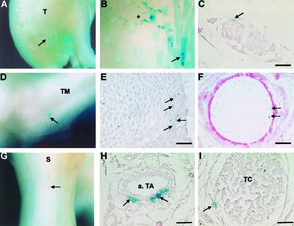
The differentiation and patterning of tendon fibroblasts is at present a poorly understood aspect of musculoskeletal development in the vertebrate limb. Precursors of tendon fibroblasts originate in the somatic mesoderm adjacent to the early limb bud and gradually become incorporated into the limb mesenchyme as development proceeds. It is unclear whether these progenitor cells are committed to the tendon lineage at this early stage, or whether cells become committed only as they are incorporated into a developing tendon. Following a review of our current knowledge of early tendon development, we present recent results from our preliminary studies looking at tendon cell commitment. Using a lacZ encoding replication-deficient retrovirus, we have mapped regions of the early limb bud that contain presumptive tendon progenitor cells, and later used these sites for implanting labelled fetal tendon fibroblasts into developing limbs. Following implantation, we found that these cells successfully re-incorporated into developing proximal and distal tendons, but also surprisingly contributed to other tissue lineages within the limb. Our results suggest that fetal tendon fibroblasts may not be irreversibly committed to a tendon cell fate in the limb and may be somewhat plastic in their ability to integrate into other tissue lineages during development.
Figures


Similar articles
-
Development and maintenance of tendons and ligaments.Development. 2021 Apr 15;148(8):dev186916. doi: 10.1242/dev.186916. Epub 2021 Apr 16. Development. 2021. PMID: 33913478 Free PMC article. Review.
-
Muscle and tendon morphogenesis in the avian hind limb.Development. 1998 Oct;125(20):4019-32. doi: 10.1242/dev.125.20.4019. Development. 1998. PMID: 9735363
-
TGFβ and FGF promote tendon progenitor fate and act downstream of muscle contraction to regulate tendon differentiation during chick limb development.Development. 2016 Oct 15;143(20):3839-3851. doi: 10.1242/dev.136242. Epub 2016 Sep 13. Development. 2016. PMID: 27624906
-
Musculoskeletal integration at the wrist underlies the modular development of limb tendons.Development. 2015 Jul 15;142(14):2431-41. doi: 10.1242/dev.122374. Epub 2015 Jun 10. Development. 2015. PMID: 26062940 Free PMC article.
-
Signals regulating tendon formation during chick embryonic development.Dev Dyn. 2004 Mar;229(3):449-57. doi: 10.1002/dvdy.10481. Dev Dyn. 2004. PMID: 14991700 Review.
Cited by
-
A novel mechanism for the protection of embryonic stem cell derived tenocytes from inflammatory cytokine interleukin 1 beta.Sci Rep. 2019 Feb 26;9(1):2755. doi: 10.1038/s41598-019-39370-4. Sci Rep. 2019. PMID: 30808942 Free PMC article.
-
Stimulatory effects of distinct members of the bone morphogenetic protein family on ligament fibroblasts.Ann Rheum Dis. 2006 Feb;65(2):169-77. doi: 10.1136/ard.2004.022756. Epub 2005 Jun 23. Ann Rheum Dis. 2006. PMID: 15975973 Free PMC article.
-
A Case Report on Myotendinous Junction.Cureus. 2023 Jul 21;15(7):e42233. doi: 10.7759/cureus.42233. eCollection 2023 Jul. Cureus. 2023. PMID: 37605688 Free PMC article.
-
Indirect co-culture with tenocytes promotes proliferation and mRNA expression of tendon/ligament related genes in rat bone marrow mesenchymal stem cells.Cytotechnology. 2009 Nov;61(1-2):1-10. doi: 10.1007/s10616-009-9233-9. Epub 2009 Oct 20. Cytotechnology. 2009. PMID: 19842053 Free PMC article.
-
Development and maintenance of tendons and ligaments.Development. 2021 Apr 15;148(8):dev186916. doi: 10.1242/dev.186916. Epub 2021 Apr 16. Development. 2021. PMID: 33913478 Free PMC article. Review.
References
-
- Benjamin M, Ralphs J. The cell and developmental biology of tendons and ligaments. Int. Rev. Cytol. 2000;196:85–130. - PubMed
-
- Brand B, Christ B, Jacob HJ. An experimental analysis of the developmental capabilities of distal parts of avian leg buds. Am. J. Anat. 1985;173:321–340. - PubMed
-
- Chevallier A, Kieny M, Mauger A. Limb-somite relationship: Origin of the limb musculature. J. Embryol. Exp. Morph. 1977;41:245–258. - PubMed
-
- Chevallier A. Etude de la migration des cellules somitiques dans le mésoderme somatopleural de l’ébauche de l’aile. Roux's Arch. Dev. Biol. 1978;184:57–73. - PubMed
Publication types
MeSH terms
Grants and funding
LinkOut - more resources
Full Text Sources
