. 2003 Feb;111(4):487-95.
doi: 10.1172/JCI16804.

Enhanced growth of tumors in SPARC null mice is associated with changes in the ECM

Affiliations

Enhanced growth of tumors in SPARC null mice is associated with changes in the ECM

Rolf A Brekken et al. J Clin Invest. 2003 Feb.

Abstract

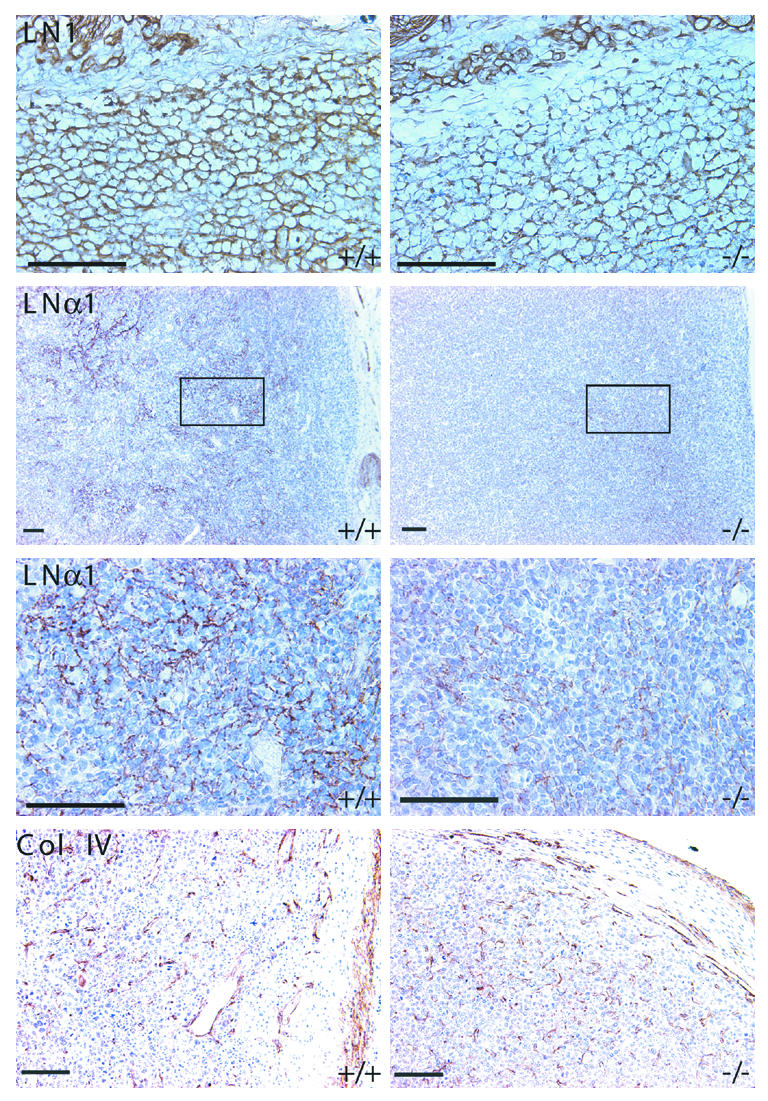
SPARC, a 32-kDa glycoprotein, participates in the regulation of morphogenesis and cellular differentiation through its modulation of cell-matrix interactions. Major functions defined for SPARC in vitro are de-adhesion and antiproliferation. In vivo, SPARC is restricted in its expression to remodeling tissues, including pathologies such as cancer. However, the function of endogenous SPARC in tumor growth and progression is not known. Here, we report that implanted tumors grew more rapidly in mice lacking SPARC. We observed that tumors grown in SPARC null mice showed alterations in the production and organization of ECM components and a decrease in the infiltration of macrophages. However, there was no change in the levels of angiogenic growth factors in comparison to tumors grown in wild-type mice, although there was a statistically significant difference in total vascular area. Whereas SPARC did inhibit the growth of tumor cells in vitro, it did not have a demonstrable effect on the proliferation or apoptosis of tumor cells in vivo. These data indicate that host-derived SPARC is important for the appropriate organization of the ECM in response to implanted tumors and highlight the importance of the ECM in regulating tumor growth.


Figures

Figure 1
Figure 1
Tumor growth is enhanced in SPARC null mice. SP+/+ and SP–/– mice were injected subcutaneously with 2 × 10⁶ LLC or 1 × 10⁶ EL4 cells (a and c, respectively). (a) Comparison of the mean final volume and weight of LLC tumors grown subcutaneously in SP+/+ and SP–/– mice. The results shown are the combination of two independent experiments, which lasted 24–29 days after tumor cell injection. (b) Comparison of the number of lung surface tumor colonies in age-matched SP+/+ and SP–/– mice 14–19 days after intravenous injection of 1 × 10⁵ LLC cells. The results shown are the combination of two independent experiments. Surface lung tumor colonies were visualized under a dissecting microscope after fixation in Bouin’s fixative. (c) Comparison of the mean final volume and weight of EL4 tumors grown subcutaneously in SP+/+ and SP–/– mice. The results shown are the combination of two independent experiments, which lasted 14–16 days after tumor cell injection. (d) Comparison of the animals’ weights after intraperitoneal injection of 1 × 10⁶ EL4 cells, and the weight of the GI tract at sacrifice. P values were determined by Student’s t test.
Figure 2
Figure 2
Tumor growth in SPARC null mice is associated with changes in collagen organization. Representative serial sections of LLC (a) and EL4 (b) tumors grown in SP+/+ or SP–/– mice and stained with hematoxylin and eosin, Masson’s trichrome, and picrosirius red. The bottom panel of (a) represents an internal highway of connective tissue in an LLC tumor from SP+/+ and SP–/– mice. Scale bars: 50 μm. (c) Immunostaining for collagen I of LLC tumors grown in SP+/+ or SP–/– mice. The top panel represents the capsule area, and the lower panel represents the interior of the tumor. Arrows denote capsular fibroblasts and arrowheads denote tumor cells.
Figure 3
Figure 3
Expression of SPARC mRNA and protein by LLC cells and tumors. (a) RT-PCR analysis of LLC, EL4, and bEnd.3 cells grown in culture. (b) RT-PCR analysis of LLC tumors grown in SP+/+ or SP–/– mice (n = 6). RNA from bEnd.3 cells grown in vitro was used as a positive control. A representative tumor from the SP+/+ and SP–/– groups is shown. (c) SDS-PAGE of tumor lysates and demonstration of SPARC expression by Western blot analyses. Two identically prepared gels were separated by SDS-PAGE; one was transferred to PVDF for Western blot analysis (left), and the other was stained with Coomassie blue (right). The latter demonstrates a similar protein profile in the lysates from tumors removed from both wild-type and SPARC null mice, and it shows equivalent protein loading among the lanes on the gel. Bars represent molecular weight standards in kDa. (d) Immunohistochemical analysis of SPARC in LLC tumors grown in SP+/+ and SP–/– mice. The rectangular insets are enlarged in the bottom panel. Arrows indicate positive tumor cells; the arrowhead indicates a positive host cell. Scale bars: 50 μm. Col I, collagen type I; rpS6, ribosomal protein S6.
Figure 4
Figure 4
Changes in the expression of LN1 are associated with increased tumor growth in SPARC null mice. Immunohistochemical analysis of LN1, LNα1, and collagen type IV in LLC tumors grown in SP+/+ or SP–/– mice. Scale bars: 50 μm. Col IV, collagen type IV.
Figure 5
Figure 5
Analysis and quantification of markers of angiogenesis in LLC tumors grown in SP+/+ and SP–/– mice. (a) Blood vessels of tumors grown in SP+/+ versus SP–/– mice stained with either anti-VEGFR2 or MECA32. (b) Representative immunohistochemical staining with F4/80, a marker of mature macrophages, showing the extent of infiltration into the LLC tumors grown in SP+/+ or SP–/– mice. Scale bars: 50 μm.

References

    1. Liotta LA, Kohn EC. The microenvironment of the tumour-host interface. Nature. 2001;411:375–379. - PubMed
    1. Hanahan D, Weinberg RA. The hallmarks of cancer. Cell. 2000;100:57–70. - PubMed
    1. Radisky D, Hagios C, Bissell MJ. Tumors are unique organs defined by abnormal signaling and context. Semin. Cancer Biol. 2001;11:87–95. - PubMed
    1. Yeo TK, Brown L, Dvorak HF. Alterations in proteoglycan synthesis common to healing wounds and tumors. Am. J. Pathol. 1991;138:1437–1450. - PMC - PubMed
    1. Bornstein P, Sage EH. Matricellular proteins: extracellular modulators of cell function. Curr. Opin. Cell Biol. 2002;14:608–614. - PubMed

Publication types

MeSH terms