. 2003 Mar;23(5):1534-45.
doi: 10.1128/MCB.23.5.1534-1545.2003.

Inhibitory and stimulatory regulation of Rac and cell motility by the G12/13-Rho and Gi pathways integrated downstream of a single G protein-coupled sphingosine-1-phosphate receptor isoform

Affiliations

Inhibitory and stimulatory regulation of Rac and cell motility by the G12/13-Rho and Gi pathways integrated downstream of a single G protein-coupled sphingosine-1-phosphate receptor isoform

Naotoshi Sugimoto et al. Mol Cell Biol. 2003 Mar.

Abstract

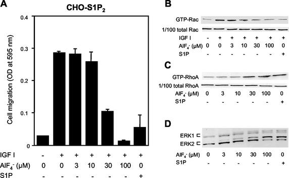
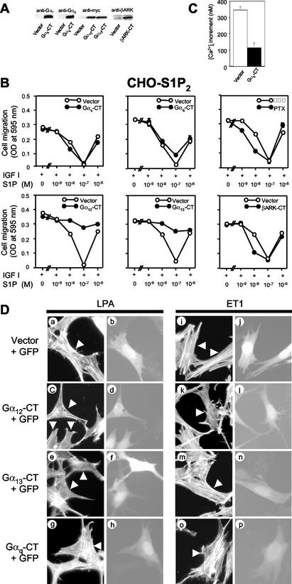
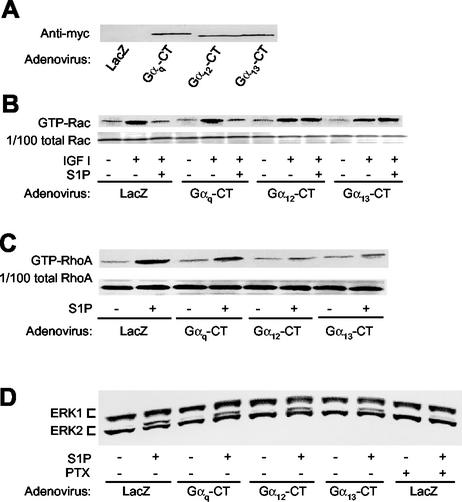
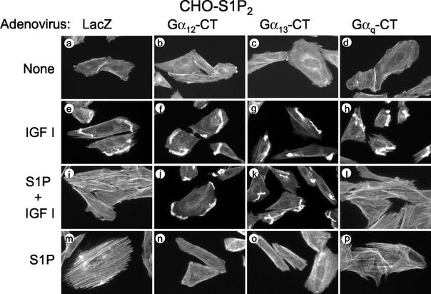
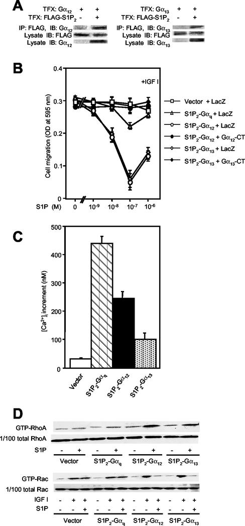
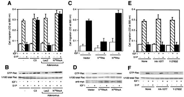
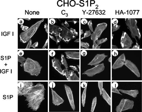
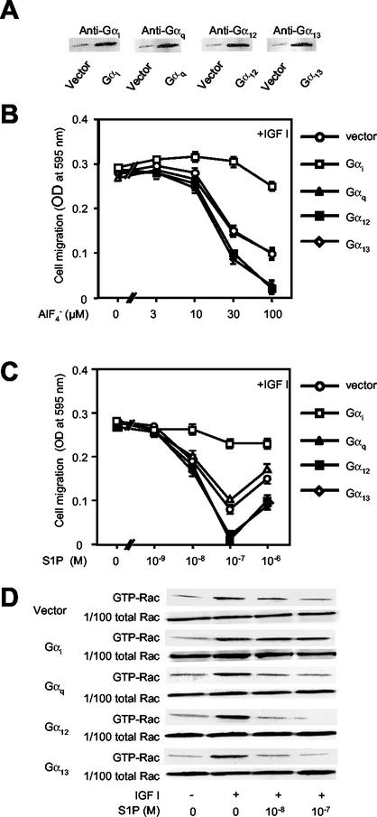
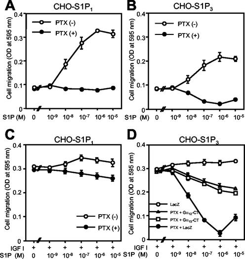
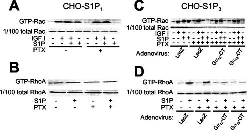
The G protein-coupled receptors S1P2/Edg5 and S1P3/Edg3 both mediate sphingosine-1-phosphate (S1P) stimulation of Rho, yet S1P2 but not S1P3 mediates downregulation of Rac activation, membrane ruffling, and cell migration in response to chemoattractants. Specific inhibition of endogenous Galpha12 and Galpha13, but not of Galphaq, by expression of respective C-terminal peptides abolished S1P2-mediated inhibition of Rac, membrane ruffling, and migration, as well as stimulation of Rho and stress fiber formation. Fusion receptors comprising S1P2 and either Galpha12 or Galpha13, but not Galphaq, mediated S1P stimulation of Rho and also inhibition of Rac and migration. Overexpression of Galphai, by contrast, specifically antagonized S1P2-mediated inhibition of Rac and migration. The S1P2 actions were mimicked by expression of V14Rho and were abolished by C3 toxin and N19Rho, but not Rho kinase inhibitors. In contrast to S1P2, S1P3 mediated S1P-directed, pertussis toxin-sensitive chemotaxis and Rac activation despite concurrent stimulation of Rho via G12/13. Upon inactivation of Gi by pertussis toxin, S1P3 mediated inhibition of Rac and migration just like S1P2. These results indicate that integration of counteracting signals from the Gi- and the G12/13-Rho pathways directs either positive or negative regulation of Rac, and thus cell migration, upon activation of a single S1P receptor isoform.


Figures

FIG. 1.
FIG. 1.
AlF4 mimicks S1P actions in inhibiting Rac and migration and stimulating Rho and ERK in S1P2 receptor-expressing cells. (A) AlF4 and S1P inhibit IGF I-directed chemotaxis. Transwell migration of CHO-S1P2 cells toward IGF I (100 ng/ml) was determined in the presence or absence of various concentrations of AlF4 and 10−7 M S1P in the lower chamber. (B) AlF4 and S1P inhibit IGF I-induced Rac stimulation. CHO-S1P2 cells were treated with various concentrations of AlF4 or 10−7 M S1P for 10 min and then stimulated with IGF I (100 ng/ml) for 1 min. Cells were then subjected to a pulldown assay for GTP-Rac as described in Materials and Methods. GTP-Rac bound to the GST-PAK1 CRIB domain immobilized onto Sepharose beads was analyzed by Western blotting using an anti-Rac antibody (top), and 1/100 of total Rac present in the cell lysate is also shown to confirm loading of equal amounts of proteins (bottom). (C) AlF4 and S1P stimulate Rho. CHO-S1P2 cells were stimulated with various concentrations of AlF4 or 10−7 M S1P for 3 min. Cells were then subjected to a pulldown assay for GTP-RhoA as described in Materials and Methods. (D) AlF4 and S1P stimulate ERK1 and -2. CHO-S1P2 cells were stimulated with various concentrations of AlF4 or 10−7 M S1P for 5 min. ERK activation was determined by band shift analysis using Western blotting.
FIG. 2.
FIG. 2.
Selective blockade of G12 and G13, but not Gs, Gi, or Gq, relieves S1P inhibition of migration in S1P2 receptor-expressing cells. (A) Western blot analysis showing expression of the Gα C-terminal peptides and βARK-CT. CHO-S1P2 cells were transfected with either expression vectors for the Gα-CTs and βARK-CT or an empty vector and were subjected to Western blot analysis using respective, specific antibodies and an anti-myc tag antibody. (B) Expression of G12-CT and G13-CT treatment relieves S1P inhibition of IGF I-directed chemotaxis.CHO-S1P2 cells were either cotransfected with LacZ and one of the expression vectors for Gαs-CT, Gαq-CT, Gα12-CT, Gα13-CT, and βARK-CT at a weight ratio of 1:2.5 or pretreated with PTX (200 ng/ml) as described in Materials and Methods. Transwell migration of transfected CHO-S1P2 cells was determined in the presence of IGF I (100 ng/ml) and various concentrations of S1P in the lower chamber. (C) Expression of Gαq-CT inhibits the S1P-induced [Ca2+]i increase in S1P2-overexpressing CHO cells. CHO-S1P2 cells stably expressing Gαq-CT and CHO-S1P2 cells expressing the vector control were stimulated by S1P at 10−8 M, and the peak increment in the [Ca2+]i was determined. (D) Inhibition of LPA- and ET1-induced stress fiber formation by expression of Gα12-CT or Gα13-CT. Swiss 3T3 cells were cotransfected with pEGFP-C1 and either an expression vector for Gα12-CT or Gα13-CT or an empty vector at a weight ratio of 1:10. Cells were stimulated with LPA or ET1 at 10−7 M for 10 min. F-actin was visualized with TRITC-labeled phalloidin. Arrowheads indicate transfected cells identified with GFP fluorescence.
FIG. 3.
FIG. 3.
Adenovirus-mediated expression of Gα12-CT and Gα13-CT abolishes S1P inhibition of Rac and stimulation of Rho in S1P2 receptor-expressing cells. (A) Western blot analysis of expression of the Gα C-terminal peptides. CHO-S1P2 cells were infected with adenoviruses encoding myc-tagged Gαq-CT, Gα12-CT, Gα13-CT, and LacZ 48 h before experiments and were subjected to Western blot analysis using an anti-myc tag antibody. (B and C) Expression of Gα12-CT and Gα13-CT, but not Gαq-CT abolishes S1P inhibition of IGF I-induced Rac stimulation and S1P stimulation of Rho. CHO-S1P2 cells that had been were infected with the adenoviruses were stimulated with IGF I (100 ng/ml) for 1 min in the presence of S1P (10−7 M) (for the Rac assay) or with S1P (10−7 M) for 3 min (for the Rho assay). For the Rac assay S1P was added 10 min before addition of IGF I. Cells were then subjected to a pulldown assay for GTP-Rac or GTP-RhoA. (D) Inhibition of S1P-induced ERK stimulation by PTX, but not by expression of Gq-CT, G12-CT, or G13-CT. CHO-S1P2 cells were either infected with the adenoviruses as described above or pretreated with PTX as for Fig. 2B. Cells were then stimulated with S1P (10−7 M) for 5 min and subjected to band shift analysis.
FIG. 4.
FIG. 4.
Expression of Gα12-CT and Gα13-CT, but not Gαq-CT, abolishes S1P inhibition of IGF I-induced membrane ruffling and also S1P stimulation of stress fiber formation in S1P2 receptor-expressing cells. CHO-S1P2 cells were infected with adenoviruses encoding Gα12-CT, Gα13-CT, Gαq-CT, or LacZ as for Fig. 3A. Cells were stimulated with IGF I (100 ng/ml) and/or S1P (10−7 M) for 30 min. Cells were fixed, permeabilized, and stained with TRITC-labeled phalloidin for F-actin.
FIG. 5.
FIG. 5.
The fusion receptors S1P2-Gα12 and S1P2-Gα13, but not S1P2-Gαq, mediate inhibition of Rac and migration. (A) Coimmunoprecipitation of S1P2 and either G12 or G13. COS7 cells were transiently cotransfected with pCAGGS-FLAG-S1P2 and either pCAGGS-Gα12 or pCAGGS-Gα13, and FLAG-tagged S1P2 was immunoprecipitated with an anti-FLAG (M2) antibody. The anti-FLAG immunoprecipitates were analyzed by Western blotting using anti-G12 or anti-G13 antibodies. Portions of cell lysates were analyzed by Western blotting using anti-FLAG, anti-G12, and anti-G13 antibodies. TFX, transfection; IP, immunoprecipitation; IB, immunoblotting. (B) S1P inhibits IGF I-directed chemotaxis in CHO cells expressing S1P2-Gα12 and S1P2-Gα13, but not S1P2-Gαq or vector, which is sensitive to expression of Gα12-CT and Gα13-CT inhibitor peptides. CHO cells stably expressing S1P2-Gα12, S1P2-Gα13, or S1P2-Gαq were infected with adenoviruses encoding Gα12-CT, Gα13-CT, or LacZ and then subjected to transwell migration as for Fig. 2B. (C) S1P-induced[Ca2+]i response in CHO cells expressing the fusion receptors. Cells were stimulated with S1P (10−7 M), and the peak increments of the [Ca2+]i responses were determined. (D) S1P inhibits IGF I-induced Rac stimulation and stimulates Rho in CHO cells expressing S1P2-Gα12 and S1P2-Gα13, but not S1P2-Gαq or vector. Cells were stimulated as described in the legend for Fig. 3B and C and were subjected to a pulldown assay for GTP-Rac and GTP-Rho.
FIG. 6.
FIG. 6.
Rho, but not Rho kinase, mediates S1P inhibition of Rac activity and migration in S1P2-expressing cells. (A and B) C3 pretreatment and N19Rho expression abolish S1P inhibition of IGF I-stimulated chemotaxis and Rac activity. CHO-S1P2 cells were either infected with adenoviruses encoding N19Rho or LacZ as for Fig. 3A, pretreated with C3 toxin (10 μg/ml) as described in Materials and Methods, or not pretreated. Transwell migration was determined in Fig. 2B. For the GTP-Rac pulldown assay, the cells were stimulated as for Fig. 3B. (C and D) Expression of V14Rho inhibits IGF I-stimulated chemotaxis and Rac activation. CHO-S1P2 cells were cotransfected with either myc-tagged N19Rho, myc-tagged V14Rho, or an empty vector and pCMV/Zeo, and they were selected in the presence of Zeocin. Zeocin-resistant cell populations, which expressed either of the myc-tagged Rho mutants, were employed in these experiments. Transwell migration was determined in the presence or absence of IGF I (100 ng/ml) in the lower chamber. For the GTP-Rac pulldown assay, cells were stimulated with IGF I (100 ng/ml) for 1 min. Expression of myc-tagged N19Rho and V14Rho proteins in the cell lysate are shown in the bottom gel of panel D. (E and F) Rho kinase inhibitors fail to prevent S1P inhibition of IGF I-stimulated chemotaxis and Rac activation. CHO-S1P2 cells were pretreated or not with HA-1077 (20 μM) or Y-27632 (10 μM) for 30 min before migration and Rac assays. Transwell migration was determined in the presence or absence of IGF I (100 ng/ml) and S1P (10−7 M) in the lower chamber. HA-1077 (20 μM) or Y-27632 (10 μM) was present in both the upper and lower chambers, where indicated. For the Rac assay, cells were stimulated with IGF I (100 ng/ml) for 1 min, with or without a 10-min pretreatment with S1P (10−7 M) and/or HA-1077 (20 μM) or Y-27632 (10 μM).
FIG. 7.
FIG. 7.
Rho kinase inhibitors do not affect S1P inhibition of IGF I-induced membrane ruffling but do abolish S1P-induced stress fiber formation. CHO-S1P2 cells were pretreated either with C3 toxin, as described in Materials and Methods, or with HA-1077 (20 μM) or Y-27632 (10 μM) for 30 min. Cells were then stimulated with IGF I (100 ng/ml) and/or S1P (10−7 M) for 30 min. Cells were stained with TRITC-labeled phalloidin for F-actin. Note that C3, but not HA-1077 or Y-27632, abolishes S1P inhibition of membrane ruffling in response to IGF I, although the Rho kinase inhibitors effectively suppress S1P-induced stress fiber formation.
FIG. 8.
FIG. 8.
Overexpression of Gαi counteracts AlF4- and S1P-induced inhibition of Rac and migration in S1P2 receptor-expressing cells. (A) Western blot analysis of expression of full-length Gα proteins. CHO-S1P2 cells stably expressing either Gαi2, Gαq, Gα12, Gα13, or an empty vector were subjected to Western blot analysis using respective, specific anti-Gα antibodies described in Materials and Methods. (B and C) Overexpression of Gαi markedly attenuates AlF4- and S1P-induced inhibition of IGF I-directed chemotaxis, but overexpression of Gα12 or Gα13 enhances such inhibition. Transwell migration of the CHO-S1P2 cells that stably express one of these Gα subunits or the empty vector was determined as for Fig. 1A and 2B. (D) Overexpression of Gαi markedly attenuates S1P inhibition of IGF I-induced Rac stimulation. CHO-S1P2 cells that stably express one of the Gα subunits or an empty vector were stimulated as for Fig. 3B and then subjected to a pulldown assay for GTP-Rac.
FIG. 9.
FIG. 9.
PTX pretreatment abolishes S1P stimulation of migration in S1P1- and S1P3-expressing cells and uncovers S1P3-mediated, G12/13-dependent inhibition of IGF I-directed chemotaxis. (A and B) PTX pretreatment abolishes chemotaxis toward S1P in CHO-S1P1 and CHO-S1P3 cells. Cells were pretreated or not with PTX as described in Materials and Methods and were then subjected to a transwell migration assay in the presence of various concentrations of S1P in the lower chamber. (C and D) PTX pretreatment unveils S1P-induced, G12/13-dependent inhibition of IGF I-directed chemotaxis in CHO-S1P3 cells, but not in CHO-S1P1 cells. CHO-S1P3 cells were infected with adenoviruses encoding Gα12-CT, Gα13-CT, and LacZ and were then pretreated or not with PTX, as for Fig. 3D. Cells were then subjected to a transwell migration assay as for Fig. 2B.
FIG. 10.
FIG. 10.
PTX pretreatment abolishes S1P stimulation of Rac in S1P1- and S1P3-expressing cells and uncovers S1P3-mediated, G12/13-dependent inhibition of IGF I-induced Rac stimulation. (A and B) Effects of PTX pretreatment on Rac and Rho activities in CHO-S1P1 cells. Cells were pretreated or not pretreated with PTX, stimulated with IGF I and/or S1P, and subjected to a pulldown assay for GTP-Rac and GTP-Rho, as described in the legends for Fig. 3B, C, and D. (C and D) Effects of PTX treatment and expression of Gα12-CT and Gα13-CT on Rac and Rho activities in CHO-S1P3 cells. Cells were infected with adenoviruses encoding Gα12-CT, Gα13-CT, and LacZ and then pretreated or not with PTX, as for Fig. 3D. Cells were stimulated in the same way as for panels A and B and were subjected to a pulldown assay for GTP-Rac and GTP-Rho.

References

    1. Akhter, S. A., L. M. Luttrell, H. A. Rockman, G. Iaccarino, R. J. Lefkowitz, and W. J. Koch. 1998. Targeting the receptor-Gq interface to inhibit in vivo pressure overload myocardial hypertrophy. Science 280:574-577. - PubMed
    1. Banyard, J., B. Anand-Apte, M. Symons, and B. R. Zetter. 2000. Motility and invasion are differentially modulated by Rho family GTPases. Oncogene 19:580-591. - PubMed
    1. Bornfeldt, K. E., L. M. Graves, E. W. Raines, Y. Igarashi, G. Wayman, S. Yamamura, Y. Yatomi, J. S. Sidhu, E. G. Krebs, S. Hakomori, and R. Ross. 1995. Sphingosine-1-phosphate inhibits PDGF-induced chemotaxis of human arterial smooth muscle cells: spatial and temporal modulation of PDGF chemotactic signal transduction. J. Cell Biol. 130:193-206. - PMC - PubMed
    1. Brinkmann, V., M. D. Davis, C. E. Heise, R. Albert, S. Cottens, R. Hof, C. Bruns, E. Prieschl, T. Baumruker, P. Hiestand, C. A. Foster, M. Zollinger, and K. R. Lynch. 2002. The immune modulator FTY720 targets sphingosine 1-phosphate receptors. J. Biol. Chem. 277:21453-21457. - PubMed
    1. Brzostowski, J. A., and A. R. Kimmel. 2001. Signaling at zero G: G-protein-independent functions for 7-TM receptors. Trends Biochem. Sci. 26:291-297. - PubMed

Publication types

MeSH terms

LinkOut - more resources