. 2003 Mar;23(5):1546-57.
doi: 10.1128/MCB.23.5.1546-1557.2003.

The C terminus of initiation factor 4E-binding protein 1 contains multiple regulatory features that influence its function and phosphorylation

Affiliations

The C terminus of initiation factor 4E-binding protein 1 contains multiple regulatory features that influence its function and phosphorylation

Xuemin Wang et al. Mol Cell Biol. 2003 Mar.

Abstract

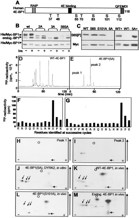
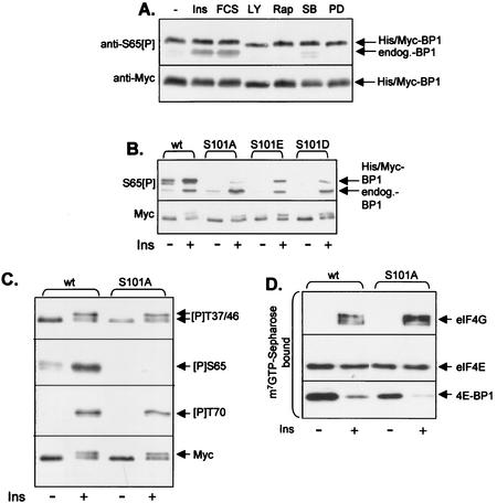
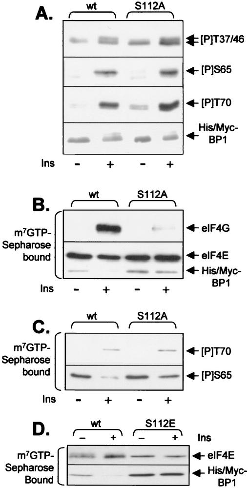
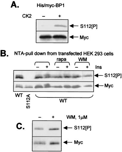
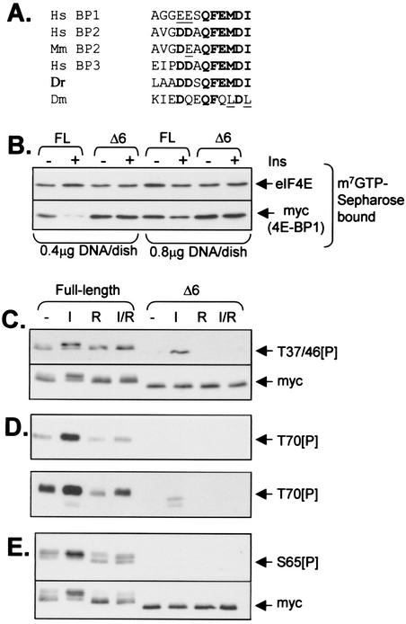
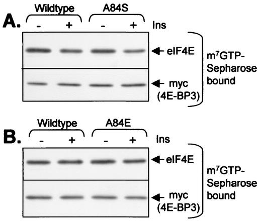
Eukaryotic initiation factor 4E (eIF4E) binds the mRNA cap structure and forms eIF4F complexes that recruit 40S subunits to the mRNA. Formation of eIF4F is blocked by eIF4E-binding proteins such as 4E-BP1, which interacts with eIF4E via a motif in the center of its 118-residue sequence. 4E-BP1 plays key roles in cell proliferation, growth, and survival. Binding of 4E-BP1 to eIF4E is regulated by hierarchical multisite phosphorylation. Here we demonstrate that three different features in the C terminus of 4E-BP1 play distinct roles in regulating its phosphorylation and function. Firstly, we identify a new phosphorylation site in its C terminus (S101). A serine or glutamate at this position is required for efficient phosphorylation at Ser65. A second C-terminal site, S112, directly affects binding of 4E-BP1 to eIF4E without influencing phosphorylation of other sites. Thirdly, a conserved C-terminal motif influences phosphorylation of multiple residues, including rapamycin-insensitive sites. These relatively long-range effects are surprising given the reportedly unstructured nature of 4E-BP1 and may imply that phosphorylation of 4E-BP1 and/or binding to eIF4E induces a more-ordered structure. 4E-BP2 and -3 lack phosphorylatable residues corresponding to both S101 and S112. However, in 4E-BP3, replacement of the alanine at the position corresponding to S112 by serine or glutamate did not confer the ability to be released from eIF4E in response to insulin.


Figures

FIG. 1.
FIG. 1.
Identification of a novel in vivo phosphorylation site in 4E-BP1. (A) Schematic illustration of wild-type human 4E-BP1, showing the sites of phosphorylation, the eIF4E-binding site, the RAIP and QFEMDI motifs, and the tags present in the fusion proteins employed in these studies. (B) HEK293 cells were transfected with vectors encoding His-Myc-tagged-versions of wild-type human 4E-BP1 or mutants in which either S65 alone (S65A), S65 and T70 (2A), or S65, T70, and S101 (3A) were mutated to A. Where indicated, cells were stimulated with insulin (100 nM; 30 min). Extracts were analyzed by SDS-PAGE and Western blotting with the anti-S65[P] antibody (top gel) or anti-Myc (bottom gel). Positions of recombinant (His/Myc) and endogenous 4E-BP1 polypeptides are shown. (C) (Left) Recombinant wild-type His-Myc-tagged 4E-BP1 or mutants in which either T37, T46, T70, and S83 are mutated to A (S65), S101 is mutated to A (S101A), or T37, T46, T70, S65, and S83 are mutated to alanine (5A) were incubated with activated Erk and unlabeled ATP. (Right) Recombinant wild-type His-Myc-tagged 4E-BP1 or the 5A mutant was incubated with DYRK2 and unlabeled ATP (+) or with ATP alone (−). Samples were then analyzed by SDS-PAGE and Western blotting using the anti-S65[P] antibody or anti-Myc, as a loading control. (D and E) Wild-type 4E-BP1 or the 5A mutant (in which T37, T46, T70, S65, and S83 are mutated to A) was radiolabeled using DYRK2 in vitro. Following cleavage by Asp-N, peptides were resolved by reverse-phase HPLC (C18 column, with in-line detection of 32P radioactivity, as described previously [46]). The two main radioactive peaks observed for 4E-BP1(5A) areindicated. (F and G) Peak 1 (F) or 2 (G) from the HPLC run for which results are shown in panel E was subjected to solid-phase Edman degradation, and the radioactivity released at each cycle was monitored. The residues identified at each cycle are also shown. (H to J) Two-dimensional phosphopeptide maps of peaks 1 (H) and 2 (I) from the HPLC run for which results are shown in panel E or of 4E-BP1(5A) phosphorylated in vitro by DYRK2 and digested with Asp-N (J). (K to M) Two-dimensional phosphopeptide maps of His-Myc-tagged 4E-BP1 (wild type [K] or S101A [L]) or endogenous 4E-BP1 (M) phosphorylated in HEK293 cells were generated by Asp-N digestion of the indicated protein that had been radiolabeled in vivo. In panels H to M, the positions of the origin (horizontal arrow), marker (dinitrophenyl-lysine) (circle), and peptides corresponding to peptides containing S101 (diagonal arrows) are shown to facilitate comparisons between maps, as are the direction of chromatography (vertical arrow) and the polarity of electrophoresis (plus and minus signs).
FIG. 2.
FIG. 2.
S101 is constitutively phosphorylated in vivo, and this affects phosphorylation at S65. (A) HEK293 cells were transfected with a vector encoding 4E-BP1(S65A) and 48 h later were treated (for 30 min) with either insulin (100 nM) (Ins), fetal calf serum (10% [vol/vol]) (FCS), LY294002 (30 μM) (LY), rapamycin (100 nM) (Rap), SB202190 (10 μM) (SB), or PD184352 (10 μM) (PD). The signal for the lower band, which corresponds to the endogenous 4E-BP1, is mainly due to phosphorylation of this protein at S65 and is accordingly increased by insulin and serum treatment. (B) HEK293 cells were transfected with wt 4E-BP1 or mutants in which S101 was mutated to A or to the acidic residue D or E. Where indicated, cells were stimulated with insulin (100 nM; 30 min). Extracts were analyzed by SDS-PAGE and Western blotting with the anti-S65[P] antibody (upper panel) or anti-Myc (lower panel). (C) Conditions were as described for panel B, but only the wild-type (wt) and S101A vectors were used. Blots were developed with anti-Myc or the indicated phosphospecific antisera for 4E-BP1. The signals for the endogenous 4E-BP1 in these extracts are too faint to be seen at this exposure of the immunoblots. (D) Conditions were as described for panel B, but extracts were subjected to affinity chromatography on m7GTP-Sepharose and SDS-PAGE followed by immunoblotting with the indicated antisera.
FIG. 3.
FIG. 3.
S112 directly modulates release of 4E-BP1 from eIF4E. HEK293 cells were transfected with a vector encoding His-Myc-tagged wild-type (wt) 4E-BP1 or the S112A or S112E mutant. (A and B) Samples were processed exactly as for Fig. 2C and D. (C) Samples were processed as for Fig. 2D but were subjected to immunoblotting using the anti-T70[P] or anti-S65[P] antisera as indicated. (D) HEK293 cells were transfected with a vector encoding His-Myc-tagged wild-type 4E-BP1 or the S112E mutant. Forty-eight hours later, after overnight serum starvation, some plates were treated with insulin, as indicated. After lysis, equal amounts of cell protein were subjected to affinity chromatography on m7GTP-Sepharose, and the bound material was analyzed by SDS-PAGE and Western blotting using anti-eIF4E or anti-Myc (as indicated).
FIG. 4.
FIG. 4.
Ser112 is constitutively phosphorylated in a wortmannin-insensitive manner. (A) Recombinant 4E-BP1 (expressed in E. coli with His and Myc tags) was incubated with or without CK2 and ATP-MgCl2, as indicated. Samples were then analyzed by SDS-PAGE and Western blotting using either anti-Myc (loading control) or an antibody raised against a peptide corresponding to the sequence around Ser112 of 4E-BP1 and containing a phosphoseryl residue as the position equivalent to Ser112 (designated S112[P]). (B) HEK293 cells were transfected with vectors encoding Myc-His-tagged forms of wild-type 4E-BP1 or an S112A mutant (as indicated). Thirty-six hours later, cells were starved of serum overnight. Where shown, cells were pretreated with rapamycin (100 nM; 30 min) (rapa) or wortmannin (100 nM; 30 min) (WM) prior to addition of insulin (where indicated; 15 min; 100 nM). Samples were subjected to affinity chromatography on NTA-agarose (to isolate the His-tagged 4E-BP1), and the bound material was analyzed as for panel A. (C) Conditions were the same as for panel B, but cells expressing wild-type 4E-BP1 were treated with wortmannin at 1 μM for 45 min prior to lysis (where indicated) and analysis by SDS-PAGE and Western blotting using anti-S112[P] or anti-Myc (loading control), as indicated.
FIG. 5.
FIG. 5.
The C-terminal tail of 4E-BP1 is dispensable for binding to eIF4E but plays a key role in the phosphorylation of the protein at multiple sites in vivo. (A) Sequence alignments of the extreme C termini of 4E-BPs from Homo sapiens (Hs), Mus musculus (Mm), Danio rerio (Dr) (zebra fish), and Drosophila melanogaster (Dm). Corresponding sequences in rat and mouse 4E-BP1 are identical to the human sequence shown. Residues conserved in all, or almost all, sequences are boldfaced. Conservative replacements are underlined. (B to E) Full-length 4E-BP1 and the Δ6 mutant were expressed in HEK293 cells as His-Myc-tagged polypeptides. Cells were starved of serum for 16 h prior to further analysis. In some cases (I), cells were treated with insulin (100 nM; 60 min). Where indicated (R), cells were pretreated with rapamycin (100 μM; 30 min) prior to addition of insulin and/or lysis. (B) Equal amounts of cell lysate protein were subjected to affinity chromatography on m7GTP-Sepharose, and the bound material was analyzed by SDS-PAGE and Western blotting using antibodies for eIF4E or Myc as indicated; cells were transfected with 0.4 or 0.8 μg of DNA (as indicated). These data exemplify the importance of low-level transfection in order to be able to observe release of wild-type 4E-BP1 in response to insulin (it is clear that little release is seen at the higher transfection/expression levels). (C to E) Amounts of cell extract containing equal amounts of recombinant wild-type and Δ6 mutant protein (see Myc blot, panel C, lower section) were analyzed directly (C and D) by SDS-PAGE and Western blotting using anti-T37/46[P] (C) or anti-T70[P] (D) phosphospecific antisera or were subjected to affinity chromatography on NTA-agarose (E) prior to analysis by SDS-PAGE and Western blotting using the anti-S65[P] phosphospecific antibody. The lower part of panel D shows a longer exposure of the same immunoblot. The signal for the endogenous 4E-BP1 is now also visible as faint bands below those due to the overexpressed His-Myc-tagged 4E-BP1. The lower part of panel E shows the results of reprobing the blot shown in the upper part with anti-Myc.
FIG. 6.
FIG. 6.
Effect of mutating A84 on the behavior of 4E-BP3. Mutants were created in which A84 in 4E-BP3 was replaced by serine (A84S, panel A) or glutamate (A84E, panel B). These variants were expressed in HEK293 cells in parallel with the wild-type protein. Cells were starved of serum overnight and in some cases were treated for 60 min with 100 nM insulin. Extracts were subjected to affinity chromatography on m7GTP-Sepharose followed by analysis of the bound material by SDS-PAGE and immunoblotting with the indicated antibodies.

References

    1. Aoki, M., E. Blazek, and P. K. Vogt. 2001. A role for the kinase mTOR in cellular transformation induced by the oncoproteins P3K and Akt. Proc. Natl. Acad. Sci. USA 98:136-141. - PMC - PubMed
    1. Avruch, J., C. Belham, Q. Weng, K. Hara, and K. Yonezawa. 2001. The p70 S6 kinase integrates nutrient and growth signals to control translational capacity. Prog. Mol. Subcell. Biol. 26:115-154. - PubMed
    1. Campbell, L. E., and C. G. Proud. 2002. Differing substrate specificities of members of the DYRK family of arginine-directed protein kinases. FEBS Lett. 510:31-36. - PubMed
    1. Chan, D. W., S. C. Son, W. Block, R. Ye, K. K. Khanna, M. S. Wold, P. Douglas, A. A. Goodarzi, J. Pelley, Y. Taya, M. F. Lavin, and S. P. Lees-Miller. 2000. Purification and characterization of ATM from human placenta. A manganese-dependent, wortmannin-sensitive serine/threonine protein kinase. J. Biol. Chem. 275:7803-7810. - PubMed
    1. Dajani, R., E. Fraser, S. M. Roe, N. Young, V. Good, T. C. Dale, and L. H. Pearl. 2001. Crystal structure of glycogen synthase kinase 3 beta: structural basis for phosphate-primed substrate specificity and autoinhibition. Cell 105:721-732. - PubMed

Publication types

MeSH terms

LinkOut - more resources