. 2003 Mar;23(5):1633-46.
doi: 10.1128/MCB.23.5.1633-1646.2003.

Identification of a structural determinant necessary for the localization and function of estrogen receptor alpha at the plasma membrane

Affiliations

Identification of a structural determinant necessary for the localization and function of estrogen receptor alpha at the plasma membrane

Mahnaz Razandi et al. Mol Cell Biol. 2003 Mar.

Abstract

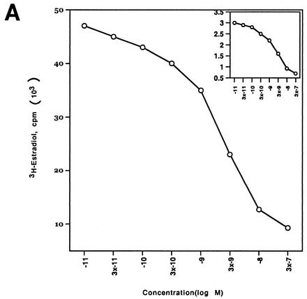
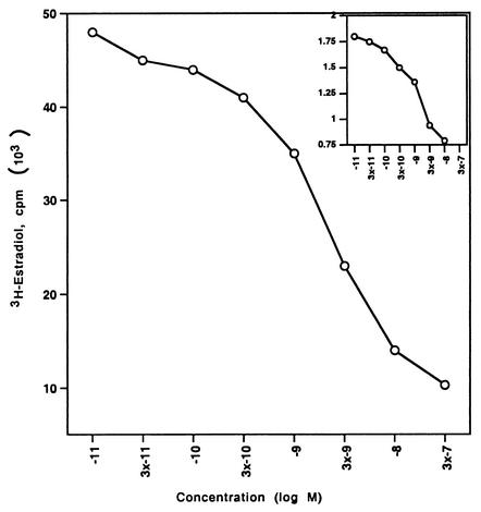
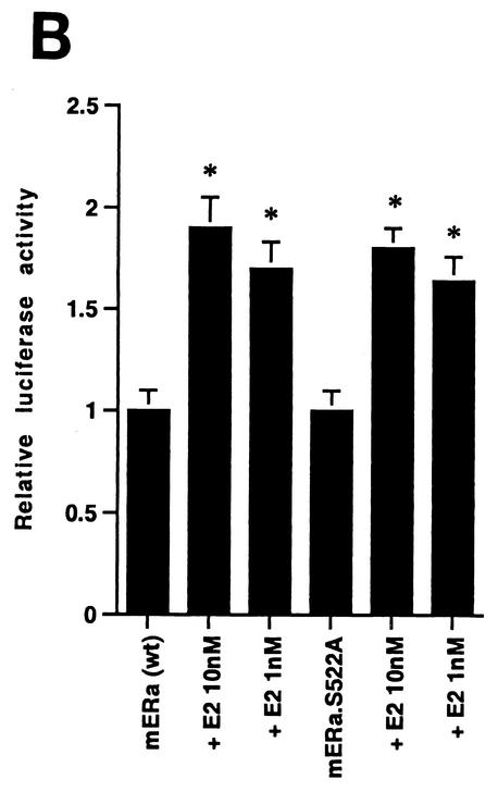
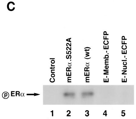
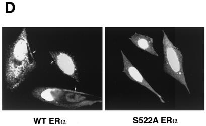
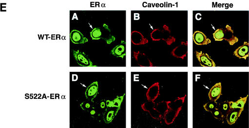
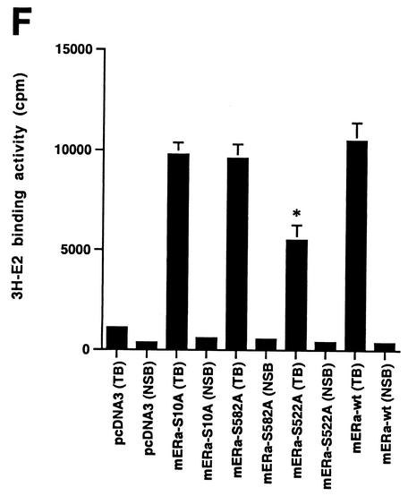
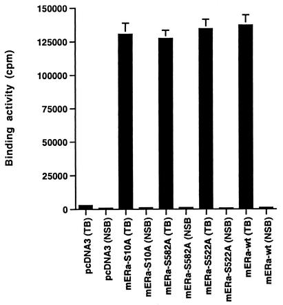
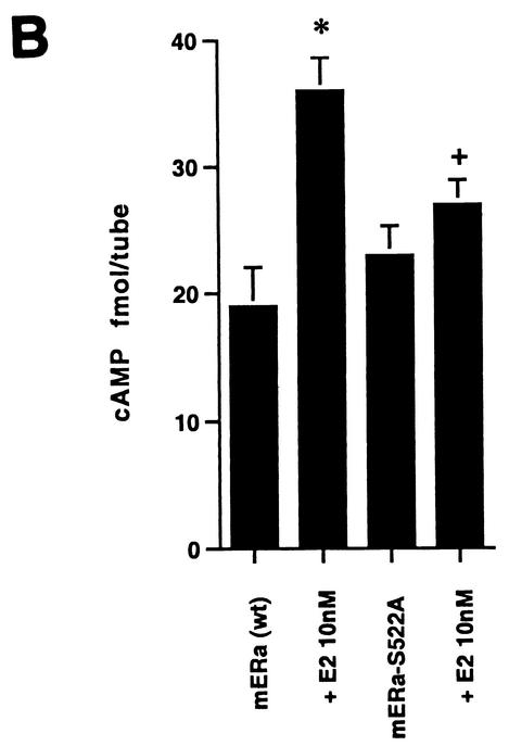
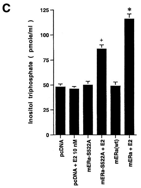
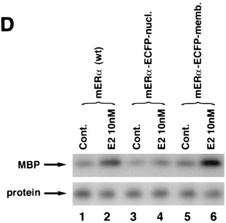
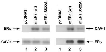
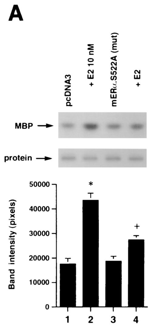
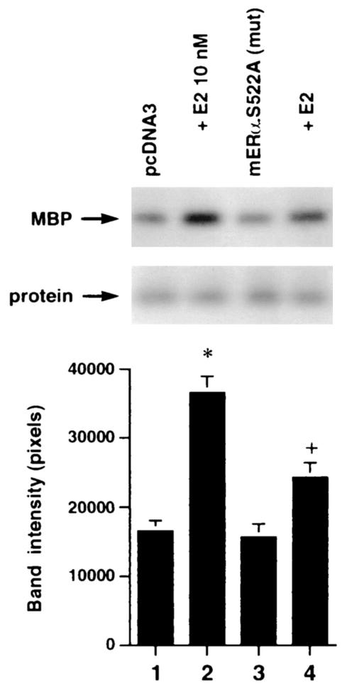
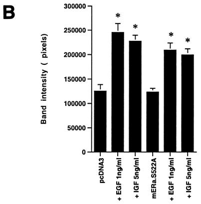
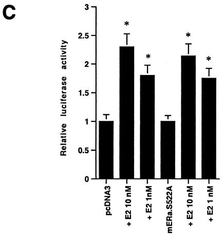
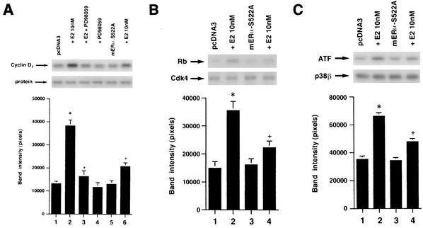
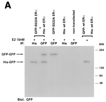
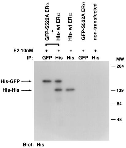
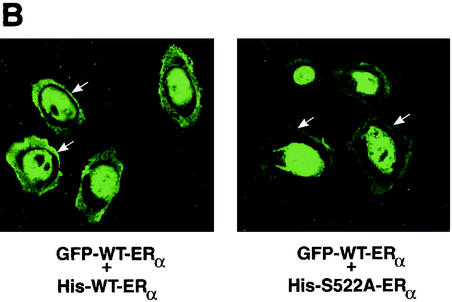
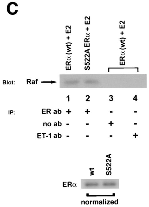
Estrogen receptors (ER) have been localized to the cell plasma membrane (PM), where signal transduction mediates some estradiol (E2) actions. However, the precise structural features of ER that result in membrane localization have not been determined. We obtained a partial tryptic peptide/mass spectrometry analysis of membrane mouse ERalpha protein. Based on this, we substituted alanine for the determined serine at amino acid 522 within the E domain of wild-type (wt) ERalpha. Upon transfection in CHO cells, the S522A mutant ERalpha resulted in a 62% decrease in membrane receptor number and reduced colocalization with caveolin 1 relative to those with expression of wt ERalpha. E2 was significantly less effective in stimulating multiple rapid signals from the membranes of CHO cells expressing ERalpha S522A than from those of CHO cells expressing wt ERalpha. In contrast, nuclear receptor expression and transcriptional function were very similar. The S522A mutant was also 60% less effective than wt ERalpha in binding caveolin 1, which facilitates ER transport to the PM. All functions of ERalpha mutants with other S-to-A substitutions were comparable to those of wt ER, and deletion of the A/B or C domain had little consequence for membrane localization or function. Transfection of ERalpha S522A into breast cancer cells that express native ER downregulated E2 binding at the membrane, signaling to ERK, and G1/S cell cycle events and progression. However, there was no effect on the E2 transactivation of an ERE-luciferase reporter. In summary, serine 522 is necessary for the efficient translocation and function of ERalpha at the PM. The S522A mutant also serves as a dominant-negative construct, identifying important functions of E2 that originate from activating PM ER.


Figures

FIG. 1.
FIG. 1.
(A) Competition binding of 17β-[3H]E2 to nuclear wt (left) or S522A mutant (right) ERα transfected into CHO-K1 cells. (Inset) Binding to cell membrane ERα. Data are transformed for Scatchard analysis by using the LIGAND program. Data shown here are from a representative study; results from three separate experiments were combined to create Table 1. (B) Transactivation of an ERE-luciferase reporter construct coexpressed in CHO cells with mouse wt ERα or the S522A mutant. Data were determined at 6 h after incubation with either 1 nM E2, 10 nM E2, or no steroid. *, P < 0.05 for the wt or the S522A mutant alone versus the same construct plus E2 (for data combined from three experiments). (C) Serine/threonine residues in the full-length receptor, but not serine 522 in the E domain of ERα, are phosphorylated. Western blotting utilized a specific antibody to serine/threonine residues (Sigma) from lysates of CHO cells transfected to express either the full-length receptor or the E domain targeted to the PM or the nucleus. (D) Membrane localization of GFP-tagged wt ERα or S522A mutant ERα expressed in CHO cells. Arrows indicate a greater membrane localization for wt ERα. Dense nuclear populations for both receptors are seen. (E) Colocalization of wt ERα with caveolin 1 at the membrane, but markedly less colocalization of ERα S522A. Results of a representative study are shown. Arrows indicate differential ER expression (green) at the membrane (panels A and D) and equal caveolin 1 expression (red) (panels B and E). The strong colocalization of wt ERα and caveolin 1 (yellow) (arrow, panel C) is not seen for ERα S522A (arrow, panel F). (F) Total specific binding of labeled E2 to membranes (left) or nuclei (center) in CHO cells expressing either S10A, S582A, or S522A mutant ERα or wt ERα. Data are combined from three experiments. *, P < 0.05 for ERα S522A versus wt ERα or other S-to-A mutant receptors. (Right) Protein blot demonstrating the purity of the membrane preparation. Caveolin 1 (Cav1) and 5′ nucleotidase (5′NT) are integral membrane proteins, while transportin and NTF-2 are nuclear proteins. β-COP is a Golgi protein.
FIG. 1.
FIG. 1.
(A) Competition binding of 17β-[3H]E2 to nuclear wt (left) or S522A mutant (right) ERα transfected into CHO-K1 cells. (Inset) Binding to cell membrane ERα. Data are transformed for Scatchard analysis by using the LIGAND program. Data shown here are from a representative study; results from three separate experiments were combined to create Table 1. (B) Transactivation of an ERE-luciferase reporter construct coexpressed in CHO cells with mouse wt ERα or the S522A mutant. Data were determined at 6 h after incubation with either 1 nM E2, 10 nM E2, or no steroid. *, P < 0.05 for the wt or the S522A mutant alone versus the same construct plus E2 (for data combined from three experiments). (C) Serine/threonine residues in the full-length receptor, but not serine 522 in the E domain of ERα, are phosphorylated. Western blotting utilized a specific antibody to serine/threonine residues (Sigma) from lysates of CHO cells transfected to express either the full-length receptor or the E domain targeted to the PM or the nucleus. (D) Membrane localization of GFP-tagged wt ERα or S522A mutant ERα expressed in CHO cells. Arrows indicate a greater membrane localization for wt ERα. Dense nuclear populations for both receptors are seen. (E) Colocalization of wt ERα with caveolin 1 at the membrane, but markedly less colocalization of ERα S522A. Results of a representative study are shown. Arrows indicate differential ER expression (green) at the membrane (panels A and D) and equal caveolin 1 expression (red) (panels B and E). The strong colocalization of wt ERα and caveolin 1 (yellow) (arrow, panel C) is not seen for ERα S522A (arrow, panel F). (F) Total specific binding of labeled E2 to membranes (left) or nuclei (center) in CHO cells expressing either S10A, S582A, or S522A mutant ERα or wt ERα. Data are combined from three experiments. *, P < 0.05 for ERα S522A versus wt ERα or other S-to-A mutant receptors. (Right) Protein blot demonstrating the purity of the membrane preparation. Caveolin 1 (Cav1) and 5′ nucleotidase (5′NT) are integral membrane proteins, while transportin and NTF-2 are nuclear proteins. β-COP is a Golgi protein.
FIG. 2.
FIG. 2.
(A) Binding of E2 to the nuclei and membranes of CHO cells expressing either HE11G (A/B domain deleted), HE19G (C domain deleted), or HEG0-537 (helix 12 and F domain deleted) ERα or wt ERα. The study was repeated. (B) ERK activation in response to E2 in CHO cells expressing either wt ERα or a deletion mutant. Activity was determined after 8 min of incubation with 10 nM E2. MBP was used as a substrate for ERK activity. Total ERK protein is shown on the immunoblot beneath the activity results.
FIG. 3.
FIG. 3.
(A) (Left) ERK activity is stimulated by E2 in CHO cells expressing wt ERα but less so in CHO cells expressing S522A mutant ERα. *, P < 0.05 for E2 versus the control (mouse ERα [mERα] without E2) in three combined experiments; +, P < 0.05 for ERK response to E2 in CHO cells expressing wt ERα (lane 2) versus ERα S522A (lane 4) in three combined experiments. (Right) Comparable stimulatory effects by E2 on ERK activity in CHO cells expressing wt ERα or the S10A or S582A mutant (lanes 3 to 8). Lanes 1 and 2 show that the intrinsic ERK activity of CHO cells expressing the empty vector, pcDNA3, cannot be stimulated by E2, due to a lack of endogenous ER. *, P < 0.05 for control versus E2. (B) Generation of cAMP in response to E2 in CHO cells expressing wt or S522A mutant ER. (C) IP3 generation in response to E2 in the above cells. Bar graph data are means ± standard errors of the means from triplicate determinations per experiment and are from two (cAMP) or three (IP3) combined studies. (D) Targeting ER to the membrane but not the nucleus allows E2 to rapidly activate ERK. CHO cells were transfected to express either nontargeted wt mERα or wt mERα targeted to the nucleus (mERα-ECFP-nucl.) or the membrane (mERα-ECFP-memb.). Cont., control. The study was repeated.
FIG. 3.
FIG. 3.
(A) (Left) ERK activity is stimulated by E2 in CHO cells expressing wt ERα but less so in CHO cells expressing S522A mutant ERα. *, P < 0.05 for E2 versus the control (mouse ERα [mERα] without E2) in three combined experiments; +, P < 0.05 for ERK response to E2 in CHO cells expressing wt ERα (lane 2) versus ERα S522A (lane 4) in three combined experiments. (Right) Comparable stimulatory effects by E2 on ERK activity in CHO cells expressing wt ERα or the S10A or S582A mutant (lanes 3 to 8). Lanes 1 and 2 show that the intrinsic ERK activity of CHO cells expressing the empty vector, pcDNA3, cannot be stimulated by E2, due to a lack of endogenous ER. *, P < 0.05 for control versus E2. (B) Generation of cAMP in response to E2 in CHO cells expressing wt or S522A mutant ER. (C) IP3 generation in response to E2 in the above cells. Bar graph data are means ± standard errors of the means from triplicate determinations per experiment and are from two (cAMP) or three (IP3) combined studies. (D) Targeting ER to the membrane but not the nucleus allows E2 to rapidly activate ERK. CHO cells were transfected to express either nontargeted wt mERα or wt mERα targeted to the nucleus (mERα-ECFP-nucl.) or the membrane (mERα-ECFP-memb.). Cont., control. The study was repeated.
FIG. 4.
FIG. 4.
ERα and caveolin 1 (CAV-1) association in the cytoplasm. CHO-K1 cell cultures (100-mm-diameter dishes) were transfected with 10 μg of wt or S522A mutant ERα plasmid DNA. The cells were lysed, and immunoprecipitation for caveolin 1 was carried out, followed by immunoblotting for ERα (left panels); or the order was reversed (right panels). Results shown are representative of three experiments. Caveolin 1 and ERα immunoblots are shown (lower panels) to demonstrate equal gel protein loading and equal expression of the two ER. mERα, mouse ERα.
FIG. 5.
FIG. 5.
(A) Expression of ERα S522A inhibits E2 activation of ERK. MCF-7 (left) or ZR-75-1 (right) breast cancer cells (which express endogenous ER) were transfected transiently to express either pcDNA3 (control) or S522A mutant ERα. The cells were then exposed to 10 nM E2 for 8 min, after which they were lysed, and immunoprecipitated ERK was assayed for activity by using MBP as a substrate. Precipitated ERK protein is shown in the lower gels, and the bar graphs each reflect three experiments combined. *, P < 0.05 for pcDNA3 in the absence versus the presence of E2; +, P < 0.05 for comparison of E2 treatments of pcDNA3-expressing versus ERα S522A-expressing cells. (B) ERα S522A does not impair EGF or IGF-1 activation of ERK. Data from three experiments are combined. *, P < 0.05 for pcDNA3-transfected or ERα S522A-expressing MCF-7 cells in the absence versus the presence of EGF or IGF-1. (C) E2 comparably activates an ERE-luciferase reporter in untransfected MCF-7 cells and MCF-7 cells transfected to express ERα S522A. Bar graph shows results for three experiments combined. *, P < 0.05 for pcDNA3- or ERα S522A-transfected MCF-7 cells without versus with E2.
FIG. 5.
FIG. 5.
(A) Expression of ERα S522A inhibits E2 activation of ERK. MCF-7 (left) or ZR-75-1 (right) breast cancer cells (which express endogenous ER) were transfected transiently to express either pcDNA3 (control) or S522A mutant ERα. The cells were then exposed to 10 nM E2 for 8 min, after which they were lysed, and immunoprecipitated ERK was assayed for activity by using MBP as a substrate. Precipitated ERK protein is shown in the lower gels, and the bar graphs each reflect three experiments combined. *, P < 0.05 for pcDNA3 in the absence versus the presence of E2; +, P < 0.05 for comparison of E2 treatments of pcDNA3-expressing versus ERα S522A-expressing cells. (B) ERα S522A does not impair EGF or IGF-1 activation of ERK. Data from three experiments are combined. *, P < 0.05 for pcDNA3-transfected or ERα S522A-expressing MCF-7 cells in the absence versus the presence of EGF or IGF-1. (C) E2 comparably activates an ERE-luciferase reporter in untransfected MCF-7 cells and MCF-7 cells transfected to express ERα S522A. Bar graph shows results for three experiments combined. *, P < 0.05 for pcDNA3- or ERα S522A-transfected MCF-7 cells without versus with E2.
FIG. 6.
FIG. 6.
(A) Cyclin D1 expression is increased in response to E2 and is dependent on ERK activation in wt MCF-7 cells. MCF-7 cells transfected to express ERα S522A show a lower response to 100 nM E2. Data are representative of three experiments, which were combined for the bar graph. *, P < 0.05 for pcDNA3-expressing cells without versus with E2; +, P < 0.05 for cells incubated with pcDNA3 plus E2 versus the same condition plus 10 μM PD 98059, or versus cells cotransfected with ERα S522A. (B) cdk4 activity is significantly downregulated by ERα S522A in MCF-7 cells. Cells were transfected with pcDNA3 or the mutant ER and exposed to 10 nM E2 for 6 h. cdk4 kinase was immunoprecipitated, and an in vitro assay of activity was accomplished by using Rb protein as a substrate. Bar graph data are from three experiments combined. (C) E2-stimulation of p38β activity in endothelial cells is inhibited by ERα S522A. Transfected endothelial cells were incubated with E2 for 20 min, and p38β activity was determined against the substrate protein ATF-1. Data are from three experiments.
FIG. 7.
FIG. 7.
(A) Homo- and heterodimerization of wt and ERα S522A after expression in CHO cells. Doubly or singly transfected cells were first exposed to 10 nM E2 for 10 min and then lysed, and the lysate underwent immunoprecipitation (IP) with an antibody to His or GFP, as indicated, eventually followed by blotting with an antibody to GFP (left) or to His (right). Proteins were separated by SDS-PAGE under nonreducing conditions (native gel). Molecular weight markers indicate the different sizes of the GFP-tagged wt ERα and His-tagged ERα S522A homodimers and the intermediate size of the heterodimer. (B) Expression of S522A prevents wt ERα localization at the membrane. CHO cells were transfected with equal amounts of plasmids encoding GFP-tagged wt ERα plus His-tagged wt ERα (10 μg of total DNA/100-mm-diameter dish) or with GFP-tagged wt ERα plus His-tagged ERα S522A. Localization of receptors was determined by confocal microscopy. (C) wt and S522A mutant ERα equally associate with Ras, Raf, or Src at the membrane. (Left) CHO cells were transfected to express either receptor, and after normalization, equal receptor protein aliquots were confirmed by Western blotting. Equal protein aliquots were used for immunoblots to determine wt or mutant ERα association with Ras or Raf. The study was carried out in the presence of 10 nM E2 for 10 min and was repeated. Immunoprecipitation with no antibody (ab) or an irrelevant antibody to the endothelin-1 peptide did not yield a protein band. (Right) MCF-7 cells were transfected with His-tagged ERα S522A (lanes 2 and 3) or with His-tagged pcDNA3 (lane 1) and were incubated or not with 10 nM E2 for 10 min. The cells were lysed and immunoprecipitated for ERα (lane 1) or for His (lanes 2 and 3), then immunoblotted for Src. Expression of His-tagged pcDNA3 did not coprecipitate Src (data not shown).
FIG. 7.
FIG. 7.
(A) Homo- and heterodimerization of wt and ERα S522A after expression in CHO cells. Doubly or singly transfected cells were first exposed to 10 nM E2 for 10 min and then lysed, and the lysate underwent immunoprecipitation (IP) with an antibody to His or GFP, as indicated, eventually followed by blotting with an antibody to GFP (left) or to His (right). Proteins were separated by SDS-PAGE under nonreducing conditions (native gel). Molecular weight markers indicate the different sizes of the GFP-tagged wt ERα and His-tagged ERα S522A homodimers and the intermediate size of the heterodimer. (B) Expression of S522A prevents wt ERα localization at the membrane. CHO cells were transfected with equal amounts of plasmids encoding GFP-tagged wt ERα plus His-tagged wt ERα (10 μg of total DNA/100-mm-diameter dish) or with GFP-tagged wt ERα plus His-tagged ERα S522A. Localization of receptors was determined by confocal microscopy. (C) wt and S522A mutant ERα equally associate with Ras, Raf, or Src at the membrane. (Left) CHO cells were transfected to express either receptor, and after normalization, equal receptor protein aliquots were confirmed by Western blotting. Equal protein aliquots were used for immunoblots to determine wt or mutant ERα association with Ras or Raf. The study was carried out in the presence of 10 nM E2 for 10 min and was repeated. Immunoprecipitation with no antibody (ab) or an irrelevant antibody to the endothelin-1 peptide did not yield a protein band. (Right) MCF-7 cells were transfected with His-tagged ERα S522A (lanes 2 and 3) or with His-tagged pcDNA3 (lane 1) and were incubated or not with 10 nM E2 for 10 min. The cells were lysed and immunoprecipitated for ERα (lane 1) or for His (lanes 2 and 3), then immunoblotted for Src. Expression of His-tagged pcDNA3 did not coprecipitate Src (data not shown).

Similar articles

Cited by

References

    1. Albanese, C., J. Johnson, G. Watanabe, N. Eklund, D. Vu, A. Arnold, and R. G. Pestell. 1995. Transforming p21 ras mutants and c-Ets-2 activate the cyclin D1 promoter through distinguishable regions. J. Biol. Chem. 270:23589-23597. - PubMed
    1. Alton, G., M. Hasilik, R. Niehues, K. Paneerselvam, J. R. Etchison, F. Fana, and H. H. Freeze. 1998. Direct utilization of mannose for mammalian glycoprotein biosynthesis. Glycobiology 8:285-295. - PubMed
    1. Anderson, R. G. 1998. The caveolae membrane system. Annu. Rev. Biochem. 67:199-225. - PubMed
    1. Aronica, S. M., W. L. Kraus, and B. S. Katzenellenbogen. 1994. Estrogen action via the cAMP signaling pathway: stimulation of adenylate cyclase and cAMP-regulated gene transcription. Proc. Natl. Acad. Sci. USA 91:8517-8521. - PMC - PubMed
    1. Catoria, G., M. V. Barone, M. Di Domenico, A. Bilancio, D. Ametrano, A. Migliaccio, and F. Auricchio. 1999. Non-transcriptional action of oestradiol and progestin triggers DNA synthesis. EMBO J. 18:2500-2510. - PMC - PubMed

Publication types

MeSH terms

LinkOut - more resources