. 2003 Mar 4;100(5):2468-73.
doi: 10.1073/pnas.0434312100. Epub 2003 Feb 14.

Rnf2 (Ring1b) deficiency causes gastrulation arrest and cell cycle inhibition

Affiliations

Rnf2 (Ring1b) deficiency causes gastrulation arrest and cell cycle inhibition

Jan Willem Voncken et al. Proc Natl Acad Sci U S A. .

Abstract

The highly homologous Rnf2 (Ring1b) and Ring1 (Ring1a) proteins were identified as in vivo interactors of the Polycomb Group (PcG) protein Bmi1. Functional ablation of Rnf2 results in gastrulation arrest, in contrast to relatively mild phenotypes in most other PcG gene null mutants belonging to the same functional group, among which is Ring1. Developmental defects occur in both embryonic and extraembryonic tissues during gastrulation. The early lethal phenotype is reminiscent of that of the PcG-gene knockouts Eed and Ezh2, which belong to a separate functional PcG group and PcG protein complex. This finding indicates that these biochemically distinct PcG complexes are both required during early mouse development. In contrast to the strong skeletal transformation in Ring1 hemizygous mice, hemizygocity for Rnf2 does not affect vertebral identity. However, it does aggravate the cerebellar phenotype in a Bmi1 null-mutant background. Together, these results suggest that Rnf2 or Ring1-containing PcG complexes have minimal functional redundancy in specific tissues, despite overlap in expression patterns. We show that the early developmental arrest in Rnf2-null embryos is partially bypassed by genetic inactivation of the Cdkn2a (Ink4aARF) locus. Importantly, this finding implicates Polycomb-mediated repression of the Cdkn2a locus in early murine development.


Figures

Figure 1
Figure 1
Null mutation of the Rnf2 locus. (a) Rnf2 targeting construct. A ±3.0-kb EcoRI–XhoI 129/Ola genomic fragment containing exons (black boxes) encoding the major part of the Ring finger (RF) domain was replaced by a neomycin selection cassette. Shaded boxes indicate the approximate position of additional exons. (b) Early detection of the Rnf2-KO genotype. Rnf2-KO embryos are present during early development until E9, but not beyond E10. Genomic DNA was analyzed by BamHI digestion and hybridization to an external 5′ probe indicated by the white box in a. (c) Functional null mutation of Rnf2 gene. Total RNA isolated from an Rnf2-HE embryo was reverse transcribed and PCR sequenced. A shorter RT-PCR product (see Fig. 3d) potentially encodes a small peptide of 41 aa, of which only the N-terminal-most 28 residues correspond to Rnf2; arrowhead indicates the aberrant fusion point within the Rnf2-KO mRNA.
Figure 2
Figure 2
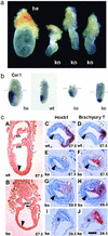
Morphological abnormalities of Rnf2-KO embryos. (a) A normal day-7.5 embryo (Left) at the late headfold stage; neural folds are visible. Three smaller Rnf2-KO embryos (Right) manifest a characteristic phenotype. (b) Gene expression analysis in pre/early streak stage embryos (E6.2). The Rnf2-HE embryo is at early streak stage; the WT and KO embryos are prestreak. The early anterior visceral endoderm (AVE) marker Cer1 is expressed in all Rnf2 genotypes. Cer1 expression is normally restricted in the KO embryo on the left, whereas it is abnormally localized in the KO on the right. Posterior is to the right; marker line: boundary extraembryonal (up)/embryonal tissue. (c) Histological and gene expression analysis of Rnf2-KO embryos confirms a significant delay in embryonic development. Posterior is to the right (AD) or to the upper right (EJ); embryonic mesoderm (white arrowhead), embryonic ectoderm (black arrowhead), and allantois (asterisk) are indicated. (A and B) Sagittal sections of embryos at E7.5. Mesoderm has expanded all around the WT egg cylinder (A). am, amnion; ch, chorion. Rnf2-KO embryos show improper epiblast elongation and accumulation of posterior mesoderm (B). amf, posterior amniotic fold. (CJ), Transverse sections of embryos analyzed for Hoxb1 (C, E, G, and I) and Brachyury T (D, F, H, and J) gene expression at E7.5 (CF) and E8.5 (GJ). Sections GJ are from the same embryo; sections H, G, J, and I are progressively more proximal, with J being at a level passing through the base of the allantois and transecting the proximo-posterior part of the amniotic cavity (the amnion is closed). Hoxb1 expression at E7.5 (C and E) is not yet initiated in the homozygous mutant embryo (E), whereas it is expressed in the posterior epiblast along the primitive streak and in nascent mesoderm in the WT (C; ref. 61). Brachyury T (D and F) is expressed in the mesoderm and ectoderm of the primitive streak in both WT and KO embryos, although it is weaker in the latter. (GJ) In E8.5 Rnf2-KO embryos (G and I) Hoxb1 is expressed in the accumulated posterior mesoderm and in the ectoderm of the primitive streak (G) but not in extraembryonic mesoderm of the allantois (I); Hoxb1 expression does not extend until the anterior end of the primitive streak. A weaker Brachyury T expression is visible in the epiblast and nascent mesoderm of the primitive streak (H) and in extraembryonic mesoderm at the base of the allantois (J) of the Rnf2-KO embryo. Note the posterior accumulation of mesoderm in the KO embryo (G and H). (Bar in J, ±0.1 mm.)
Figure 3
Figure 3
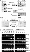
Rnf2 interactions in whole cells and early embryonic PcG expression profiles. (a) Cotransfection experiments demonstrate interaction of Rnf2-TAP-tag with HA-tagged Cbx2 (M33) and Cbx4 (MPc2) in COS-7 cells (Right) and with endogenous Bmi1 and Edr (Mph) in human U2-OS osteosarcoma cells (Left). Size markers: 97, 66, and 46 kDa. (b) Coimmunoprecipitation of myc-Ezh2 with HA-Eed from transfected U2-OS cell extracts shows interactions between class I PcG members. (c) TAP-tagged Rnf2 does not interact with HA-Eed or HA-Ezh2 in U2-OS cells (Left). Size markers: 97 and 66 kDa. (d) PcG gene expression profiles during early murine development. RT-PCR was done on total RNA from WT blastocysts (E3.5), ES cells (ESC), and E6.5, E7.5, and E12.5 embryos (Left). The expression profiles of Rnf2, Ring1 (Ring1a), Bmi1, Eed, Ezh1, and Ezh2 genes are shown. β-Actin serves as a qualitative control. (Right) PcG gene expression is not altered in Rnf2-KO embryos. Expression analysis in WT, HE, and KO E7.5 embryos. The shorter Rnf2-KO transcript (arrow) is the result of a deletion of exons encoding the major part of the Ring finger (see Fig. 1c).
Figure 4
Figure 4
Genetic interaction of Rnf2 and Bmi1 mutations. (a) Poor postnatal growth of Rnf2-HE/Bmi1-KO mice. Gross appearance of Rnf2-HE/Bmi1-KO (Upper) and Bmi1-KO (Lower) mice at 4 weeks of age as compared with WT and double HE littermates. (Right) Postnatal growth curves of Bmi1-KO (n = 2) and Rnf2-HE/Bmi1-KO (n = 2) mice versus double HE (n = 4) and WT (n = 2) male littermates and body weight at 4 weeks (Inset). Body weight was arbitrarily set at 1 at the time of genotyping (i.e., 10–11 days postpartum). Age at death of Rnf2-HE/Bmi1-KO was 4–5 weeks. (b) Aggravated cerebellar neuropathology in Rnf2-HE/Bmi1-KO versus Bmi1-KO mice. The cerebellum of Rnf2-HE/Bmi1-KO is remarkably smaller (A). Both the size and cellularity of the molecular and granular layers are significantly reduced [B, hematoxylin and eosin (H&E) staining; D, NeuN staining; white arrowheads demarcate the granular layer]. Although reduced in absolute numbers (overall reduction of the cerebellar size), Purkinje cells (PC) fully differentiate as judged by calbindin expression (C, calbindin staining) and align well (compare with WT Purkinje cells; C Left). Although the orientation of neurites and dendrites is correct, arborization of PC cells in the molecular layer is different: dendrites are thicker and less branched (C, black arrowhead). The abnormal arborization, already prevalent in the Bmi1-KO, is more pronounced in the Rnf2-HE/Bmi1-KO cerebellum. Abnormal arborization may be explained by absence of contact and cross talk with molecular neurons during development, which are severely reduced in the Bmi1-KO and virtually absent in some cerebellar sections in the Rnf2-HE/Bmi1-KO (D). Both Bmi1-KO and Rnf2-HE/Bmi1-KO cerebellum display distinct gliosis [asterisk in E, glial fibrillary acidic protein (GFAP) staining; the asterisk in B is an approximate reference point to E].
Figure 5
Figure 5
Partial bypass of the Rnf2 null mutant developmental arrest by genetic inactivation of the Cdkn2a locus. (a) Up-regulation of P16INK4a expression in E7.5 embryos as detected by semiquantitative RT-PCR. (b Upper) Compared with Rnf2-KO embryos, E8 Rnf2/Cdkn2a-dKO embryos clearly develop further; they are hardly delayed compared with WTs at a similar age; note the well developed neural folds (nf), neural tube (nt), and allantois (al); note also an abnormal frontal neural vesicle (fv) frequently observed in dKO embryos (compare with Fig. 2a). Rnf2-KO/Cdkn2a-KO embryos, however, still do not develop beyond 10–11 days in utero. The phenotypes of dKO and of Rnf2-HE/Cdkn2a-KO embryos (indicated by he/ko) are compared. DNA from a double heterozygote animal served as a positive control for both the Cdkn2a-WT and KO alleles and the Rnf2-WT allele (Lower). (c) Abnormal histology of Rnf2-KO/Cdkn2a-KO E8.5 embryos. Sagittal sections of dKO embryos confirming abnormal forebrain development in dKO embryos, with a near complete lack of head mesenchyme; the wall of the abnormal frontal vesicle has a neurectoderm-like epithelial structure (ne). Several discernable somites (so), although clearly malformed, indicate paraxial mesoderm formation and segmentation in E8 Rnf2/Cdkn2a-dKO embryos; cardiac structures were not visible in dKO embryos.

Similar articles

Cited by

References

    1. Paro R. Trends Genet. 1990;6:416–421. - PubMed
    1. Kennison J A. Annu Rev Genet. 1995;29:289–303. - PubMed
    1. Simon J. Curr Opin Cell Biol. 1995;7:376–385. - PubMed
    1. Pirrotta V. Cell. 1998;93:333–336. - PubMed
    1. Lewis E B. Nature. 1978;276:565–570. - PubMed

Publication types

MeSH terms

Substances