. 2003 Feb;14(2):349-60.
doi: 10.1091/mbc.e02-07-0382.

Integrin-mediated tyrosine phosphorylation of Shc in T cells is regulated by protein kinase C-dependent phosphorylations of Lck

Affiliations

Integrin-mediated tyrosine phosphorylation of Shc in T cells is regulated by protein kinase C-dependent phosphorylations of Lck

Shi Niu et al. Mol Biol Cell. 2003 Feb.

Abstract

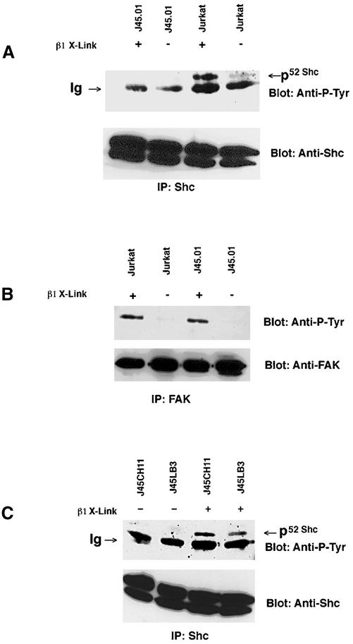
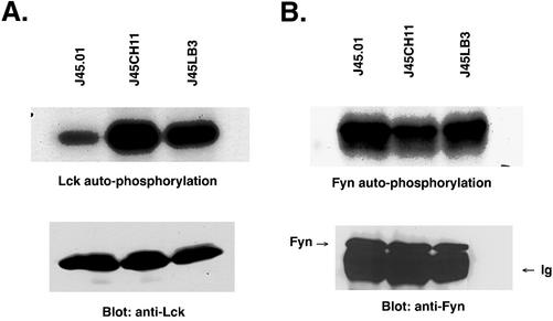
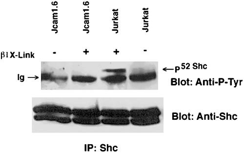
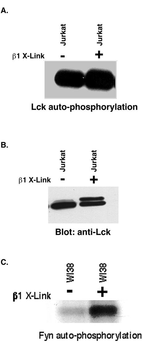
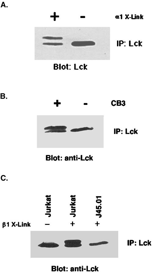
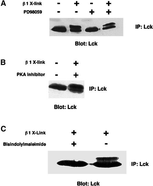
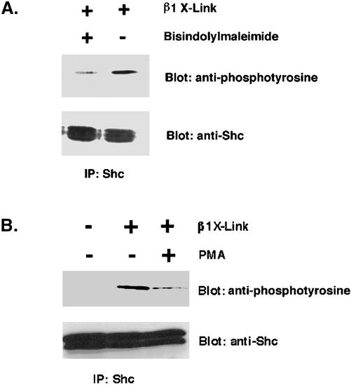
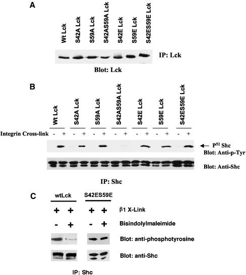
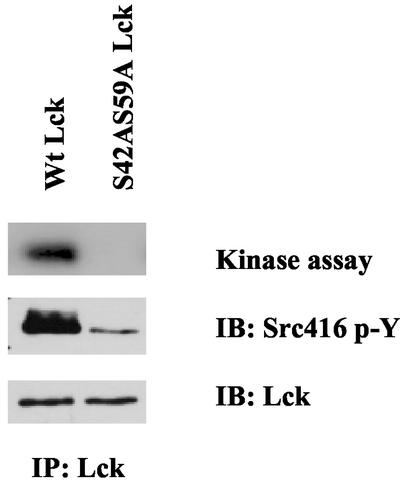
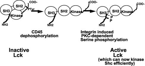
Integrin receptor signals are costimulatory for mitogenesis with the T-cell receptor during T-cell activation. A subset of integrin receptors can link to the adapter protein Shc and provide a mitogenic stimulus. Using a combination of genetic and pharmacological approaches, we show herein that integrin signaling to Shc in T cells requires the receptor tyrosine phosphatase CD45, the Src family kinase member Lck, and protein kinase C. Our results suggest a model in which integrin-dependent serine phosphorylation of Lck is the critical step that determines the efficiency of Shc tyrosine phosphorylation in T cells. Serine phosphorylation of Lck is dependent on PKC and is also linked to CD45 dephosphorylation. Mutants of Lck that cannot be phosphorylated on the critical serine residues do not signal efficiently to Shc and have greatly reduced kinase activity. This signaling from integrins to Lck may be an important step in the costimulation with the T-cell receptor during lymphocyte activation.


Figures

Figure 1
Figure 1
Integrin-dependent Shc phosphorylation in Jurkat cells: requirement for CD45. (A) Wild-type Jurkat cells were compared with CD45-deficient Jurkat cells (J45.01). Cells (107) for each cell line were either clustered with anti-β1 antibody beads (+) or left unclustered (−) and incubated for 15 min at 37°C followed by extraction. Extracts were immunoprecipitated using rabbit anti-Shc antibodies and separated by SDS-PAGE. After transfer to nitrocellulose, blots were stained for phosphotyrosine by using peroxidase-conjugated anti-P-Tyr (RC-20) and visualized by ECL. Blots were then stripped and reprobed with rabbit anti-Shc and peroxidase-conjugated protein A. Arrow points to the position of the 52-kDa isoform of Shc. The band below, which is present in all lanes, is related to the polyclonal anti-Shc, because it remains present even in the absence of cell extracts (our unpublished data). Note the cross-linking–dependent phosphorylation of Shc in Jurkat, but in the CD45-deficient cell line J45.01. (B) FAK phosphorylation in Jurkat cells. Wild-type Jurkat cells were compared with CD45-dependent Jurkat cells (J45.01). Cells (107) for each cell line were either clustered with anti-β1 antibody beads (+) or left unclustered (−) and incubated for 15 min at 37°C followed by extraction. Extracts were immunoprecipitated using rabbit anti-FAK antibodies and separated by SDS-PAGE. After transfer to nitrocellulose, blots were stained for phosphotyrosine by using peroxidase-conjugated anti-P-Tyr (RC-20) and visualized by ECL. Blots were then stripped and reprobed with rabbit anti-FAK and peroxidase-conjugated protein A. Note the cross-linking–dependent phosphorylation of FAK in both cell lines, demonstrating that FAK phosphorylation does not require CD45 and that β1 cross-linking in J45.01 is effective. (C) Integrin-dependent Shc phosphorylation: requirement of the presence of CD45 phosphatase. CD45 cDNA (J45LB3) or a chimera consisting of the extracellular domain of HLA A2 connected to the intracellular domain of CD45 (J45CH11) was expressed in previously CD45-deficient J45.01 cells. Cells (107) for each line were either clustered with anti-β1 antibody beads (+) or left unclustered (−) and incubated for 15 min at 37°C followed by extraction. Extracts were immunoprecipitated using rabbit anti-Shc antibodies and separated by SDS-PAGE. After transfer to nitrocellulose, blots were stained for phosphotyrosine by using peroxidase-conjugated anti-P-Tyr (RC-20) and visualized by ECL. Blots were then stripped and reprobed with rabbit anti-Shc and peroxidase-conjugated protein A. Note the cross-linking–dependent phosphorylation of Shc is restored with both CD45 (J45LB3) and with the chimeric molecule (J45CH11).
Figure 2
Figure 2
(A) Determination of Lck activity via autophosphorylation in Jurkat cell lines. CD45 cDNA (J45LB3) or a chimera consisting of the extracellular domain of HLA A2 connected to the intracellular domain of CD45 (J45CH11) was expressed in previously CD45-deficient J45.01 cells. Extracts of 107 cells of each of the three lines were immunoprecipitated with monoclonal anti-Lck, followed by incubation in [32P]ATP in kinase buffer for 2 min at 30°C and then washed and separated by SDS-PAGE. The gels were soaked in KOH for 2 h at 65°C and then dried before autoradiography. Extracts of same number of each of the three lines also were immunoprecipitated with Lck antibodies and separated by SDS-PAGE. After transfer to nitrocellulose, the blot was stained with monoclonal Lck antibodies and peroxidase-conjugated anti-mouse light chain antibodies, and visualized by ECL. Note the restored activity in both cell lines with CD45 (J45LB3) and with the chimera molecule (J45CH11). (B) Lack of regulation of Fyn kinase activity by CD45 phosphatase. J45LB3, J45CH11, or J45.01 cells were used. Extracts of 107 cells of each of the three lines were immunoprecipitated with polyclonal anti-Fyn, followed by incubation in [32P]ATP in kinase buffer for 2 min at 30°C, and then washed and separated by SDS-PAGE. The gels were soaked in KOH and then dried before autoradiography. Extracts of same number of each of the three lines also were immunoprecipitated with Fyn antibodies and separated by SDS-PAGE. After transfer to nitrocellulose, the blot was stained with polyclonal Fyn antibodies and peroxidase-conjugated protein A and visualized by ECL. Note the very similar levels of kinase activity in all cell lines, in contrast to Lck.
Figure 3
Figure 3
Requirement of Lck for integrin-dependent phosphorylation of Shc. Wild-type Jurkat cells were compared with Lck-deficient Jurkat (Jcam 1.6). Cells (107) for each cell line were either clustered with anti-β1 antibody beads (+) or left unclustered (−) and incubated for 15 min at 37°C followed by extraction. Extracts were immunoprecipitated using rabbit anti-Shc antibodies and separated by SDS-PAGE. After transfer to nitrocellulose blots were stained for phosphotyrosine by using peroxidase-conjugated anti-P-Tyr (RC-20) and visualize by ECL. Blots were then stripped and reprobed with rabbit anti-Shc and peroxidase-conjugated protein A. Note the cross-linking–dependent phosphorylation of Shc in Jurkat, but not in the Lck-deficient cell line Jcam 1.6.
Figure 4
Figure 4
Lack of stimulation of Lck activity by antibody-mediated β1 integrin clustering. Cells (107) were either clustered with anti-β1 antibody beads (+) or left unclustered (−) and incubated for 15 min at 37°C followed by extraction. Extracts were immunoprecipitated using monoclonal anti-Lck antibodies. Half the immune complex from each treatment was incubated in [32P]ATP in kinase buffer for 2 min at 30°C and then washed and separated by SDS-PAGE (A). The gels were soaked in KOH and then dried before autoradiography. The other half of the immune complex from each treatment was separated by SDS-PAGE. After transfer to nitrocellulose, the blot was stained with monoclonal Lck antibodies and peroxidase-conjugated anti-mouse light chain antibodies and visualized by ECL (B). Note there is no significant change in the kinase activity after cross-linking, but the mobility of a portion of the Lck is slower. (C) Fyn activation by integrin cross-linking in WI 38 cells. WI 38 cells were cross-linked in suspension for 10 min with beads coated either with or without anti-β1 antibodies. Lysates (600 μg) from each treatment were immunoprecipitated with polyclonal anti-Fyn antibody (Santa Cruz Biotechnology) and subjected to kinase assay. The gel was treated with 1 M NaOH at 56°C for 2 h before autoradiography.
Figure 5
Figure 5
Modification of Lck electrophoretic mobility upon integrin cross-linking. (A) Jurkat cells (107) were either clustered with anti-α1 antibody beads (+) or left unclustered (−) and incubated for 15 min at 37°C followed by extraction. (B) Jurkat cells (107) were either clustered with the cell binding fragment CB3 of collagen IV-coated beads (+) or left unclustered (−) and incubated for 15 min at 37°C followed by extraction. (C) Jurkat or J45.01 cells (107) were either clustered with anti-β1 antibody beads (+) or left unclustered (−) and incubated for 15 min at 37°C followed by extraction. Note the requirement of CD45 for the integrin-dependent modification of Lck. In all cases, extracts were immunoprecipitated using monoclonal anti-Lck antibodies. The immune complexes from each treatment were separated by SDS-PAGE. After transfer to nitrocellulose the blot was stained with monoclonal Lck antibodies and peroxidase-conjugated anti-mouse light chain antibodies and visualized by ECL. Note the shift in mobility of the Lck upon cross-linking.
Figure 6
Figure 6
Effect of protein kinase inhibitors on the Lck gel mobility shift. (A) Jurkat cells were cultured in the presence or absence of 50 μM of PD98059, a potent inhibitor of MEK1, for 1 d. Treated or nontreated cells (107) were clustered with either anti-β1 antibody-coated beads or with noncoated beads for 10 min at 37°C followed by extraction. Extracts were immunoprecipitated with Lck antibodies and separated by 10% SDS PAGE. After transfer to nitrocellulose, the blot was stained with monoclonal Lck antibodies and peroxidase-conjugated anti-mouse light chain antibodies and visualized by ECL. (B) Jurkat cells were treated with 5 μM PKA inhibitor 14-22 for 1 h at 37°C. The treated or nontreated cells were clustered with either anti-β1 antibody-coated beads or -noncoated beads. (C) Jurkat cells were incubated with 5 μM of bisindolylmaleimide, a specific inhibitor of PKCα, βI, βII, and γ subtypes, for 1 h at 37°C. The cells were either clustered with anti-β1 antibody-coated beads or noncoated beads. Gel mobility of Lck from these cells was detected as described above.
Figure 7
Figure 7
Requirement of PKC activity for integrin-dependent Shc phosphorylation in T cells. (A) Jurkat cells were incubated with 5 μM bisindolylmaleimide for 1 h at 37°C. Cells (107) were then clustered with either anti-β1 antibody-coated beads or noncoated beads for 10 min at 37°C, followed by extraction. Extracts were immunoprecipitated with an anti-Shc polyclonal antibody and separated by SDS-PAGE. After transfer to nitrocellulose, the blot was stained with RC-20 and visualized by ECL. (B) Effect of PMA on integrin cross-linking induced tyrosine phosphorylation of Shc. Jurkat cells were incubated with 70 nM of PMA for 24 h at 37°C. Cells (107) were then clustered with either anti-β1 antibody-coated or -noncoated beads for 10 min for at 37°C, followed by extraction. Extracts were immunoprecipitated with an anti-Shc polyclonal antibody and separated by SDS-PAGE. After transfer to nitrocellulose, the blot was stained with RC-20 and visualized with ECL. The blot then was stripped and reprobed with polyclonal anti-Shc and peroxidase-conjugated protein A. Quantitation of the inhibitory effect showed approximately a fourfold reduction in Shc phosphorylation in both A and B with blockade of PKC.
Figure 8
Figure 8
Serine 42 and serine 59 of the Lck N-terminal unique region are critical for integrin-dependent Shc phosphorylation. (A) Determination of the expression levels of Wt Lck and single and double mutants of serine 42 and serine 59 to alanine (S42A, S59A and S42AS59A) or glutamic acid (S42E, S59E and S42ES59E) of Lck in Jcam 1.6 cells. Equal amounts of protein from the extracts of these stably transfected Jcam 1.6 cell lines were immunoprecipitated with monoclonal anti-Lck antibody. The resulting immune complexes were separated by SDS-PAGE. After transfer to nitrocellulose, the blot was stained with monoclonal Lck antibodies and peroxidase-conjugated anti-light chain antibodies, and visualized by ECL. Similar levels of Lck are expressed in all of the lines used. (B) Integrin-dependent Shc phosphorylation requires both the Ser 42 or Ser 59 sites in the Lck N-terminal unique region. The cell lines shown above were either left unclustered (−) or clustered with antibody beads (+) and incubated for 15 min at 37°C followed by extraction. Equal amounts of protein from the extracts were immunoprecipitated with rabbit anti-Shc antibody and separated by SDS-PAGE. After transfer to nitrocellulose, the blot was stained for phosphotyrosine by using peroxidase-conjugated anti-p-Tyr (RC-20) and visualized by ECL. Blots were then stripped and reprobed with rabbit anti-Shc antibodies and peroxidase-conjugated protein A. Note the markedly reduced Shc phosphorylation in S42AS59A double mutant Lck Jcam 1.6 cell line upon integrin activation, whereas the S42ES59E mutant had normal amounts of Shc phosphorylation. (C) S42E S59E Lck is resistant to inhibition of PKC. Wt Lck and S42ES59E Lck-transfected Jcam1.6 cell lines were incubated with 5 μM bisindolylmaleimide, a specific inhibitor of PKCα, βI, βII, and γ subtypes, for 1 h at 37°C or without bisindolylmaleimide. The cells were then clustered with antibody beads (+) and incubated for 15 min at 37°C followed by extraction. Equal amounts of protein from the extracts were immunoprecipitated with rabbit anti-Shc antibody and separated by SDS-PAGE. After transfer to nitrocellulose, the blot was stained for phosphotyrosine by using peroxidase-conjugated anti-p-Tyr (RC-20) and visualized by ECL. Blots were then stripped and reprobed with rabbit anti-Shc antibody and peroxidase-conjugated protein A. Note that the S42E S59E form of Lck produced high levels (>90% of untreated) of Shc despite pretreatment with a PKC inhibitor, whereas the Wt shows a fivefold reduction (as determined by NIH Image software) in Shc phosphorylation with the same treatment.
Figure 9
Figure 9
Reduced kinase activity of Lck S42AS59A. Stably transfected Jcam1.6 cell lines expressing either Wt or S42AS59A Lck were analyzed. Equal amounts of detergent extracts from the two cell lines were immunoprecipitated using monoclonal anti-Lck antibodies. Half the immune complex from each treatment was incubated in [32P]ATP in kinase buffer for 2 min at 30°C and then washed and separated by SDS-PAGE. The gels were soaked in KOH and then dried before autoradiography. The other half of the immune complex from each treatment was separated by SDS-PAGE. After transfer to nitrocellulose, the blot was stained with a polyclonal anti-phospho-Src family Tyr 416 antibody and peroxidase-conjugated protein A. The blot was then stripped and reprobed with monoclonal Lck antibodies and peroxidase-conjugated anti-light chain antibodies, and visualized by ECL. Note that despite equal amounts of Lck recovered, both the labeling by 32P and the blot measuring the activation loop tyrosine phosphorylation show severely reduced kinase activity (8- to 10-fold) in the S42AS59A Lck compared with the wild type.
Figure 10
Figure 10
Model of Integrin signaling through Lck to Shc.

Similar articles

Cited by

References

    1. Anderson SJ, Perlmutter RM. A signaling pathway governing early thymocyte maturation. Immunol Today. 1995;16:99–105. - PubMed
    1. Brown DA, London E. Functions of lipid rafts in biological membranes. Annu Rev Cell Dev Biol. 1998;14:111–136. - PubMed
    1. Byth KF, Conroy LA, Howlett S, Smith AJ, May J, Alexander DR, Holmes N. CD45-null transgenic mice reveal a positive regulatory role for CD45 in early thymocyte development, in the selection of CD4 + CD8 + thymocytes, and B cell maturation. J Exp Med. 1996;183:1707–1718. - PMC - PubMed
    1. Cahir McFarland ED, Hurley TR, Pingel JT, Sefton BM, Shaw A, Thomas ML. Correlation between Src family member regulation by the protein-tyrosine-phosphatase CD45 and transmembrane signaling through the T-cell receptor. Proc Natl Acad Sci USA. 1993;90:1402–1406. - PMC - PubMed
    1. Casnellie JE, Lamberts RJ. Tumor promoters cause changes in the state of phosphorylation and apparent molecular weight of a tyrosine protein kinase in T lymphocytes. J Biol Chem. 1986;261:4921–4925. - PubMed

Publication types

MeSH terms