. 2003 Feb;14(2):396-406.
doi: 10.1091/mbc.e02-08-0458.

Actin filament polymerization regulates gliding motility by apicomplexan parasites

Affiliations

Actin filament polymerization regulates gliding motility by apicomplexan parasites

D M Wetzel et al. Mol Biol Cell. 2003 Feb.

Abstract

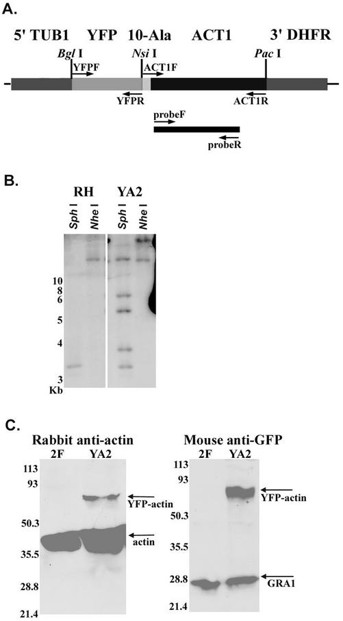
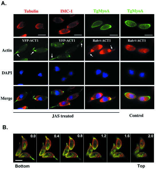
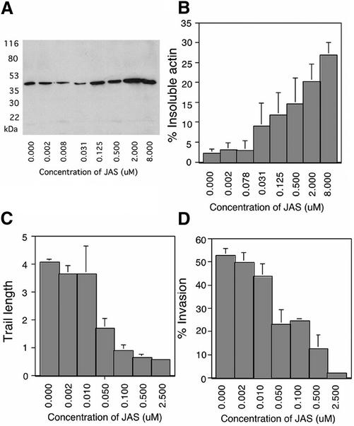
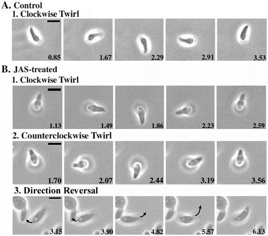
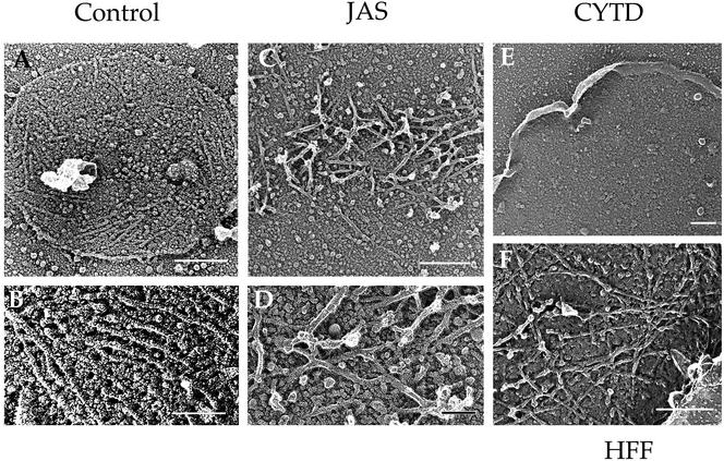
Host cell entry by Toxoplasma gondii depends critically on actin filaments in the parasite, yet paradoxically, its actin is almost exclusively monomeric. In contrast to the absence of stable filaments in conventional samples, rapid-freeze electron microscopy revealed that actin filaments were formed beneath the plasma membrane of gliding parasites. To investigate the role of actin filaments in motility, we treated parasites with the filament-stabilizing drug jasplakinolide (JAS) and monitored the distribution of actin in live and fixed cells using yellow fluorescent protein (YFP)-actin. JAS treatment caused YFP-actin to redistribute to the apical and posterior ends, where filaments formed a spiral pattern subtending the plasma membrane. Although previous studies have suggested that JAS induces rigor, videomicroscopy demonstrated that JAS treatment increased the rate of parasite gliding by approximately threefold, indicating that filaments are rate limiting for motility. However, JAS also frequently reversed the normal direction of motility, disrupting forward migration and cell entry. Consistent with this alteration, subcortical filaments in JAS-treated parasites occurred in tangled plaques as opposed to the straight, roughly parallel orientation observed in control cells. These studies reveal that precisely controlled polymerization of actin filaments imparts the correct timing, duration, and directionality of gliding motility in the Apicomplexa.


Figures

Figure 1
Figure 1
Expression of YFP actin in transgenic T. gondii. (A) Diagram of the YFP-actin construct (YFP-ACT1) used to transfect the RH strain of T. gondii, yielding strain YA2. A 10-alanine linker joins the YFP and actin (ACT-1) sequences. Expression of YFP-ACT1 is controlled by the tubulin promoter (5′-TUB1) and the 3′-DHFR sequence. Restriction sites used during construction of YFP-ACT1 are indicated above the diagram. PCR primers are indicated by arrows. The probe used in B is illustrated by the black bar. (B) Southern blot illustrating the copy number of YFP-ACT1 in YA2. Left, RH-strain (wild-type) genomic DNA digested with SphI and NheI and probed with a partial actin sequence. Right, YA2 genomic DNA visualized with the same probe after digestion with the same restriction enzymes. (C) Western blot demonstrates that YFP-ACT1 is expressed in strain YA2 but not in 2F (RH transfected with β-galactosidase). Left, probed with anti-ACT1 polyclonal sera. Right, probed with monoclonal anti-GFP antibody 3E6. Mouse mAb Tg17–43 against an unrelated protein, GRA1, was used as a loading control. Numbers refer to molecular mass in kDa.
Figure 2
Figure 2
Distribution of cytoskeletal elements in JAS-treated T. gondii. (A) Wide-field fluorescence microscopy demonstrates that JAS alters the distribution of actin in T. gondii but does not affect microtubules (tubulin), reticular network filaments (IMC1), or myosin (TgMyoA tail). Arrows indicate the apical extensions of actin that form with JAS treatment. Strongly staining posterior localization of actin is also seen in JAS-treated but not control cells. Bar, 5 μm. (B) Confocal microscopy shows the posterior actin spiral that is induced beneath the parasite plasma membrane by JAS treatment. SAG1 (red) was visualized with mAb DG52, followed by goat anti-mouse Alexa 594. Actin (green) was visualized by staining of rabbit polyclonal anti-ACT1 followed by goat anti-rabbit Alexa 488. Each panel represents a 0.4-μm section starting from the edge of the parasite attached to the bottom coverslip. Pinhole, 0.4 μm. Bar, 5 μm.
Figure 3
Figure 3
Effects of JAS treatment on T. gondii actin polymerization, trail formation, and invasion. In control parasites, the vast majority of actin was detergent-soluble (monomeric), and the amount of F-actin increased proportionally with JAS treatment. (A) Western blot analysis of the filamentous actin in 2F (RH-strain parasites expressing β-galactosidase). Parasites were treated with JAS or DMSO and lysed with detergent, then filamentous actin was pelleted by centrifugation. Equal cell equivalents of insoluble fractions (F-actin) were loaded in each lane; all lanes were probed with anti-actin antibody. (B) Phosphoimager quantification of Western blots demonstrated that the percentage of F-actin increased with increasing concentrations of JAS. Values were normalized for cell lysis as determined by release of β-galactosidase from the cytosol. Bars represent the average of four separate experiments (mean ± SEM). (C) The average length of the trails deposited during gliding decreased with increasing concentrations of JAS. Parasites were treated with JAS or DMSO and allowed to glide on serum-coated glass. Trails were visualized with anti-SAG1 antibody. Bars show average trail length in parasite body lengths (7 μm) from five randomly selected fields that contained ∼50 parasites per field in each of three separate experiments (mean ± SEM). (D) Percentage of parasites invading host cells decreased with increasing concentrations of JAS. Parasites were treated with JAS or DMSO and allowed to invade HFF cells. A two-color immunofluorescence assay was used to distinguish between intracellular and extracellular parasites. Bars show the average percentage of intracellular parasites (green but not red) from five randomly selected high-power microscope fields that contained ∼50 parasites per field from three separate experiments (mean ± SEM).
Figure 4
Figure 4
Videomicroscopy analysis of gliding motility in JAS-treated T. gondii. JAS treatment increased the gliding speed of parasites and caused frequent reversal of direction. Extracellular parasites were treated with 2 μm JAS or DMSO, allowed to glide, and recorded with time-lapse videomicroscopy. Each panel shows a single still frame taken from the supplemental videos. Elapsed time is indicated in seconds in the lower right of each frame. Bar, 5 μm. (A) Upright twirling in control gliding T. gondii always occurs in a clockwise direction. Example is from Video 1. (B) JAS treatment increased the speed of normal clockwise twirling (1) and often caused direction reversal, resulting in counterclockwise spirals (2). Example in B1 is taken from Video 2, example in B2 is taken from Video 3. JAS treatment also led to frequent direction reversals in gliding parasites (3). In the example shown (from Video 4), the parasite in the center first moves toward the left and then reverses direction, moving upward and to the right. Polarity is indicated by a prominent vacuole; arrow illustrates direction of movement between successive frames.
Figure 5
Figure 5
Freeze-dried platinum replicas of actin-like filaments in gliding T. gondii. Parasites were allowed to glide on coverslips, sonicated, and prepared for platinum-replica EM (Håkansson et al., 1999). After sonication, the surface membrane of the gliding parasite remained attached to the substrate. The surface of the membrane that was originally exposed to the cytosol was decorated by filaments ranging from 0.2 to >1 μm in length. (A and B) In control parasites, short unbranched filaments were found on the cytoplasmic face of plasma membrane patches. (C) After treatment with JAS, parasite filaments were clustered in dense interwoven mats. (D) Parasite filaments formed in JAS were bundled in dense mats (enlarged from C). (E) Parasite filaments were disrupted in the presence of CytD, leaving only the bare membrane. (F) In control HFF cells, actin filaments under the membrane formed branched arrays. Bars in A, C, E, and F, 0.25 μm; in B and D, 0.1 μm.

References

    1. Allen LH, Dobrowolski JM, Muller H, Sibley LD, Mansour TE. Cloning and characterization of actin depolymerizing factor from Toxoplasma gondii. Mol Biochem Parasitol. 1997;88:43–52. - PubMed
    1. Arrowood MJ, Sterling CR, Healey MC. Immunofluorescent microscopical visualization of trails left by gliding Cryptosporidium parvum sporozoites. J Parasitol. 1991;77:315–317. - PubMed
    1. Barragan A, Sibley LD. Transepithelial migration of Toxoplasma gondii is linked to parasite motility and virulence. J Exp Med. 2002;195:1625–1633. - PMC - PubMed
    1. Bubb MR, Senderowicz AMJ, Sausville EA, Duncan KLK, Korn ED. Jasplakinolide, a cytotoxic natural product, induces actin polymerization and completely inhibits the binding of phalloidin to F-actin. J Biol Chem. 1994;269:14869–14871. - PubMed
    1. Bubb MR, Spector I, Beyer BB, Fosen KM. Effects of jasplakinolide on the kinetics of actin polymerization. J Biol Chem. 2000;275:5163–5170. - PubMed

Publication types

MeSH terms