. 2003 Feb;14(2):417-31.
doi: 10.1091/mbc.02-04-0053.

Convergence of non-clathrin- and clathrin-derived endosomes involves Arf6 inactivation and changes in phosphoinositides

Affiliations

Convergence of non-clathrin- and clathrin-derived endosomes involves Arf6 inactivation and changes in phosphoinositides

Naava Naslavsky et al. Mol Biol Cell. 2003 Feb.

Abstract

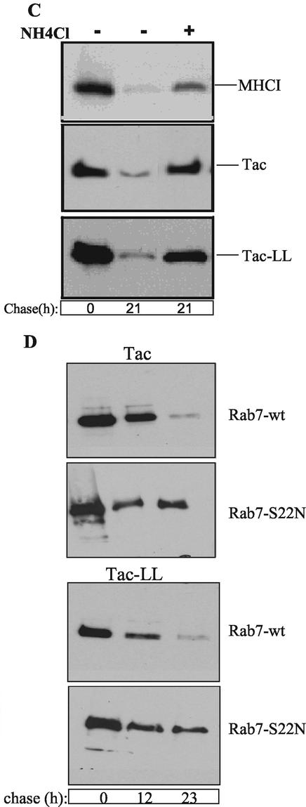
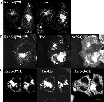
The trafficking of two plasma membrane (PM) proteins that lack clathrin internalization sequences, major histocompatibility complex class I (MHCI), and interleukin 2 receptor alpha subunit (Tac) was compared with that of PM proteins internalized via clathrin. MHCI and Tac were internalized into endosomes that were distinct from those containing clathrin cargo. At later times, a fraction of these internalized membranes were observed in Arf6-associated, tubular recycling endosomes whereas another fraction acquired early endosomal autoantigen 1 (EEA1) before fusion with the "classical" early endosomes containing the clathrin-dependent cargo, LDL. After convergence, cargo molecules from both pathways eventually arrived, in a Rab7-dependent manner, at late endosomes and were degraded. Expression of a constitutively active mutant of Arf6, Q67L, caused MHCI and Tac to accumulate in enlarged PIP(2)-enriched vacuoles, devoid of EEA1 and inhibited their fusion with clathrin cargo-containing endosomes and hence blocked degradation. By contrast, trafficking and degradation of clathrin-cargo was not affected. A similar block in transport of MHCI and Tac was reversibly induced by a PI3-kinase inhibitor, implying that inactivation of Arf6 and acquisition of PI3P are required for convergence of endosomes arising from these two pathways.


Figures

Figure 1
Figure 1
MHCI and Tac enter cells in endosomes distinct from those carrying clathrin-derived cargo and then later merge. (A) Untransfected HeLa cells and those transfected with Tac were allowed to internalize surface-bound anti-MHCI or anti-Tac antibodies, respectively, along with soluble DiI-LDL (10 μg/ml) for 5 and 20 min at 37°C in complete media. Black and white inset at 20 min shows fluorescently labeled structures for each cargo with colocalized structures indicated by yellow arrows. (B) Untransfected and transfected cells expressing either Tac-LL or Tac were allowed to internalize soluble transferrin or DiI-LDL with antibodies to MHCI or Tac for 5 min at 37°C. For both A and B, at the end of the 37°C incubation, surface associated antibodies and DiI-LDL or transferrin were then removed with acid wash, cells were fixed, and internalized antibodies were labeled with fluorescently conjugated secondary antibodies. Bar, 10 μm
Figure 2
Figure 2
Surface MHCI and Tac are internalized independently of dynamin, are not in rafts, and do not colocalize with clathrin at the PM. (A) HeLa cells were transfected with either Tac (squares) or Tac-LL (circles) and either the wild-type (open symbols) or K44A mutant (filled symbols) of dynamin-2. Tac internalization was monitored by Cell ELISA using biotinylated anti-Tac antibody as described in MATERIALS AND METHODS, and the fraction of Tac antibody remaining on the surface at different time points was recorded. A representative experiment is shown with the mean and SD for triplicates of each time point. (B) HeLa cells were transfected with either Tac, Tac-LL, or Tac-GPI, treated with 1.0% Triton X-100 at 4°C for 3 min, and then fixed. Surface Tac proteins were assessed by incubation with anti-Tac antibodies (without saponin). (C) HeLa cells were transiently transfected with Tac or Tac-LL and fixed. Surface labeling was performed using rabbit anti-Tac antibody in the absence of saponin, followed by a second fixation. Cells were then labeled in the presence of saponin with mouse anticlathrin antibody and with secondary antibodies. Background, surface fluorescence staining was diminished in these images to enhance the punctate pattern of the proteins. Magnified insets are designated. Bar, 10 μm.
Figure 3
Figure 3
MHCI and LDL are internalized in separate endosomes, acquire EEA1, and then fuse. Untransfected HeLa cells were incubated with mouse anti-MHCI at 4°C and then warmed to 37°C in media containing DiI-LDL for 5 min. Surface MHCI antibody was removed by low pH treatment, and the cells were either fixed (pulse) or warmed at 37°C in fresh media for a further 5 and 15 min (chase). Cells were then fixed, and internalized MHCI antibody was visualized with isotype-specific Alexa 546 goat anti-mouse IgG2a. EEA1 was localized with monoclonal anti-EEA1 followed by Alexa 647 goat anti-mouse IgG1. (A) Yellow merged image indicates colocalization between MHCI (green) and DiI-LDL (red). (B) Turquoise merged image indicates colocalization between MHCI (green) and EEA1 (blue) (C) Magenta merged image indicates colocalization between DiI-LDL (red) and EEA1 (blue). (D) White triple-merged image indicates colocalization between all three fluorescent dyes. (E) Quantitative analysis of colocalization as described in MATERIALS AND METHODS. Yellow and turquoise bars indicate percentage of MHCI-area that overlap with LDL and EEA1, respectively. Magenta bar indicates percentage of LDL-area that overlaps with EEA1. The mean and SD are indicated for each condition. Analysis of variance using ANOVA test indicates that the amount of convergence of MHCI and LDL at 5 min was different from that after 5-min chase (p < 0.05) and 15-min chase (p < 0.01) and that the 5-min chase was different from the 15-min chase (p < 0.01).
Figure 4
Figure 4
The activity of PI3kinase is necessary for fusion of clathrin-dependent and -independent endosomes. Untransfected cells were labeled with mouse anti-MHCI and DiI-LDL as in Figure 3 and shifted to 37°C for 10 min. Remaining surface MHCI antibody was immediately removed by low pH treatment, and the cells were either fixed (pulse) or warmed at 37°C in fresh media with or without 50 μM LY294002 for 1 h (chase) and fixed. Internalized MHCI antibody was visualized with isotype-specific Alexa 546 goat anti-mouse IgG2a (green). EEA1 was localized with monoclonal anti-EEA1 followed by Alexa 647 goat anti-mouse IgG1 (blue). For washout, after the chase treatment, LY294002 was replaced by fresh media for additional 15 min recovery at 37°C. (AD) Merged images as in Figure 3. (E) Quantitative analysis of area overlap as in Figure 3. The mean and SD are indicated for each condition. Statistical tests using ANOVA indicate that the amount of convergence of MHCI and LDL after 1-h chase was different from that observed for 1-h chase in the presence of LY (p < 0.01) and that the association of EEA1 with MHCI was different at 1-h chase in the absence as compared with the presence of LY (p < 0.05). Importantly, upon removal of LY and recovery, there were no significant differences between these values and those obtained for the 1-h chase in the absence of LY.
Figure 5
Figure 5
MHCI and Tac get access to late endosomes and lysosomes where they are degraded. (A) HeLa cells cotransfected with GFP-Rab7 and either Tac or Tac-LL were incubated with anti-Tac for 30 min at 4°C. Cells were then incubated at 37°C for 7 h to allow endocytosis of PM-bound antibody to late endosomes. Remaining PM-anti-Tac was removed with acid wash, and cells were fixed. Late endosomes were visualized with GFP-Rab7 and internalized anti-Tac was detected with secondary antibody. (B) Cells transfected with lgp120-GFP, Tac, or Tac-LL were cooled on ice and incubated with anti-MHCI or rabbit anti-Tac for 30 min to label the surface and then incubated at 37°C with complete media containing 15 mM NH4Cl for 11 h to allow delivery and accumulation of antibody to lysosomes. After fixation, lysosomes were visualized with lgp120-GFP (in the case of MHCI) or using mouse anti-Lamp1, followed by the appropriate secondary antibodies. (C) Untransfected cells or cells transfected with either Tac or Tac-LL were surface biotinylated on ice for 30 min and then chased for 21 h with or without 15 mM NH4Cl in complete media. Lysates were immunoprecipitated with anti-MHCI or anti-Tac antibody and analyzed by SDS-PAGE. Biotinylated proteins were visualized using HRP-conjugated streptavidin. (D) Cells were cotransfected with either Tac or Tac-LL and either wild-type or T22N mutant of Rab7, and after surface biotinylation and internalization, proteins were immunoprecipitated with Tac antibodies as in C above. Bar, 10 μm.
Figure 5
Figure 5
MHCI and Tac get access to late endosomes and lysosomes where they are degraded. (A) HeLa cells cotransfected with GFP-Rab7 and either Tac or Tac-LL were incubated with anti-Tac for 30 min at 4°C. Cells were then incubated at 37°C for 7 h to allow endocytosis of PM-bound antibody to late endosomes. Remaining PM-anti-Tac was removed with acid wash, and cells were fixed. Late endosomes were visualized with GFP-Rab7 and internalized anti-Tac was detected with secondary antibody. (B) Cells transfected with lgp120-GFP, Tac, or Tac-LL were cooled on ice and incubated with anti-MHCI or rabbit anti-Tac for 30 min to label the surface and then incubated at 37°C with complete media containing 15 mM NH4Cl for 11 h to allow delivery and accumulation of antibody to lysosomes. After fixation, lysosomes were visualized with lgp120-GFP (in the case of MHCI) or using mouse anti-Lamp1, followed by the appropriate secondary antibodies. (C) Untransfected cells or cells transfected with either Tac or Tac-LL were surface biotinylated on ice for 30 min and then chased for 21 h with or without 15 mM NH4Cl in complete media. Lysates were immunoprecipitated with anti-MHCI or anti-Tac antibody and analyzed by SDS-PAGE. Biotinylated proteins were visualized using HRP-conjugated streptavidin. (D) Cells were cotransfected with either Tac or Tac-LL and either wild-type or T22N mutant of Rab7, and after surface biotinylation and internalization, proteins were immunoprecipitated with Tac antibodies as in C above. Bar, 10 μm.
Figure 6
Figure 6
Arf6-Q67L affects trafficking of clathrin-independent cargo but not clathrin-derived cargo. (A) Cos cells were transfected with Arf6 Q67L and Tac (first column), Tac-LL (second column), or Arf6 Q67L alone (third column). Cells were allowed to internalize mouse anti-Tac (first and second column) or DiI-LDL (10 μg/ml; third column) for 1 h at 37°C. (B) Cos cells were cotransfected with Arf6 Q67L and PH-GFP or Q67L alone, and EEA1 was detected in fixed cells using monoclonal anti-EEA1. Cells in A and B were labeled with polyclonal anti-ARF6 followed by the appropriate secondary antibodies. (C) HeLa cells coexpressing Arf6 Q67L and Tac or Tac-LL were incubated with polyclonal anti-Tac antibody for 30 min at 4°C and then incubated for 11 h at 37°C (in the presence of 15 mM NH4Cl) to allow delivery of PM-bound antibody to lysosomes. After acid wash, cells were fixed, and lysosomes were visualized with monoclonal anti-Lamp1 followed by secondary antibodies. Bar, 10 μm. (D) HeLa cells coexpressing Arf6 wt or Q67L and Tac or Tac-LL were subjected to biotinylation pulse-chase as described in MATERIALS AND METHODS. Lysates were immunoprecipitated with anti-Tac mAb and analyzed as described in Figure 5C.
Figure 7
Figure 7
Rab5 and Arf6 affect different internalization pathways. HeLa cells coexpressing GFP-Rab5 Q79L and Tac (A); GFP-Rab5 Q79L, Arf6 Q67L and Tac (B); or GFP-Rab5 Q79L, Arf6 Q67L, and Tac-LL (C) were allowed to internalize monoclonal anti-Tac for 1 h, followed by an acid wash. After fixation, Arf6 was labeled with rabbit anti-Arf6 and Alexa 594 goat anti-rabbit and internalized Tac antibody was detected with Alexa 647 goat anti-mouse. Bar, 10 μm.
Figure 8
Figure 8
Arf6 inactivation and PI3P synthesis are required for fusion between non–clathrin- and clathrin-derived endosomes. PM proteins (e.g., MHCI, Tac), lacking cytoplasmic clathrin/AP2-targeting sequences enter cells in Arf6-GTP endosomes independent of clathrin-coated pits. These endosomes may undergo homotypic fusion followed by Arf6-GAP stimulated inactivation of Arf6 and loss or modification of PIP2. In cells expressing Arf6Q67L, inactivation of Arf6 is inhibited, preventing Arf6-derived endosomes from fusing with clathrin-derived endosomes. Instead, extensive homotypic fusion occurs. We speculate that Arf6 endosomes mature or fuse to form a hypothetical Arf6-associated sorting endosome (shown in brackets), where a fraction of the cargo along with Arf6 is recycled back to the PM. Another fraction of the cargo, destined to be degraded, is retained on the membrane that contains the fluid of the endosome. Acquisition of PI3P and recruitment of EEA1 on these endosomes then allows fusion with Rab5/EEA1 early endosomes, thus merging the clathrin-independent and clathrin-derived (LDL-R and Tac-LL) cargo compartments. Inhibition of PI3-kinase with LY294002 (LY) blocks fusion between the two compartments; the exact site of the block and the intermediate that accumulates is not known.

References

    1. Amyere M, Payraste B, Krause U, Van Der Smissen P, Veithen A, Courtoy PJ. Constitutive macropinocytosis in oncogene-transformed fibroblasts depend on sequential permanent activation of phosphoinositide 3-kinase and phospholipase C. Mol Biol Cell. 2000;11:3453–3467. - PMC - PubMed
    1. Brown D, Rose J. Sorting of GPI-anchored proteins to glycolipid-enriched membrane subdomains during transport to the apical cell surface. Cell. 1992;68:533–544. - PubMed
    1. Brown FD, A.L. Rozelle AL, Yin HL, Balla T, Donaldson JG. Phosphatidylinositol 4,5-biphosphate and Arf6-regulated membrane traffic. J Cell Biol. 2001;154:1007–1017. - PMC - PubMed
    1. Bucci C, Thomsen P, Nicoziani P, McCarthy J, van Deurs B. Rab7: a key to lysosome biogenesis. Mol Biol Cell. 2000;11:467–480. - PMC - PubMed
    1. Caplan S, Naslavsky N, Hartnell LM, Lodge R, Polishchuk RS, Donaldson JG, Bonifacino JS. A tubular EHD1-containing compartment involved in the recycling of major histocompatibility complex class I molecules to the plasma membrane. EMBO J. 2002;21:2557–2567. - PMC - PubMed

MeSH terms

LinkOut - more resources