. 2003 Feb;14(2):477-90.
doi: 10.1091/mbc.e02-07-0413.

Chemical genetic analysis of Apg1 reveals a non-kinase role in the induction of autophagy

Affiliations

Chemical genetic analysis of Apg1 reveals a non-kinase role in the induction of autophagy

Hagai Abeliovich et al. Mol Biol Cell. 2003 Feb.

Abstract

Macroautophagy is a catabolic membrane trafficking phenomenon that is observed in all eukaryotic cells in response to various stimuli, such as nitrogen starvation and challenge with specific hormones. In the yeast Saccharomyces cerevisiae, the induction of autophagy involves a direct signal transduction mechanism that affects membrane dynamics. In this system, the induction process modifies a constitutive trafficking pathway called the cytoplasm-to-vacuole targeting (Cvt) pathway, which transports the vacuolar hydrolase aminopeptidase I, from the formation of small Cvt vesicles to the formation of autophagosomes. Apg1 is one of the proteins required for the direct signal transduction cascade that modifies membrane dynamics. Although Apg1 is required for both the Cvt pathway and autophagy, we find that Apg1 kinase activity is required only for Cvt trafficking of aminopeptidase I but not for import via autophagy. In addition, the data support a novel role for Apg1 in nucleation of autophagosomes that is distinct from its catalytic kinase activity and imply a qualitative difference in the mechanism of autophagosome and Cvt vesicle formation.


Figures
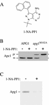
Figure 1
Figure 1
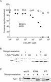
Precursor Ape1 maturation in the apg1M102A mutant is selectively inhibited by 1-NA-PP1. (A) Structure of 1-NA-PP1. (B) apg1Δ (HAY395) yeast cells carrying a wild-type APG1 or a mutant apg1M102A gene on pRS415 were grown to an A600 of 0.5 and treated with or without 20 μM 1-NA-PP1. The cells were incubated an additional 3 h and then harvested and processed for Western blotting as described in MATERIALS AND METHODS. (C) Kinase activity of Apg1M102A is inhibited by 20 μM 1-NA-PP1. Cell extract (2 mg per reaction) was immunoprecipitated with anti-Apg1 antiserum and assayed for autophosphorylation activity in the presence or absence of 20 μM 1-NA-PP1 as described in MATERIALS AND METHODS.
Figure 2
Figure 2
Inhibition of Apg1 kinase by 1-NA-PP1 blocks the Cvt pathway but allows autophagic maturation of prApe1. (A) Dose response for 1-NA-PP1 under standard growth conditions and under starvation. apg1Δ (HAY395) yeast cells expressing Apg1M102A from a plasmid were grown to midlog (A600 of 0.5), collected and resuspended in SD medium with the indicated concentrations of 1-NA-PP1. After 1 h incubation, one half of the cell culture was collected, washed with distilled water, and resuspended in nitrogen starvation medium (SD-N) in the presence of the same concentration of 1-NA-PP1. After a further incubation of 3 h, the cells were harvested and cells extracts were prepared. 0.5 A600 equivalents of each extract were resolved by SDS-PAGE and Ape1 was identified by Western blotting. The relative amount of prApe1 and mApe1 was quantified using a Bio-Rad Fluor-S MAX as described in MATERIALS AND METHODS. (B) Western blot of a subset of the samples described in A. (C) The kinase inactive apg1K54A mutant is blocked for prApe1 maturation in rich medium but not under starvation conditions. apg1Δ (HAY395) cells or these cells harboring a plasmid borne wild-type APG1 gene or apg1K54A allele were grown to midlog and either harvested immediately or transferred to nitrogen starvation medium for 3 h before extract preparation. Ape1 was detected by SDS-PAGE and Western blotting.
Figure 3
Figure 3
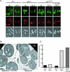
Inhibition of Apg1 kinase activity in the apg1M102A mutant does not block autophagy. (A) Wild-type (HAY75, WT), apg1Δ (HAY395) and apg1Δ cells harboring apg1M102A on pRS415 were transformed with a centromeric plasmid (pRS414) expressing Aut7-GFP under the control of the AUT7 promoter. Cells were grown to midlog and stained with FM 4–64 (0.8 μM) for 20 min, washed and incubated with or without 30 μM 1-NA-PP1 for 1 h in SD medium. Cells were then either viewed directly or washed with distilled water and transferred to nitrogen starvation medium for 4 h before viewing by light and fluorescence microscopy. Top panels: GFP fluorescence. Middle panels: FM 4–64 fluorescence. Bottom panels: DIC, differential interference contrast. (B) The vam3ts apg1Δ strain (HAY571) harboring apg1M102A on pRS415 was grown to midlog at 26°C, transferred for 1 h into medium containing 30 μM 1-NA-PP1 or mock-treated, and then transferred for 20 min to 37.5°C. Rapamycin was added to a final concentration of 0.2 μg/ml and the cells were incubated for a further 90 min before permanganate fixation and electron microscopy as described in MATERIALS AND METHODS. AP, autophagosome; M, mitochondrion; N, nucleus; V, vacuole. (C) The pho13Δ pho8Δ60 apg1Δ strain (HAY572) or HAY572 cells harboring apg1M102A on a plasmid were subjected to the same treatments as in (A) and alkaline phosphatase activity was measured in extracts as described in MATERIALS AND METHODS. Results represent duplicate measurements from two independent experiments.
Figure 4
Figure 4
The C terminus of Apg1 is engaged in a Cvt pathway-specific interaction. (A) The pep4Δ Apg1-prA strain (HAY437) expressing a full-length Apg1-prA fusion, or the pep4Δ apg1Δ880-prA strain (HAY478) expressing an Apg1Δ880 truncation fused to prA were grown to midlog in YPD medium and converted to spheroplasts. The spheroplasts were treated with or without 0.2 μg/ml rapamycin for 15 min before cell lysis, and the Apg1-prA fusions were purified from the extracts using IgG sepharose as described in MATERIALS AND METHODS. Apg1-prA was identified and quantified in the extracts by SDS-PAGE and immunodetection with anti-prA antibodies. (B) Truncation of the last 18 amino acids of Apg1 results in a Cvt pathway-specific phenotype. The apg1Δ850-prA (HAY454), apg1Δ880-prA (HAY455), apg1Δ886-prA (HAY518) and full-length (FL) Apg1-prA (HAY370) strains were grown to midlog. One-half of each culture was transferred to nitrogen starvation medium for 3 h before extract preparation and the other half was harvested directly. Precursor Ape1 maturation and the levels of the fusion proteins were assessed by Western blotting (0.5 A600 equivalents per lane). Survival of yeast in nitrogen starvation conditions was determined by patching cells on phloxine B plates (Tsukada and Ohsumi, 1993); nd, not determined. (C) A leucine to glycine mutation at position 886 results in a Cvt pathway-specific phenotype. WT (HAY75), apg1N884A, and apg1L886G yeast cells were grown to midlog in SD and either harvested directly or first transferred to nitrogen starvation medium for 3 h before extract preparation. Precursor Ape1 maturation was assayed by Western blotting. (D) apg1L886G cells do not lose viability in nitrogen starvation. The apg1L886G (HAY602) and apg1Δ (HAY395) cells were grown to midlog and transferred to nitrogen starvation medium. At indicated time points, viable cell counts were performed by removing an aliquot and plating an appropriate dilution on YPD.
Figure 5
Figure 5
Apg13 has an autophagy-specific function. (A) Logarithmically growing apg1Δ880 (HAY455), apg13Δ (ResGen) or apg13Δ apg1Δ880 (HAY487) cells either containing or not containing the APG13 gene on a multicopy plasmid were either directly harvested or first transferred to nitrogen starvation medium and then harvested. Protein extracts were prepared and Ape1 was detected by Western blotting. (B) Starvation-dependent maturation of prApe1 in the presence of 20 μM 1-NA-PP1 requires Apg13. apg1Δ (HAY395) or apg1Δ apg13Δ (HAY603) cells carrying the apg1M102A gene on pRS415 were grown to midlog and treated with or without 20 μM 1-NA-PP1 for 1 h, and either starved for nitrogen in the presence of 1-NA-PP1 or allowed to remain in SD medium, for a further 3 h before preparation of protein extracts and SDS-PAGE. Ape1 was detected by Western blotting.
Figure 6
Figure 6
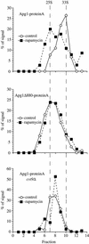
Structural changes occur in Apg1 upon induction of autophagy and depend on Cvt9 and the C terminus of Apg1. The Apg1-protein A pep4Δ (HAY437), Apg1Δ880-protein A pep4Δ (HAY478), and Apg1-protein A cvt9Δ pep4Δ (HAY591) strains were grown to midlog in YPD and converted to spheroplasts. Spheroplasts were treated with or without rapamycin (0.2 μg/ml) for 15 min before lysis. Lysates were precleared by differential centrifugation at 100,000 × g for 15 min and loaded onto a 5–20% sucrose gradient. The gradients were centrifuged at 259,000 × g for 7 h, fractions were precipitated with 10% TCA and Apg1-prA was identified by Western blotting with anti-prA antiserum and quantified using a Bio-Rad Fluor-S MAX. The migration of the two peaks observed in wild-type cells is denoted by the vertical dashed lines (calculated to be 25S and 33S for the slow and fast moving components, respectively).
Figure 7
Figure 7
The Apg1Δ880 mutant is hypophosphorylated. Cells expressing full-length prA-tagged Apg1 (HAY370) or the Δ880 truncation fused to prA (HAY455) were grown to midlog, and labeled with 150 μCi 32P for 1 h before treatment with 0.2 ng/ml rapamycin or mock solution. After 20 min exposure to rapamycin, protein extracts were prepared and Apg1 was immunoprecipitated as in MATERIALS AND METHODS. One half of the immunoprecipitate (IP) was separated by SDS-PAGE and exposed for phosphorimaging while the other half was separated, transferred to nitrocellulose, and immunoblotted for Apg1.
Figure 8
Figure 8
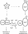
Model for mechanistic coupling between kinase activity and nucleation of autophagosomes. Under normal growing conditions Tor kinase activity induces Apg1 kinase autophosphorylation, either through modulation of Apg13 phosphorylation or through a direct effect on Apg1. Inhibition of Tor results in a partial dephosphorylation of Apg13, and a decrease of Apg1 kinase autophosphorylation, leading to dephosphorylation and a structural change in Apg1. This structural change is correlated with an increased interaction with Apg13, and involves Cvt9. The dephosphorylated form of Apg1 directs autophagosome formation when associated with Apg13, whereas the phosphorylated form directs nucleation of Cvt vesicles.

Similar articles

Cited by

References

    1. Abeliovich H, Darsow T, Emr SD. A t-SNARE/Sec1p complex composed of Tlg2p and Vps45p is required for cytoplasm to vacuole targeting of aminopeptidase I. EMBO J. 1999;18:6005–6016. - PMC - PubMed
    1. Abeliovich H, Dunn WA, Jr, Kim J, Klionsky DJ. Dissection of autophagosome biogenesis into distinct nucleation and expansion steps. J Cell Biol. 2000;151:1025–1033. - PMC - PubMed
    1. Abeliovich H, Grote E, Novick P, Ferro-Novick S. Tlg2p, a yeast syntaxin homolog that resides on the Golgi and Endocytic structures. J Biol Chem. 1998;273:11719–11727. - PubMed
    1. Abeliovich H, Klionsky DJ. Autophagy in yeast: mechanistic insights and physiological function. Microbiol Mol Biol Rev. 2001;65:463–479. - PMC - PubMed
    1. Baba M, Osumi M, Scott SV, Klionsky DJ, Ohsumi Y. Two distinct pathways for targeting proteins from the cytoplasm to the vacuole/lysosome. J Cell Biol. 1997;139:1687–1695. - PMC - PubMed

Publication types

MeSH terms