. 2003 Feb;14(2):529-44.
doi: 10.1091/mbc.02-03-0037.

Transforming growth factor-beta1 (TGF-beta)-induced apoptosis of prostate cancer cells involves Smad7-dependent activation of p38 by TGF-beta-activated kinase 1 and mitogen-activated protein kinase kinase 3

Affiliations

Transforming growth factor-beta1 (TGF-beta)-induced apoptosis of prostate cancer cells involves Smad7-dependent activation of p38 by TGF-beta-activated kinase 1 and mitogen-activated protein kinase kinase 3

Sofia Edlund et al. Mol Biol Cell. 2003 Feb.

Abstract

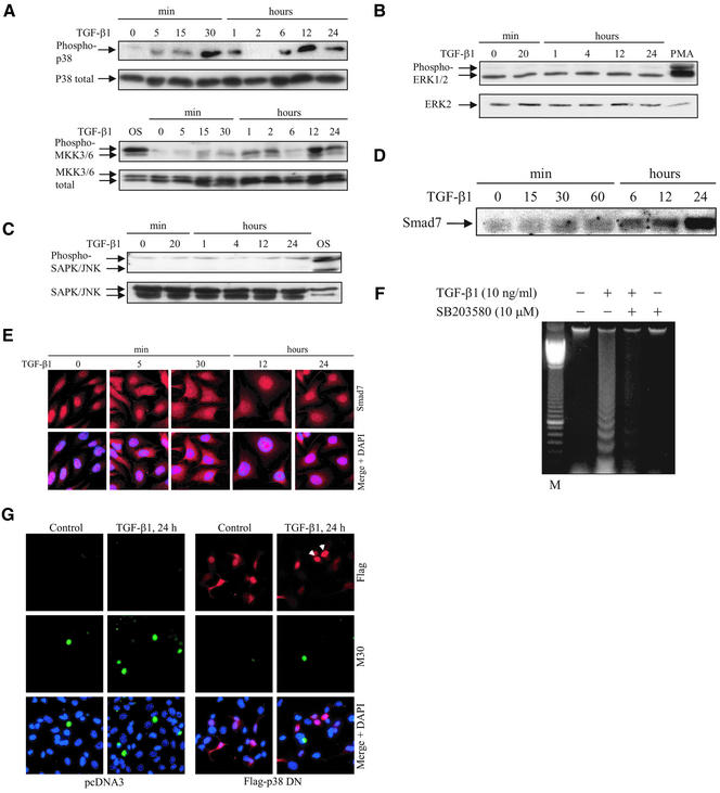
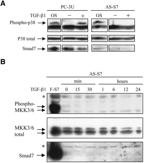
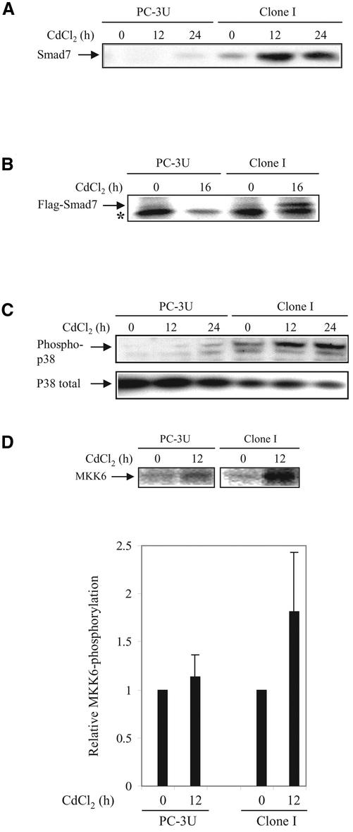
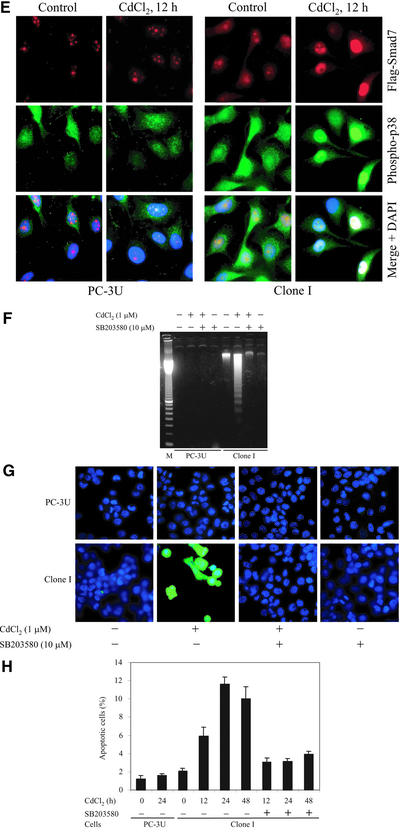
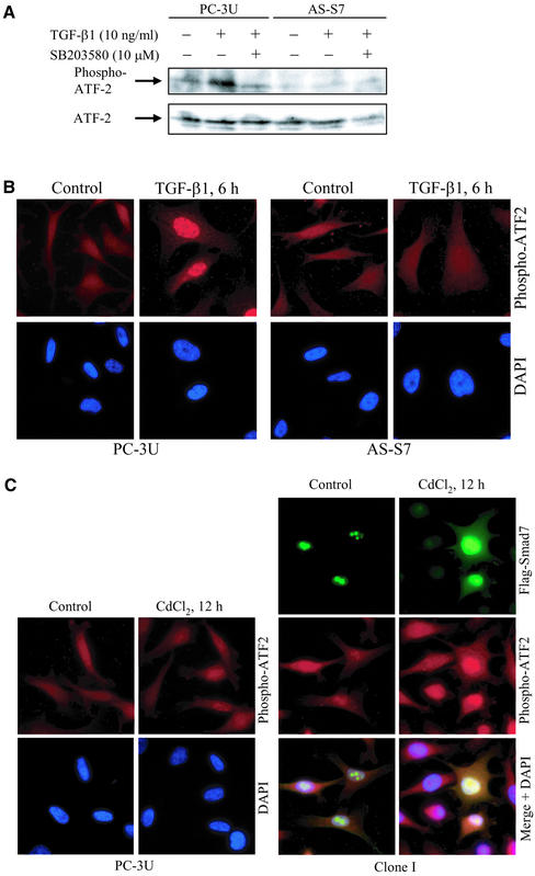
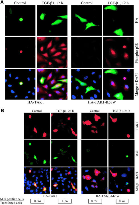
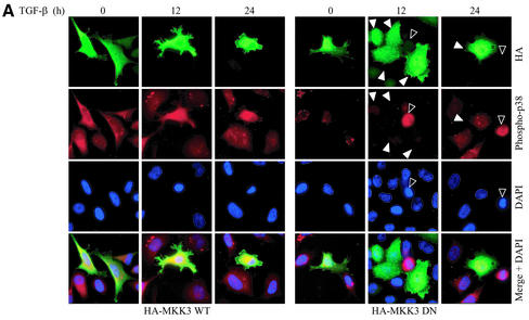
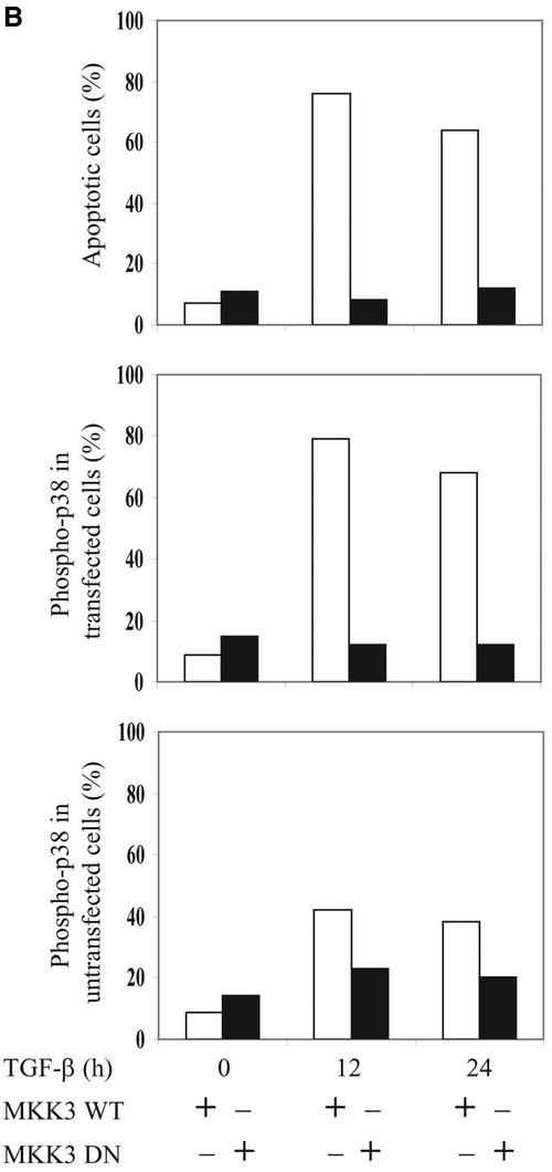
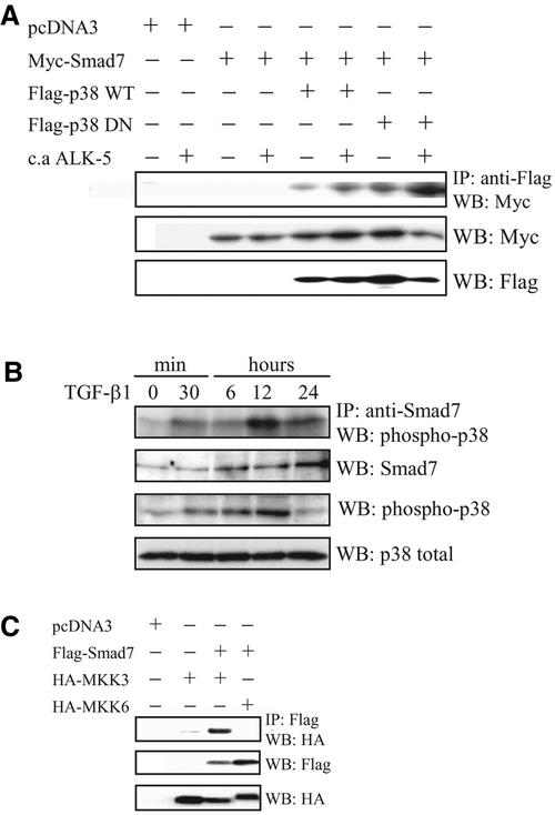
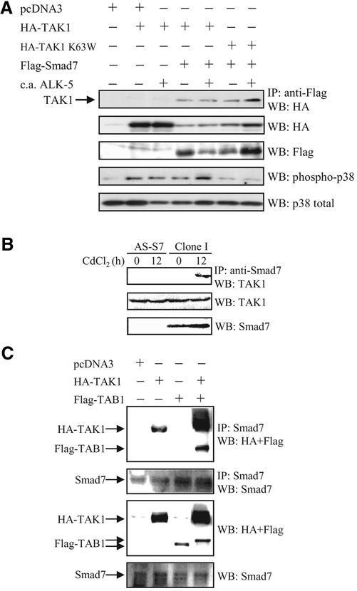
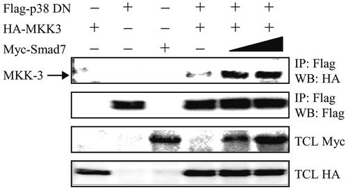
The inhibitory Smad7, a direct target gene for transforming growth factor-beta (TGF-beta), mediates TGF-beta1-induced apoptosis in several cell types. Herein, we report that apoptosis of human prostate cancer PC-3U cells induced by TGF-beta1 or Smad7 overexpression is caused by a specific activation of the p38 mitogen-activated protein kinase pathway in a TGF-beta-activated kinase 1 (TAK1)- and mitogen-activated protein kinase kinase 3 (MKK3)-dependent manner. Expression of dominant negative p38, dominant negative MKK3, or incubation with the p38 selective inhibitor [4-(4-fluorophenyl)-2-(4-methylsulfinylphenyl)-5-(4-pyridyl)1H-imidazole], prevented TGF-beta1-induced apoptosis. The expression of Smad7 was required for TGF-beta-induced activation of MKK3 and p38 kinases, and endogenous Smad7 was found to interact with phosphorylated p38 in a ligand-dependent manner. Ectopic expression of wild-type TAK1 promoted TGF-beta1-induced phosphorylation of p38 and apoptosis, whereas dominant negative TAK1 reduced TGF-beta1-induced phosphorylation of p38 and apoptosis. Endogenous Smad7 was found to interact with TAK1, and TAK1, MKK3, and p38 were coimmunoprecipitated with Smad7 in transiently transfected COS1 cells. Moreover, ectopically expressed Smad7 enhanced the coimmunoprecipitation of HA-MKK3 and Flag-p38, supporting the notion that Smad7 may act as a scaffolding protein and facilitate TAK1- and MKK3-mediated activation of p38.


Figures

Figure 1
Figure 1
Specific activation of p38 by TGF-β1 causes apoptosis. Time courses of phosphorylation of endogenous p38 and MKK3/6 (A), ERK1/2 (B), and SAPK/JNK (C), and expression levels of endogenous Smad7 (D) after TGF-β stimulation. Total cell lysates were prepared from TGF-β1–treated or untreated PC-3U cells and used for immunoblotting. In the top and bottom panels, the phosphorylated and the nonphosphorylated forms, respectively, of p38, MKK3/6, ERK1/2, and SAPK/JNK are indicated. Cell lysates from PC-3U cells exposed to 0.7 M NaCl for 30 min (OS) or 10 nM PMA for 20 min (PMA) were used as positive controls. (E) Subcellular localization of endogenous Smad7 in untreated PC-3U cells or PC-3U cells treated for 5 min, 30 min, 12 h, or 24 h with TGF-β, as investigated by immunostainings with antibodies against Smad7 and additional staining of the nuclei by DAPI. An overlay of both pictures (merge + DAPI) shows that Smad7 is predominantly localized in nuclei of untreated cells, whereas 5 min after TGF-β treatment an export to the cytoplasm is observed, followed by accumulation in the nucleus after 12 and 24 h. (F) Analysis of fragmentation of DNA isolated from PC-3U cells, TGF-β1–treated or not for 24 h in the presence or absence of SB203580. (G) Apoptosis of PC-3U cells, transiently transfected with pcDNA3 (control) and dominant negative Flag p38 (Flag-p38 DN), treated with TGF-β1 for 24 h, as analyzed by immunostainings with antibodies against Flag and the apoptotic marker M30. An overlay of both pictures with additional staining of nuclei with DAPI (merge + DAPI) showed a reduced number of M30-positive cells per field in cells expressing Flag-p38 DN after treatment with TGF-β1, compared with control cells. Note that cells expressing Flag-p38 DN, indicated by arrowheads, do not undergo apoptosis.
Figure 2
Figure 2
Smad7 is necessary for TGF-β–induced phosphorylation of p38 and MKK3/6. (A) Phosphorylation of endogenous p38 in total cell lysates prepared from PC-3U and antisense Smad7 PC-3U cells (AS-S7), untreated or treated with TGF-β1 for 1 h, was analyzed by immunoblotting. The phosphorylated and nonphosphorylated forms of p38 are shown in the top and bottom panels, respectively. Cell lysates from PC-3U cells exposed to 0.7 M NaCl for 30 min (OS) were used as positive control. The same filter was stripped, blocked, and reprobed with antibodies against Smad7. Note the lowered Smad7 expression and the lack of phosphorylated p38 in AS-S7 cells. (B) Phosphorylation of endogenous MKK3/6 in total cell lysates prepared from AS-S7 cells, untreated or treated with TGF-β1 for 15 min to 24 h, was analyzed by immunoblotting. The phosphorylated and nonphosphorylated forms of MKK3/6 are shown in the top and bottom panels, respectively. Cell lysates from PC-3U cells infected with adenoviral Flag-Smad7 (F-S7) was used as positive control. The same filter was stripped, blocked, and reprobed with antibodies against Smad7. Note the lowered Smad7 expression and the lack of phosphorylated MKK3/6 in AS-S7 cells. A background band is indicated by an asterisk (*).
Figure 3
Figure 3
Ectopic expression of Smad7 induces increased levels of p38 phosphorylation and apoptosis. (A) Time course of Smad7 levels in PC-3U and PC-3U cells stably transfected with pMEP-4 F-Smad7 (Clone I cells). Total cell lysates were prepared from wild-type PC-3U cells and Clone I cells, treated or not with CdCl2 and subjected to immunoblotting. (B) Ectopic expression of Flag-Smad7 is induced by CdCl2 stimulation (16 h) of Clone I cells as shown by metabolic labeling and immunoprecipitation with Flag antibody. A background band is indicated by an asterisk (*). (C) Time course of phosphorylation of endogenous p38. Total cell lysates were prepared from wild-type PC-3U cells and Clone I cells, treated or not with CdCl2 and subjected to immunoblotting. The phosphorylated p38 (phospho-p38) and the total p38 are shown in the top and bottom panels, respectively. (D) Activation of MKK6 by Smad7 overexpression is shown. PC-3U and Clone I cells were treated or not with CdCl2 for 12 h. Cell lysates were immunoprecipitated with anti-TAK1 antibodies and immunoprecipitates were subjected to in vitro kinase assay by using His-MKK6 as substrate. The phosphorylated proteins were resolved by SDS-PAGE and visualized by autoradiography (top). The mean relative values from three independently performed experiments including scanning electron microscopy are presented (bottom). (E) Immunofluorescence analyses of phosphorylated p38 in PC-3U cells and Clone I cells treated or not with CdCl2 for 12 h. Cells were also analyzed for Flag-Smad7 expression by immunofluorescence staining by using Flag antibodies. An overlay of both pictures with additional staining of nuclei by DAPI (merge + DAPI) reveals several nuclei with a colocalization of Smad7 and phospho-p38 in Clone I cells. (F) Analysis of fragmentation of DNA isolated from control PC-3U cells and Clone I cells treated or not for 24 h with CdCl2, in the presence or absence of SB203580. (G) Stainings with DAPI and TUNEL (fluorescein isothiocyanate) of control PC-3U cells and Clone I cells treated or not for 24 h with CdCl2 in the presence or absence of SB203580. Note the enhanced TUNEL staining of nuclei showing morphological criteria for apoptosis in cells overexpressing Flag-Smad7, which is inhibited by SB203580. (H) Apoptosis of PC-3U cells and Clone I cells treated or not with CdCl2, alone or together with SB203580, as revealed by immunostaining with the apoptotic marker M30; staining was quantified as described in MATERIALS AND METHODS. Note that the increase of M30 staining in Clone I cells treated with CdCl2 is reduced by SB203580, whereas only a minor apoptotic effect was detected in PC-3U cells after 24 h treatment with CdCl2.
Figure 3
Figure 3
Ectopic expression of Smad7 induces increased levels of p38 phosphorylation and apoptosis. (A) Time course of Smad7 levels in PC-3U and PC-3U cells stably transfected with pMEP-4 F-Smad7 (Clone I cells). Total cell lysates were prepared from wild-type PC-3U cells and Clone I cells, treated or not with CdCl2 and subjected to immunoblotting. (B) Ectopic expression of Flag-Smad7 is induced by CdCl2 stimulation (16 h) of Clone I cells as shown by metabolic labeling and immunoprecipitation with Flag antibody. A background band is indicated by an asterisk (*). (C) Time course of phosphorylation of endogenous p38. Total cell lysates were prepared from wild-type PC-3U cells and Clone I cells, treated or not with CdCl2 and subjected to immunoblotting. The phosphorylated p38 (phospho-p38) and the total p38 are shown in the top and bottom panels, respectively. (D) Activation of MKK6 by Smad7 overexpression is shown. PC-3U and Clone I cells were treated or not with CdCl2 for 12 h. Cell lysates were immunoprecipitated with anti-TAK1 antibodies and immunoprecipitates were subjected to in vitro kinase assay by using His-MKK6 as substrate. The phosphorylated proteins were resolved by SDS-PAGE and visualized by autoradiography (top). The mean relative values from three independently performed experiments including scanning electron microscopy are presented (bottom). (E) Immunofluorescence analyses of phosphorylated p38 in PC-3U cells and Clone I cells treated or not with CdCl2 for 12 h. Cells were also analyzed for Flag-Smad7 expression by immunofluorescence staining by using Flag antibodies. An overlay of both pictures with additional staining of nuclei by DAPI (merge + DAPI) reveals several nuclei with a colocalization of Smad7 and phospho-p38 in Clone I cells. (F) Analysis of fragmentation of DNA isolated from control PC-3U cells and Clone I cells treated or not for 24 h with CdCl2, in the presence or absence of SB203580. (G) Stainings with DAPI and TUNEL (fluorescein isothiocyanate) of control PC-3U cells and Clone I cells treated or not for 24 h with CdCl2 in the presence or absence of SB203580. Note the enhanced TUNEL staining of nuclei showing morphological criteria for apoptosis in cells overexpressing Flag-Smad7, which is inhibited by SB203580. (H) Apoptosis of PC-3U cells and Clone I cells treated or not with CdCl2, alone or together with SB203580, as revealed by immunostaining with the apoptotic marker M30; staining was quantified as described in MATERIALS AND METHODS. Note that the increase of M30 staining in Clone I cells treated with CdCl2 is reduced by SB203580, whereas only a minor apoptotic effect was detected in PC-3U cells after 24 h treatment with CdCl2.
Figure 4
Figure 4
Smad7 is necessary for TGF-β–induced phosphorylation of ATF-2. (A) PC-3U and AS-S7 cells were treated or not with TGF-β for 6 h in the absence or presence of SB203580. Total cell lysates were then prepared and subjected to immunoblotting, by using antibodies against ATF-2 phosphorylated at Thr69 and Thr71 (phospho-ATF-2) (top) or total ATF-2 (bottom). (B) Immunofluorescence investigations of phospho-ATF-2 in PC-3U and AS-S7 cells treated with TGF-β1 for 6 h. Note the lack of staining for phospho-ATF-2 in AS-S7 cells compared with control PC-3U cells. (C) Coimmunofluorescence stainings of Flag-Smad7 and phospho-ATF-2 in Clone I cells treated for 12 h with CdCl2. An overlay of both pictures with additional stainings of nuclei with DAPI (merge + DAPI) shows colocalization of Smad7 and phospho-ATF-2 in the nucleus. Immunofluorescence stainings of phospho-ATF-2 in PC-3U cells untreated or treated for 12 h with CdCl2 are shown as control. Immunofluorescence staining with Flag antibodies of PC-3U cells treated for 12 h with CdCl2 as control for Flag, is shown in Figure 3E.
Figure 5
Figure 5
TAK1 enhances TGF-β1–induced phosphorylation of p38 and apoptosis in PC-3U cells. PC-3U cells were transfected with either wild-type HA-TAK1 or kinase inactive HA-TAK1-K63W and treated with TGF-β1 for 12 or 24 h. (A) Coimmunofluorescence stainings for HA, detected by fluorescein isothiocyanate and phosphorylated p38 (phospho-p38) by tetramethylrhodamine B isothiocyanate. An overlay of both pictures with additional stainings of nuclei with DAPI (merge + DAPI) reveals a high degree of colocalization of HA-TAK1 and phospho-p38 in PC-3U cells expressing wild-type HA-TAK1, both before and after TGF-β treatment for 12 h. In contrast, PC-3U cells expressing HA-TAK1-K63W, do not express phospho-p38 after TGF-β1 treatment, as shown in the overlay of both pictures with additional stainings of nuclei with DAPI (merge + DAPI). (B) Coimmunofluorescence stainings for endogenous TAK1 (by using polyclonal antibodies against TAK1) as shown by tetramethylrhodamine B isothiocyanate and the apoptotic marker M30, as shown by fluorescein isothiocyanate, was used to identify apoptotic cells after TGF-β treatment for 24 h. An overlay of both pictures with additional stainings of nuclei with DAPI (merge + DAPI) reveals a high degree of colocalization of TAK1 and M30, in PC-3U cells expressing wild-type HA-TAK1. In contrast, in PC-3U cells expressing HA-TAK1-K63W, no colocalization of mutant TAK1 and M30 was observed, as shown in the overlay of both pictures with additional stainings of nuclei with DAPI (merge + DAPI). The number of transfected cells, expressing high levels of TAK1, and the number of M30-positive cells, were counted in several, randomly chosen fields. At total of at least 1000 cells was counted. The ratio between M30-positive cells and transfected cells is shown below the picture.
Figure 6
Figure 6
MKK3 enhances TGF-β1–induced phosphorylation of p38 and apoptosis in PC-3U cells. PC-3U cells were transfected with either wild-type HA-MKK3 or DN HA-MKK3 and treated with TGF-β1 for 12 or 24 h. (A) Coimmunofluorescence stainings for HA, detected by fluorescein isothiocyanate and phospho-p38 by tetramethylrhodamine B isothiocyanate. An overlay of both pictures with additional stainings of nuclei with DAPI (merge + DAPI) reveals a high degree of colocalization of wild-type HA-MKK3 and phospho-p38 in PC-3U cells expressing wild-type HA-MKK3 both before and after TGF-β treatment for 12 h and 24 h. In contrast, in PC-3U cells expressing DN HA-MKK3 indicated by arrowheads, no colocalization of mutant MKK3 and phospho-p38, indicated by an open arrowhead, is observed, as shown in the overlay of both pictures with additional stainings of nuclei with DAPI (merge + DAPI). The number of transfected cells, expressing high levels of MKK3, the number of phospho-p38-positive cells and apoptotic nuclei indicated by an open arrowhead (identified by DAPI), were counted in several, randomly chosen fields. A total of at least 500 cells was counted. The percentage of MKK3-expressing cells, positively stained for HA and phospho-p38 and apoptotic cells are shown in Figure 6B.
Figure 6
Figure 6
MKK3 enhances TGF-β1–induced phosphorylation of p38 and apoptosis in PC-3U cells. PC-3U cells were transfected with either wild-type HA-MKK3 or DN HA-MKK3 and treated with TGF-β1 for 12 or 24 h. (A) Coimmunofluorescence stainings for HA, detected by fluorescein isothiocyanate and phospho-p38 by tetramethylrhodamine B isothiocyanate. An overlay of both pictures with additional stainings of nuclei with DAPI (merge + DAPI) reveals a high degree of colocalization of wild-type HA-MKK3 and phospho-p38 in PC-3U cells expressing wild-type HA-MKK3 both before and after TGF-β treatment for 12 h and 24 h. In contrast, in PC-3U cells expressing DN HA-MKK3 indicated by arrowheads, no colocalization of mutant MKK3 and phospho-p38, indicated by an open arrowhead, is observed, as shown in the overlay of both pictures with additional stainings of nuclei with DAPI (merge + DAPI). The number of transfected cells, expressing high levels of MKK3, the number of phospho-p38-positive cells and apoptotic nuclei indicated by an open arrowhead (identified by DAPI), were counted in several, randomly chosen fields. A total of at least 500 cells was counted. The percentage of MKK3-expressing cells, positively stained for HA and phospho-p38 and apoptotic cells are shown in Figure 6B.
Figure 7
Figure 7
Interaction between Smad7, p38, and MKK3/6 in vivo. (A) Smad7 binds to p38 in vivo. COS1 cells were transfected with Myc-tagged Smad7 alone or together with wild-type (WT) or DN Flag-tagged p38, with or without constitutively active TGF-β type I receptor (c.a. ALK-5). Lysates were immunoprecipitated with anti-Flag antibodies and the immunoblots were incubated with antibodies specific for Myc or Flag. (B) Endogenous Smad7 binds to phosphorylated p38 in vivo. PC-3U cells were treated or not with TGF-β1 for the indicated time periods. Cell lysates were immunoprecipitated with anti-Smad7 antibodies and the immunoblots were incubated with antibodies specific for phospho-p38. Immunoblots from total cell lysates were incubated with antibodies specific for Smad7, phosphorylated p38, and p38. Smad7 binds to p38 in vivo. (C) COS1 cells were transfected with Flag-tagged Smad7 alone or together with wild-type HA MKK3 or HA-MKK6. Lysates were immunoprecipitated with anti-Flag antibodies and the immunoblots were probed with antibodies specific for HA. Immunoblots from total cell lysates were incubated with antibodies specific for HA and Flag. A clear MKK3 band is seen; upon prolonged exposure, a faint, specific MKK6 band is also seen.
Figure 8
Figure 8
Smad7 interacts with TAK1 in vivo. (A) COS1 cells were transfected with Flag-tagged Smad7 alone or together with wild-type TAK1 or kinase inactive (K63W) HA-tagged TAK1, with or without constitutively active TGF-β type I receptor (c.a. ALK-5). Lysates were immunoprecipitated with anti-Flag antibodies and the immunoblots were probed with antibodies specific for HA. Immunoblots from total cell lysates were incubated with antibodies specific for HA, Flag, phosphorylated p38, and p38. (B) Antisense Smad7 PC-3U cells (AS-S7) cells and PC-3U cells stably transfected with pMEP-4 F-Smad7 (Clone I cells), treated or not with CdCl2 for 12 h to induce Smad7 expression in Clone I cells. Lysates were immunoprecipitated with anti-Smad7 antibodies and the immunoblots were probed with antibodies specific for TAK1. Immunoblots from total cell lysates were incubated with antibodies specific for Smad7 and TAK1. (C) PC-3U cells were transiently transfected with HA-TAK1, Flag-TAB1, either alone or together. Lysates were immunoprecipitated with anti-Smad7 antibodies and the immunoblots were probed with antibodies specific for HA and Flag. Immunoblots from total cell lysates were incubated with antibodies specific for Smad7, Flag, and HA.
Figure 9
Figure 9
Expression of Smad7 facilitates the interaction between MKK3 and p38 MAP kinase. COS1 cells were transfected with wild-type HA-MKK3, DN Flag-tagged p38, and Myc-tagged Smad7 either alone or together. Increasing amount of Myc-tagged Smad7 (0.5 or 1.5 μg) was used for cotransfections. Lysates were immunoprecipitated with anti-Flag M2 antibodies and the immunoblots were incubated with antibodies specific for HA and Flag. Immunoblots from total cell lysates were incubated with antibodies specific for HA and Myc.

References

    1. Adachi-Yamada T, Nakamura M, Irie K, Tomoyasu Y, Sano Y, Mori E, Goto S, Ueno N, Nishida Y, Matsumoto K. p38 mitogen-activated protein kinase can be involved in transforming growth factor-β superfamily signal transduction in Drosophila wing morphogenesis. Mol Cell Biol. 1999;19:2322–2329. - PMC - PubMed
    1. Afrakhte M, Morén A, Jossan S, Itoh S, Sampath K, Westermark B, Heldin C-H, Heldin N-E, ten Dijke P. Induction of inhibitory Smad6 and Smad7 mRNA by TGF-β family members. Biochem Biophys Res Commun. 1998;249:505–511. - PubMed
    1. Atfi A, Buisine M, Mazars A, Gespach C. Induction of apoptosis by DPC4, a transcriptional factor regulated by transforming growth factor-β through stress-activated protein kinase/c-Jun N-terminal kinase (SAPK/JNK) signaling pathway. J Biol Chem. 1997a;272:24731–24734. - PubMed
    1. Atfi A, Djelloul S, Chastre E, Davis R, Gespach C. Evidence for a role of Rho-like GTPases and stress-activated protein kinase/c-Jun N-terminal kinase (SAPK/JNK) in transforming growth factor β-mediated signaling. J Biol Chem. 1997b;272:1429–1432. - PubMed
    1. Attisano L, Wrana JL. Smads as transcriptional co-modulators. Curr Opin Cell Biol. 2000;12:235–243. - PubMed

Publication types

MeSH terms