. 2003 Feb;14(2):585-99.
doi: 10.1091/mbc.02-06-0101.

IKKalpha regulates mitogenic signaling through transcriptional induction of cyclin D1 via Tcf

Affiliations

IKKalpha regulates mitogenic signaling through transcriptional induction of cyclin D1 via Tcf

Chris Albanese et al. Mol Biol Cell. 2003 Feb.

Abstract

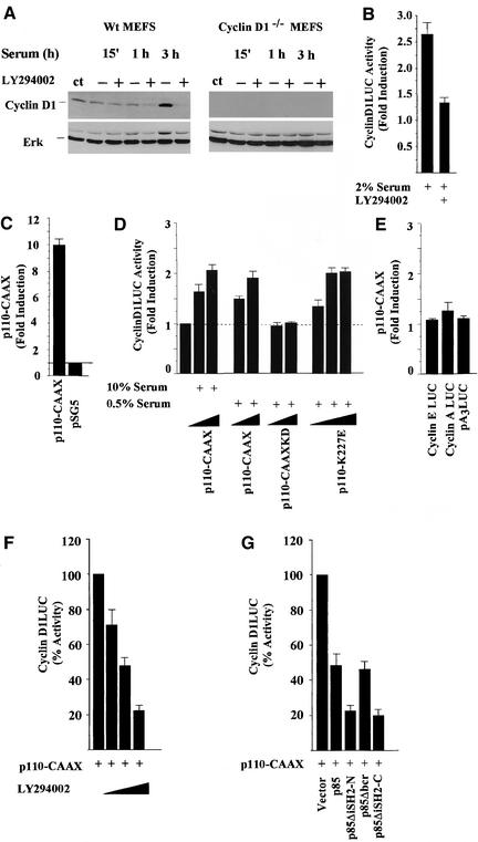
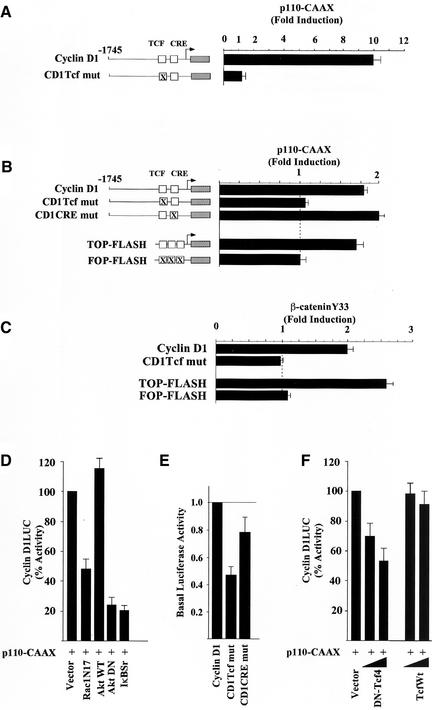
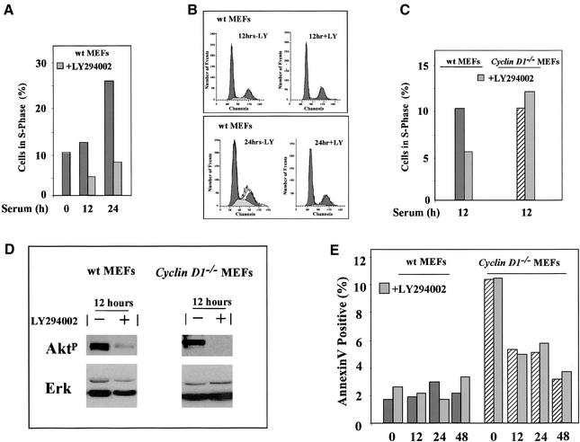
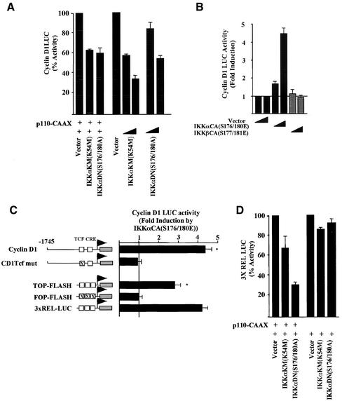
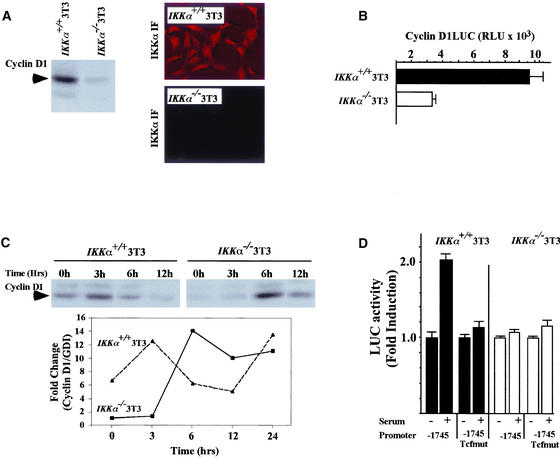
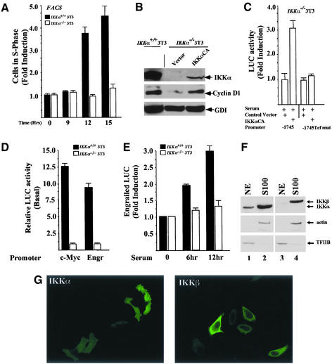
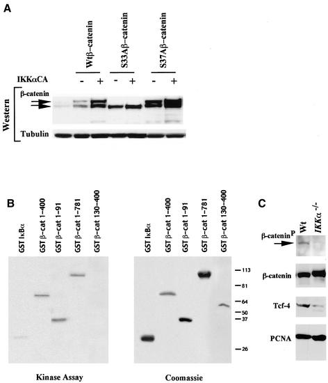
The Wnt/beta-catenin/Tcf and IkappaB/NF-kappaB cascades are independent pathways involved in cell cycle control, cellular differentiation, and inflammation. Constitutive Wnt/beta-catenin signaling occurs in certain cancers from mutation of components of the pathway and from activating growth factor receptors, including RON and MET. The resulting accumulation of cytoplasmic and nuclear beta-catenin interacts with the Tcf/LEF transcription factors to induce target genes. The IkappaB kinase complex (IKK) that phosphorylates IkappaB contains IKKalpha, IKKbeta, and IKKgamma. Here we show that the cyclin D1 gene functions as a point of convergence between the Wnt/beta-catenin and IkappaB pathways in mitogenic signaling. Mitogenic induction of G(1)-S phase progression and cyclin D1 expression was PI3K dependent, and cyclin D1(-/-) cells showed reduced PI3K-dependent S-phase entry. PI3K-dependent induction of cyclin D1 was blocked by inhibitors of PI3K/Akt/IkappaB/IKKalpha or beta-catenin signaling. A single Tcf site in the cyclin D1 promoter was required for induction by PI3K or IKKalpha. In IKKalpha(-/-) cells, mitogen-induced DNA synthesis, and expression of Tcf-responsive genes was reduced. Reintroduction of IKKalpha restored normal mitogen induction of cyclin D1 through a Tcf site. In IKKalpha(-/-) cells, beta-catenin phosphorylation was decreased and purified IKKalpha was sufficient for phosphorylation of beta-catenin through its N-terminus in vitro. Because IKKalpha but not IKKbeta induced cyclin D1 expression through Tcf activity, these studies indicate that the relative levels of IKKalpha and IKKbeta may alter their substrate and signaling specificities to regulate mitogen-induced DNA synthesis through distinct mechanisms.


Figures

Figure 1
Figure 1
PI3K-induction of cyclin D1. (A) Western blot analysis of MEFs for cyclin D1 derived from wild-type (cyclin D1 wt) or cyclin D1−/− mice and treated with serum either with or without the PI3K inhibitor LY294002. The Western blot was probed for cyclin D1 and total ERK. (B) The serum-induced activity of the cyclin D1 promoter in the presence or absence of the PI3 kinases inhibitor LY294002 (20 μM). (C) DU145 cells at either >90% or (D) 30% confluence were transfected with a cyclin D1 promoter luciferase reporter plasmid (−1745CD1LUC) and either the p110α-CAAX, or (D) the p110α kinase dead mutant (p110α-CAAX-KD) or the constitutively active p110α-K227E mutant expression plasmid in the presence of either 10% or 0.5% serum. The fold induction of the luciferase reporter activity is shown for nine separate experiments as mean ± SEM throughout. (E) The effect of p110-CAAX on reporter plasmids for cyclin A and the cyclin E promoter, and the luciferase reporter pA3LUC. (F) The p110α-CAAX induction of the cyclin D1 promoter activity was inhibited by LY294002 (using 2, 20, and 100 μM). (G) The cyclin D1 promoter activity in the presence of p110α-CAAX is shown as 100% and is compared with the effect of cotransfected dominant negative inhibitors of PI3K including p85α, p85ΔiSH2-N, 85ΔiSH2-C, or p85ΔBCR (Rodriguez-Viciana et al., 1997). The results are shown compared with equal amounts of empty control vector for each expression vector plasmid.
Figure 2
Figure 2
PI3K-induction of cyclin D1 requires the Tcf site and is dependent upon IκB. (A) The effects of the p110α-CAAX expression plasmid on the activity of the cyclin D1 promoter (−1745 CD1LUC) or of a point mutant of the Tcf site at −81 (−1745 Tcf mut) in DU145 cells grown to either > 90% or (B) 30% confluence. Regulation of the cyclin D1 and Tcf-responsive (TOPFLASH) and mutant (FOPFLASH) reporter constructs by either the p110-CAAX plasmid or (C) the activated β-catenin point mutant (Y33). The fold induction of the luciferase reporter activity is shown for at least nine separate experiments as mean ± SEM throughout. (D) The cyclin D1 promoter activity in the presence of p110α-CAAX is shown as 100% and is compared with the effect of cotransfected dominant negative inhibitors of PI3K including RacN17, Akt wt, AktDN (K179 M), or IκBαSr. The results are shown compared with equal amounts of empty control vector for each expression vector plasmid. (E) Point mutations of the cyclin D1 promoter Tcf or CRE site were compared with the basal promoter activity of −1745 CD1LUC. The activity of the wild-type promoter construction was set as 1.0. The data are mean ± SEM of five separate transfections. (F) The cyclin D1 promoter activity in the presence of p110α-CAAX (100%) is compared with the effect of dominant negative or wild-type Tcf.
Figure 3
Figure 3
Involvement of cyclin D1 in PI3K-dependent S-phase entry in primary cells. (A) MEFs were treated either with vehicle (DMSO) or LY294002 (20 μM) and DNA synthesis assessed by FACS. (B) FACS analysis of wt MEFs in the presence and absence of LY294002 (LY; 20 μM) 12 and 24 h after serum stimulation. (C) The effects of LY294002 on S-phase wt and cyclin D1−/− MEFs are shown after serum stimulation as mean of four separate experiments. (D) Western blotting for phosphorylated Akt or total ERK of MEFs treated with serum and either vehicle or LY294002. (E) The level of apoptosis was determined by Annexin V staining in serum-starved–stimulated cyclin D1wt and cyclin D1−/− MEFs.
Figure 4
Figure 4
IKKα induces the cyclin D1 gene through the β-catenin/Tcf site. (A) DU145 cells were transfected with the cyclin D1 promoter luciferase reporter plasmid (−1745 CD1LUC) and the p110α-CAAX, with the dominant inhibitors (IKKαKM(K54 M), IKKαDN(S176/180A)) or (B) constitutively active plasmid (IKKαCA(S176/180E) or IKKβ(S177/181E)). (C) Identification of the IKKα-responsive sequences in the cyclin D1 promoter. The IKKαCA(S176/180E) expression plasmid was coexpressed with the luciferase reporters shown and fold induction determined compared with equal amounts of empty expression vector cassette. (D) Effect of IKKα kinase dead (IKKαK54 M) and dominant negative mutants (IKKα(S176/180A)) on p110α-CAAX induced NF-κB activity assessed using the 3XREL LUC reporter.
Figure 5
Figure 5
Reduced mitogen-induced cyclin D1 expression in IKKα−/− cells involves Tcf signaling. (A) 3T3 cells from either wt or IKKα−/− mice were examined by Western blotting for cyclin D1, using equal amounts of total protein. Immunostaining for IKKα in the wt or IKKα−/− 3T3 cells. (B) The cyclin D1 promoter luciferase reporter plasmid (−1745 CD1LUC) was transfected into either wt or IKKα−/− 3T3 cells along with the β-galactosidase control reporter. Relative cyclin D1 promoter activity is shown as mean ± SEM for n = 3. (C) Western blotting for cyclin D1 of wt or IKKα−/− 3T3 cells treated with serum for the time points indicated. The fold change in cyclin D1 protein levels is shown normalized to GDI as a loading control. The data is representative of three separate experiments. (D) The cyclin D1 promoter (−1745CD1LUC) or the point mutant of the Tcf site (−1745Tcfmut) were compared for relative activity in wt or IKKα−/−3T3 cells. The data are mean ± SEM, n = 9.
Figure 6
Figure 6
IKKα regulates mitogen-induced DNA synthesis and is required for Tcf signaling to natural Tcf-responsive genes. (A) The serum-induced S-phase fraction of wt or IKKα−/− 3T3 cells was compared. The data are mean ± SEM, N = 7. (B) Western blotting of wt or IKKα−/− 3T3 cells transfected with an expression vector for IKKαCA or empty expression vector. IKKα and cyclin D1 immunoblotting is shown with GDI as an internal control for loading. (C) Relative activity of the cyclin D1 promoter or the corresponding Tcf point mutant in IKKα−/− 3T3 cells transfected with either IKKα expression vector or the control vector. The data are mean ± SEM, n = 8. (D) Relative activity of the c-Myc and Engr promoters (n = 6) in randomly cycling wt or IKKα−/− 3T3 cells. The activity of the promoter is set as 1 in the IKKα−/− 3T3 cells. (E) Activity of the Engr promoter in the presence of serum stimulation. The data is mean ± SEM of n = 12 separate experiments. (F) Nuclear and cytosolic fractions of Cos-7 cells were analyzed by Western blotting for the abundance of IKKβ or IKKα in the nuclear (NE) and cytosolic (S100) fractions. Internal controls for (nuclear; TFIIB) and cytoplasmic (actin) markers are shown. Substantially more IKKα than IKKβ was found in the nuclear extracts (NE) of Cos-7 cells. (G) Immunostaining for IKKα and IKKβ. IKKβ is predominantly extranuclear, whereas IKKα was found in both nuclear and cytoplasmic compartments.
Figure 7
Figure 7
IKKα phosphorylates β-catenin and increases β-catenin abundance. (A) The IKKαCA(S/E) expression plasmid was coexpressed in cells transfected with either wild-type or mutants (S33A, S37A) of β-catenin. Western blotting analysis showed an increase the total amount of β-catenin, including a higher molecular weight form (upper arrow). The S33Aβ-catenin shows no increase in the amount of the high molecular weight form. (B) IKKα kinase assays conducted using baculovirus expressed purified IKKα and the GST-β-catenin constructs as shown. Kinase activity (left panel) and the Coomassie stained gel for the substrate are shown. (C) Western blot analysis of IKKα−/− or IKKwt 3T3 cells with antibodies to phosphospecific β-catenin, total β-catenin, Tcf-4, and PCNA.

References

    1. Aberle H, Bauer A, Stappert J, Kispert A, Kemler R. beta-Catenin is a target for the ubiquitin-proteasome pathway. EMBO J. 1997;16:3797–3804. - PMC - PubMed
    1. Albanese C, et al. Activation of the cyclin D1 gene by the E1A-associated protein p300 through AP-1 inhibits cellular apoptosis. J Biol Chem. 1999;274:34186–34195. - PubMed
    1. Albanese C, Johnson J, Watanabe G, Eklund N, Vu D, Arnold A, Pestell RG. Transforming p21ras mutants and c-Ets-2 activate the cyclin D1 promoter through distinguishable regions. J Biol Chem. 1995;270:23589–23597. - PubMed
    1. Amanatullah D, Zafonte B, Albanese C, Fu M-F, Messiers C, Hassell J, Pestell RG. Ras regulation of cyclin D1 promoter. Methods Enzymol. 2001;332:116–127. - PubMed
    1. Ashton AW, Watanabe G, Albanese C, Harrington EO, Ware JA, Pestell RG. Protein kinase Cδ inhibition of S-phase transition in capillary endothelial cells involves the cyclin dependant kinase inhibitor p27Kip1. J Biol Chem. 1999;274:20805–20811. - PubMed

Publication types

MeSH terms