. 2003 Feb;14(2):600-10.
doi: 10.1091/mbc.e02-09-0582.

Nup98 localizes to both nuclear and cytoplasmic sides of the nuclear pore and binds to two distinct nucleoporin subcomplexes

Affiliations

Nup98 localizes to both nuclear and cytoplasmic sides of the nuclear pore and binds to two distinct nucleoporin subcomplexes

Eric R Griffis et al. Mol Biol Cell. 2003 Feb.

Abstract

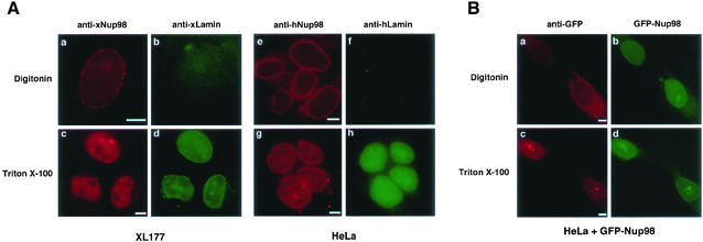
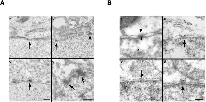
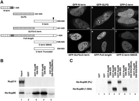
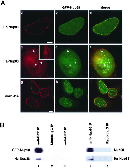
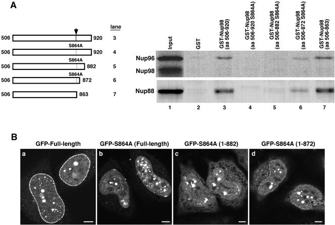
The vertebrate nuclear pore is an enormous structure that spans the double membrane of the nuclear envelope. In yeast, most nucleoporins are found symmetrically on both the nuclear and cytoplasmic sides of the structure. However, in vertebrates most nucleoporins have been localized exclusively to one side of the nuclear pore. Herein, we show, by immunofluorescence and immunoelectron microscopy, that Nup98 is found on both sides of the pore complex. Additionally, we find that the pore-targeting domain of Nup98 interacts directly with the cytoplasmic nucleoporin Nup88, a component of the Nup214, Nup88, Nup62 subcomplex. Nup98 was previously described to interact with the nuclear-oriented Nup160, 133, 107, 96 complex through direct binding to Nup96. Interestingly, the same site within Nup98 is involved in binding to both Nup88 and Nup96. Autoproteolytic cleavage of the Nup98 C terminus is required for both of these binding interactions. When cleavage is blocked by a point mutation, a minimal eight amino acids downstream of the cleavage site is sufficient to prevent most binding to either Nup96 or Nup88. Thus, Nup98 interacts with both faces of the nuclear pore, a localization in keeping with its previously described nucleocytoplasmic shuttling activity.


Figures

Figure 1
Figure 1
Nup98 localizes to the cytoplasmic side of the nuclear pore in digitonin-permeabilized cells. (A) XL177 (a–d) and HeLa cells (e–h) were fixed and then permeabilized using either digitonin or Triton X-100 before detecting Nup98 and lamins by immunofluorescence. Nup98 is visible at the nuclear rim in cells permeabilized with digitonin (a and e), whereas the intranuclear lamins are detected only when nuclei are permeabilized using Triton X-100 (d and h). (B) HeLa cells expressing GFP-Nup98 were fixed and permeabilized as described above and then GFP was detected directly (b and d) or indirectly using an anti-GFP mAb and rhodamine-isothiocyanate-labeled secondary (a and c). The GFP antibody clearly recognizes GFP-Nup98 on the nuclear pore in a digitonin permeabilized cell (a) and does not detect the intranuclear GFP-fusion protein unless the nucleus is permeabilized with Triton X-100 (c). Bars, 5 μm.
Figure 2
Figure 2
Nup98 localizes to both sides of the pore by immunoelectron microscopy. (A) Either untransfected (a and b) or GFP-Nup98 expressing cells (c and d) were treated for immunoelectron microscopy under conditions that maintained an intact nuclear envelope, and bound antibody was detected by the deposition of DAB. When primary antibody was omitted, no staining was seen at nuclear pores (a). Nup98 antibodies associated with the cytoplasmic side of the nuclear pore in both wild-type and transfected cells (b and c). In a rare nucleus that was permeabilized by the procedure, Nup98 was detected on both sides of the NPC (d). Nuclear pores are indicated by arrowheads. (B) Immunogold electron microscopy was used to detect Nup98 on both sides of the pore in HeLa cells after freeze-thaw permeabilization. Endogenous Nup98 was localized in HeLa cells by using an affinity-purified anti-hNup98 (a and b). GFP-Nup98 was also detected on both sides of the pore by using an anti-GFP monoclonal in a cell line stably expressing the protein (c and d). In all panels, orientation is with cytoplasm up, nucleus down. Bars, 250 nm.
Figure 3
Figure 3
The C terminus of Nup98 binds to the cytoplasmic nucleoporin Nup88 in vitro. (A) N-terminal (a), GLFG (b), and C-terminal (c) domains of Nup98 were expressed as GFP-fusion proteins in HeLa cells, which were then fixed and permeabilized with 2% paraformaldehyde and 0.2% Triton X-100 before imaging on an LSM 510 confocal microscope. Only the C-terminal domain produces a nuclear rim stain. A construct containing the C-terminal and GLFG domains (d) results in a rim stain as intense as the full-length protein (e). Mutation of serine 864 to alanine in the C-terminal GFP fusion completely abolishes pore targeting (f). (B) Nup214 and Ha-tagged Nup88 were in vitro transcribed and translated in the presence of [35S]methionine and then mixed with glutathione-Sepharose beads bound to GST (lane 2), GST-Nup98 C terminus (amino acids 506–920, lane 3), truncated GST-Nup98 C terminus (amino acids 506–863, lane 4), and uncleavable GST-Nup98 C terminus (amino acids 506–920 S864A, lane 5). Lane 1 represents the input translation (Ha-Nup88) or the input translation pulled down with GST-TAP (Nup214). (C) To map the domain of Nup88 required to bind to Nup98, a stop codon was inserted after amino acid 584 of Nup88 to eliminate the C-terminal coiled-coil domain. Full-length and truncated Nup88 constructs were in vitro transcribed and translated and then mixed with beads carrying GST (lanes 1 and 2), GST-Nup98 C terminus (lanes 3 and 4), the truncated GST-Nup98 C terminus (lanes 5 and 6), and uncleavable GST-Nup98 C terminus (S864A; lanes 7 and 8). Bars, 5 μm.
Figure 4
Figure 4
Nup98 and Nup88 interact in vivo. (A) HeLa cells coexpressing GFP-Nup98 and Ha-Nup88 were processed for immunofluorescence by using appropriate antibodies as detailed under MATERIALS AND METHODS. When GFP-Nup98 (b) and Ha-Nup88 (a) were expressed at low levels, they colocalized only at the nuclear rim (c). When coexpressed at high levels, (d–f) Ha-Nup88 (d) was also observed in the intranuclear foci along with GFP-Nup98 (f, arrowheads). In the absence of Nup98 overexpression, Nup88 never accumulated in the nucleus even when highly overexpressed (d, inset). Monoclonal 414 (g) did not detect any colocalization of FXFG nucleoporins in the intranuclear foci even with extremely high levels of GFP-Nup98 expression (i). Bars, 5 μm. (B) Cos1 cells expressing GFP-Nup98 and Ha-Nup88 were lysed and the lysate was immunoprecipated with anti-GFP (lane 1) or nonspecific mouse IgG (lane 2). When cells expressed only Ha-Nup88, anti-GFP antibodies did not coprecipitate any Ha-Nup88 (lane 3). Cells expressing Ha-Nup88 alone were lysed and the lysate was immunoprecipated with anti-hNup98 (lane 4) or a nonspecific rabbit IgG (lane 5).
Figure 5
Figure 5
Nup88 and Nup96 bind to the same domain of Nup98. (A) Truncations were made to the uncleavable GST-Nup98 S864A mutant to remove portions of the C-terminal tail peptide. These truncations were tested in in vitro binding assays with both translated Nup96 and Ha-Nup88. The wild-type and truncated GST fusions bound both translated proteins at the same level (lanes 3 and 7, respectively). The full-length S864A mutant and the 882 truncation mutant did not bind to either Nup96 or Nup88 (lanes 4 and 5). The uncleavable mutant truncated at amino acid 872 bound some Nup88 and Nup96 (lane 6). (B) Truncation mutations were localized as GFP-Nup98 fusions in HeLa cells fixed with 2% paraformaldehyde, 0.2% Triton X-100. The wild-type protein gives the typical strong rim stain observed under these conditions (a), whereas the S864A mutant gives no appreciable rim staining (b). The 882 (c) and 872 (d) truncation mutants give progressively stronger nuclear rim stains. Bar, 5 μm.

References

    1. Allen TD, Cronshaw JM, Bagley S, Kiseleva E, Goldberg MW. The nuclear pore complex: mediator of translocation between nucleus and cytoplasm. J Cell Sci. 2000;113:1651–1659. - PubMed
    1. Bailer SM, Balduf C, Katahira J, Podtelejnikov A, Rollenhagen C, Mann M, Pante N, Hurt E. Nup116p associates with the Nup82p-Nsp1p-Nup159p nucleoporin complex. J Biol Chem. 2000;275:23540–23548. - PubMed
    1. Bailer SM, Siniossoglou S, Podtelejnikov A, Hellwig A, Mann M, Hurt E. Nup 116p and Nup 100p are interchangeable through a conserved motif which constitutes a docking site for the mRNA transport factor Gle2p. EMBO J. 1998;17:1107–1119. - PMC - PubMed
    1. Bastos R, Lin A, Enarson M, Burke B. Targeting and function in mRNA export of nuclear pore complex protein Nup153. J Cell Biol. 1996;134:1141–1156. - PMC - PubMed
    1. Bastos R, Ribas de Pouplana L, Enarson M, Bodoor K, Burke B. Nup84, a novel nucleoporin that is associated with CAN/Nup214 on the cytoplasmic face of the nuclear pore complex. J Cell Biol. 1997;137:989–1000. - PMC - PubMed

Publication types

MeSH terms

LinkOut - more resources