. 2003 Feb;14(2):670-84.
doi: 10.1091/mbc.e02-06-0345.

Involvement of the Arp2/3 complex and Scar2 in Golgi polarity in scratch wound models

Affiliations

Involvement of the Arp2/3 complex and Scar2 in Golgi polarity in scratch wound models

Juana Magdalena et al. Mol Biol Cell. 2003 Feb.

Abstract

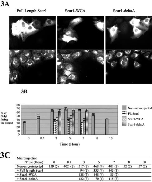
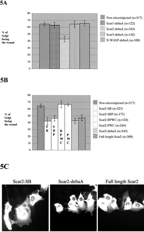
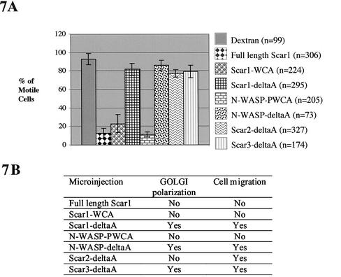
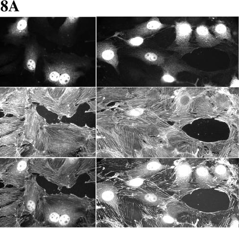
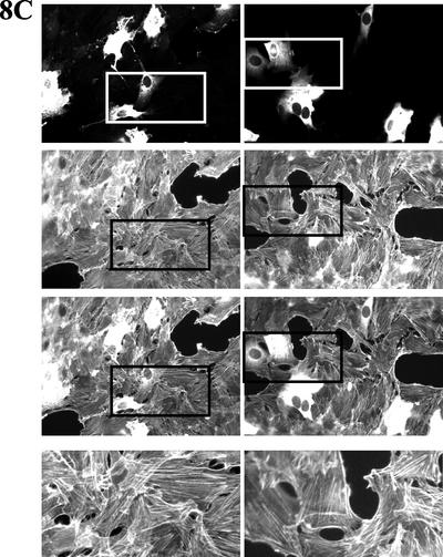
Cell motility and cell polarity are essential for morphogenesis, immune system function, and tissue repair. Many animal cells move by crawling, and one main driving force for movement is derived from the coordinated assembly and disassembly of actin filaments. As tissue culture cells migrate to close a scratch wound, this directional extension is accompanied by Golgi apparatus reorientation, to face the leading wound edge, giving the motile cell inherent polarity aligned relative to the wound edge and to the direction of cell migration. Cellular proteins essential for actin polymerization downstream of Rho family GTPases include the Arp2/3 complex as an actin nucleator and members of the Wiskott-Aldrich Syndrome protein (WASP) family as activators of the Arp2/3 complex. We therefore analyzed the involvement of the Arp2/3 complex and WASP-family proteins in in vitro wound healing assays using NIH 3T3 fibroblasts and astrocytes. In NIH 3T3 cells, we found that actin and Arp2/3 complex contributed to cell polarity establishment. Moreover, overexpression of N-terminal fragments of Scar2 (but not N-WASP or Scar1 or Scar3) interfere with NIH 3T3 Golgi polarization but not with cell migration. In contrast, actin, Arp2/3, and WASP-family proteins did not appear to be involved in Golgi polarization in astrocytes. Our results thus indicate that the requirement for Golgi polarity establishment is cell-type specific. Furthermore, in NIH 3T3 cells, Scar2 and the Arp2/3 complex appear to be involved in the establishment and maintenance of Golgi polarity during directed migration.


Figures

Figure 1
Figure 1
Expression of Arp2/3 and WASP-family proteins by NIH 3T3 cells. Analyses were performed by Western blot and by immunofluorescence. (A) NIH 3T3 cell extracts were analyzed by Western blot with the following antibodies as described from left to right: Scar1, pan-Scar, WASP, N-WASP, and p34-Arc (ARPC2) of the Arp2/3 complex. Molecular weight markers (in kDa) are shown. (B) Cells were fixed and stained for pan-Scar (top: middle and right panels) and for the p34-Arc (bottom: middle and right panels), and in both cases labeled concomitantly for actin (left panels). The right panels are enlargements of a region from the middle panels as indicated.
Figure 2
Figure 2
Scar and N-WASP constructs used in this study. Full-length Scar1 contains the following domains: Scar Homology domain (SHD), basic motif (B), a polyproline-rich region (Polypro), WASP Homology 2/central/acidic (WH2/C/A) domain also called WCA domain. N-WASP contains: WASP Homology 1 (WH1) domain, basic motif (B), GTPase binding domain (GBD), a polyproline-rich region (Polypro), WASP Homology 2/central/acidic (WH2WH2/C/A) domain. Scar1–2-3-deltaA and N-WASP-deltaA constructs are lacking the C-terminal region as shown in the Figure. Scar1-WCA and N-WASP-PWCA constructs are lacking the N-terminal region. All constructs are tagged, with the green fluorescent protein (GFP) or with the myc-tag, as indicated. The numbers at the top of the constructs give the amino acid number of the protein sequence that is at the beginning and the end of the indicated domains.
Figure 3
Figure 3
Golgi polarization, at the edge of the wound, of fulllength Scar1-, Scar1-WCA–, and Scar1-deltaA–expresisng cells. Golgi reorientation to face the wound was evaluated when NIH 3T3 cells at the edge of the wound was microinjected with the three different pEGFP-Scar1 expression constructs described in Figure 2. (A) The microinjected cells expressing the GFP-fusion protein are shown (top panels): full-length Scar1 (left), Scar1-WCA (middle), and Scar1-deltaA (right). Cells were fixed and stained for the Golgi apparatus with anti-beta-COP (bottom panels.) (B) Percentage of Golgi orientated toward the wound was determined at 3, 5, and 7 h after wounding for cells microinjected with full-length Scar1, Scar1-WCA, and Scar1-deltaA expression vectors. Golgi polarization of non-microinjected wound edge cells, over the time course of wound closure, is included as a control (see legend on the right side of the graph). (C) quantification data. The first number corresponds to the number of Golgi observed and second, in parentheses, is the number of separate experiments.
Figure 4
Figure 4
Golgi polarization, at the edge of the wound, of N-WASP-PWCA– and N-WASP-deltaA–expressing cells. Golgi reorientation to face the wound was evaluated when NIH 3T3 cells at the edge of the wound were microinjected with the two different N-WASP expression constructs (pEGFPC1- or pRK5-myc; see Figure 4). (A) Percentage of Golgi orientated toward the wound was determined at 3, 5, and 7 h after wounding for cells microinjected with N-WASP-PWCA and N-WASP-deltaA expression vectors. Golgi polarization of non-microinjected wound edge cells, over the time course of wound closure, is included as a control (see legend on the right side of the graph). (B) Quantification data. The first number corresponds to the number of Golgi observed and the second, in parentheses, is the number of separate experiments.
Figure 5
Figure 5
Scar2-deltaA inhibits Golgi polarization of NIH 3T3 fibroblasts and the SHD domain of Scar2 is necessary. Golgi polarization, at the wound edge, of cells expressing (A) the deltaA constructs of four WASP-family proteins: Scar1, Scar2, Scar3, N-WASP and (B) full-length Scar2 and Scar2-deltaA deletion constructs. Golgi reorientation to face the wound was evaluated when NIH 3T3 cells at the edge of the wound were microinjected with the appropriate pEGFPC1 expression constructs described in Figures 2 and 6. The percentage of Golgi oriented toward the wound was determined at 3 h after wounding for cells microinjected with the WASP-family protein deletion constructs. Golgi polarization of non-microinjected wound edge cells is included as a control of cell polarity (see legend on the right side of the graph; n is the number of Golgi observed; quantification was performed in at least 3 separate experiments). (C) Microinjected cells expressing the GFP-fusion proteins are shown as indicated: Scar2-SB (left), Scar2-deltaA (middle), and full-length Scar2 (right).
Figure 6
Figure 6
The five different pEGFPC1-Scar2-deltaA deletion constructs used in this study. Domains are described in the Figure 2 legend.
Figure 7
Figure 7
NIH 3T3 motility and Golgi polarization of the wound edge cells during wound closure. Wound edge cells were microinjected with the expression vectors coding for the Scar and the N-WASP constructs (described in Figure 2 and listed in A and B). (A) Motility. Percentage of microinjected cells still able to carry out wound closure and staying at the edge of the wound as it closed. These percentages were determined from quantification of data as described in MATERIALS AND METHODS; n is the number of cells observed; quantification was performed in at least three separate experiments. (B) Cell polarity of microinjected cells was determined by Golgi reorientation to face the wound. These results were described over time as % of Golgi polarization, in Figures 3, 4, and 5. Cell motility was determined as shown in section A.
Figure 8
Figure 8
Wound closure can be performed by Scar1-deltaA as well as Scar2-deltaA but not Scar1-WCA expressing wound edge NIH 3T3 cells. Cells were fixed 10 h after microinjection to look at the final stages of wound closure. Wound edge cells were microinjected with (A) Alexa fluor 594–conjugated dextran (as positive control of wound closure); (B) the Scar1-deltaA expression vector; (C) the Scar2-deltaA expression vector; and (D) the Scar1-WCA expression vector. The top panels show the labeled microinjected cells, middle panels are the corresponding actin labeling panels, and third bottom panels corresponding to the merge of top and middle ones. For sections B and C, the fourth bottom panels are enlargements of the actin labeling region as indicated above them in the corresponding column.
Figure 8
Figure 8
Wound closure can be performed by Scar1-deltaA as well as Scar2-deltaA but not Scar1-WCA expressing wound edge NIH 3T3 cells. Cells were fixed 10 h after microinjection to look at the final stages of wound closure. Wound edge cells were microinjected with (A) Alexa fluor 594–conjugated dextran (as positive control of wound closure); (B) the Scar1-deltaA expression vector; (C) the Scar2-deltaA expression vector; and (D) the Scar1-WCA expression vector. The top panels show the labeled microinjected cells, middle panels are the corresponding actin labeling panels, and third bottom panels corresponding to the merge of top and middle ones. For sections B and C, the fourth bottom panels are enlargements of the actin labeling region as indicated above them in the corresponding column.
Figure 9
Figure 9
Astrocytes do not depend on Arp2/3 and WASP-family proteins for Golgi polarization. Polarity of the wound edge cells expressing full-length Scar1, Scar1-WCA, and the deltaA constructs of the three Scar was examined. Golgi reorientation to face the wound was evaluated when astrocytes at the edge of the wound were microinjected with the corresponding pEGFP-Scar expression constructs described in Figure 2. (A) The microinjected cells expressing the GFP-fusion proteins are shown (top panels): full-length Scar1 (left) and Scar1-WCA (right). Cells were fixed and stained for the Golgi apparatus with anti–beta-COP and Hoechst was used to stain the nucleus (bottom panels). (B) Percentage of Golgi orientated toward the wound was determined at 8 h after wounding. Golgi polarization of microinjected wound edge cells expressing the GFP and the N17Cdc42 mutated protein (Etienne-Manneville and Hall, 2001) was included as positive and negative control, respectively (see legend on the right side of the graph; n is the number of Golgi observed; quantification was performed in three separate experiments per vector).

Similar articles

Cited by

References

    1. Abercrombie M, Heaysman JE, Pegrum SM. The locomotion of fibroblasts in culture. I. Movements of the leading edge. Exp Cell Res. 1970;59:393–398. - PubMed
    1. Alrutz MA, Srivastava A, Wong KW, D'Souza-Schorey C, Tang M, Ch'Ng LE, Snapper SB, Isberg RR. Efficient uptake of Yersinia pseudotuberculosis via integrin receptors involves a Rac1-Arp 2/3 pathway that bypasses N-WASP function. Mol Microbiol. 2001;42:689–703. - PubMed
    1. Bailly M, Ichetovkin I, Grant W, Zebda N, Machesky LM, Segall JE, Condeelis J. The F-actin side binding activity of the Arp2/3 complex is essential for actin nucleation and lamellipod extension. Curr Biol. 2001;11:620–625. - PubMed
    1. Brock J, Midwinter K, Lewis J, Martin P. Healing of incisional wounds in the embryonic chick wing bud: characterization of the actin purse-string and demonstration of a requirement for Rho activation. J Cell Biol. 1996;135:1097–1107. - PMC - PubMed
    1. Burns S, Thrasher AJ, Blundell MP, Machesky L, Jones GE. Configuration of human dendritic cell cytoskeleton by Rho GTPases, the WAS protein, and differentiation. Blood. 2001;98:1142–1149. - PubMed

Publication types

MeSH terms

LinkOut - more resources