

Diarrhoea in the growing pig - a comparison of clinical, morphological and microbial findings between animals from good and poor performance herds
- PMID: 12589741
- PMCID: PMC7127389
- DOI: 10.1016/s0034-5288(02)00187-x
Diarrhoea in the growing pig - a comparison of clinical, morphological and microbial findings between animals from good and poor performance herds
Abstract
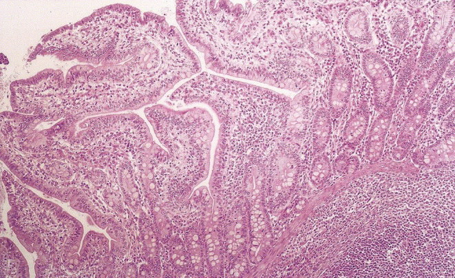
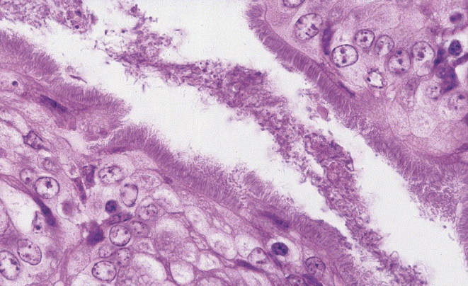
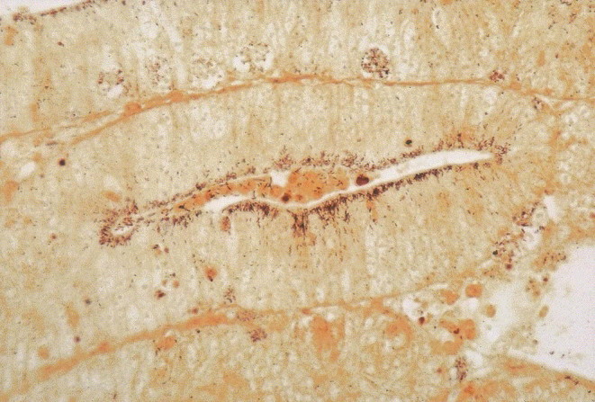
Diarrhoea among growing pigs (8-13 weeks old) is a significant problem in many herds. Nine herds with poor performance and diarrhoea among growing pigs were selected on the basis of their piglet mean age at a body weight of 25 kg, compared to the overall mean age in Swedish herds. In addition, four herds with good average performance and no problems with diarrhoea were selected. Pigs were necropsied and samples for histology and microbiology were collected. Based on the necropsy findings, the pigs from the good performing herds were all judged to be healthy. The presence of Brachyspira pilosicoli and Lawsonia intracellularis was significantly correlated to poor performing herds and the results indicate that these microbes are main pathogens involved in enteric diseases among Swedish grower pigs. In addition, concomitant infections with other presumptive pathogens were commonly found.
Figures
Similar articles
-
The prevalences of Brachyspira spp. and Lawsonia intracellularis in Swedish piglet producing herds and wild boar population.J Vet Med B Infect Dis Vet Public Health. 2005 Nov;52(9):386-91. doi: 10.1111/j.1439-0450.2005.00865.x. J Vet Med B Infect Dis Vet Public Health. 2005. PMID: 16283917
-
Analysis of bacterial load and prevalence of mixed infections with Lawsonia intracellularis, Brachyspira hyodysenteriae and/or Brachyspira pilosicoli in German pigs with diarrhoea.Berl Munch Tierarztl Wochenschr. 2011 May-Jun;124(5-6):236-41. Berl Munch Tierarztl Wochenschr. 2011. PMID: 22059295
-
Clinical utility and performance of sock sampling in weaner pig diarrhoea.Prev Vet Med. 2015 Jul 1;120(3-4):313-20. doi: 10.1016/j.prevetmed.2015.04.015. Epub 2015 Apr 30. Prev Vet Med. 2015. PMID: 25975665
-
Comparative pathology of bacterial enteric diseases of swine.Adv Exp Med Biol. 1999;473:83-101. doi: 10.1007/978-1-4615-4143-1_7. Adv Exp Med Biol. 1999. PMID: 10659346 Review.
-
Postweaning multisystemic wasting syndrome--PMWS. the first year with the disease in Sweden.Vet Q. 2004 Dec;26(4):170-87. doi: 10.1080/01652176.2004.9695179. Vet Q. 2004. PMID: 15663213 Review.
Cited by
-
Prevalences and excretion levels of Lawsonia intracellularis, Brachyspira pilosicoli and Escherichia coli F4 and F18 in fecal sock samples from Danish weaner and finisher pig batches and the association with diarrhea.Porcine Health Manag. 2022 Oct 28;8(1):44. doi: 10.1186/s40813-022-00290-x. Porcine Health Manag. 2022. PMID: 36307895 Free PMC article.
-
Putative probiotic Lactobacillus spp. from porcine gastrointestinal tract inhibit transmissible gastroenteritis coronavirus and enteric bacterial pathogens.Trop Anim Health Prod. 2010 Dec;42(8):1855-60. doi: 10.1007/s11250-010-9648-5. Epub 2010 Jul 10. Trop Anim Health Prod. 2010. PMID: 20623187 Free PMC article.
-
Zinc phosphate-based nanoparticles as alternatives to zinc oxide in diet of weaned piglets.J Anim Sci Biotechnol. 2020 Jun 9;11:59. doi: 10.1186/s40104-020-00458-x. eCollection 2020. J Anim Sci Biotechnol. 2020. PMID: 32528676 Free PMC article.
-
Inclusion of Oat in Feeding Can Increase the Potential Probiotic Bifidobacteria in Sow Milk.Animals (Basel). 2015 Jul 22;5(3):610-23. doi: 10.3390/ani5030375. Animals (Basel). 2015. PMID: 26479377 Free PMC article.
-
Pharmacokinetics and relative bioavailability of an oral amoxicillin-apramycin combination in pigs.PLoS One. 2017 Apr 20;12(4):e0176149. doi: 10.1371/journal.pone.0176149. eCollection 2017. PLoS One. 2017. PMID: 28426744 Free PMC article.
References
-
- Anon . Reference Book 418. third ed. Her Majesty’s Stationary Office; London: 1986. Manual of veterinary parasitological laboratory techniques; pp. 11–12.
-
- Asplund K., Hakkinen M., Okkonen T., Vanhala P., Nurmi E. Effects of growth- promoting antimicrobials on inhibition of Yersinina enterocolitica O:3 by porcine ileal microflora. Journal of Applied Microbiology. 1998;85:164–170. - PubMed
-
- Batte E.G., McLamb R.D., Muse K.E., Tally S.D., Vestal T.J. Pathophysiology of swine trichuriasis. American Journal of Veterinary Research. 1977;38:1075–1079. - PubMed
-
- Bertschinger H.U., Eggenberger E., Jucker H., Pfirter H.P. Evaluation of low nutrient, high fibre diets for the prevention of porcine Escherichia coli enterotoxaemia. Veterinary Microbiology. 1978/1979;3:281–290.
-
- Elvander, M., Larsson, B., Engvall, A., Klingeborn, B., Gunnarsson, A., 1997. Nation-wide surveys of TGE/PRCV, CSF, PRRS, SVD, L. pomona and B. suis in pigs in Sweden. Epidémiologie santé animale 31–32
Publication types
MeSH terms
LinkOut - more resources
Full Text Sources
Other Literature Sources
Medical
