Case Reports
. 2003 Feb;24(2):213-7.

Pseudo-reorganization of language cortical function at fMR imaging: a consequence of tumor-induced neurovascular uncoupling

Affiliations
Case Reports

Pseudo-reorganization of language cortical function at fMR imaging: a consequence of tumor-induced neurovascular uncoupling

John L Ulmer et al. AJNR Am J Neuroradiol. 2003 Feb.

Abstract

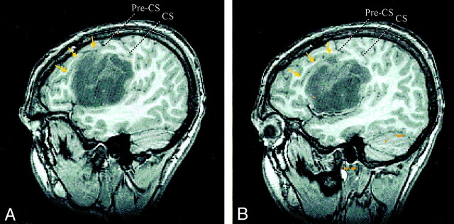
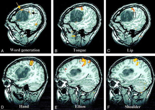
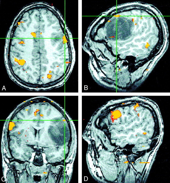
A left-handed patient with a grade II left frontal lobe astrocytoma had spontaneous seizures causing speech arrest and uncontrolled right upper extremity movements. Word-generation functional MR (fMR) imaging showed activity nearly exclusively in the right inferior frontal gyrus. The clinical history of the speech arrest and the intraoperative mapping proved left-hemisphere language dominance. Tumor involvement of the left inferior frontal gyrus caused uncoupling of the blood oxygenation level-dependent (BOLD) and neuronal response, leading to the erroneous fMR imaging appearance of right-hemisphere language dominance. Discrepancies between BOLD and intraoperative mapping in areas near lesions illustrate the complementary nature of these techniques.


Figures

F<sc>ig</sc> 1.
Fig 1.
Sagittal SPGR images of a left frontal astrocytoma centered in the left subcentral gyrus and IFG. The tumor displaces the pars opercularis of the left IFG superiorly and anteriorly, as demarcated by the displaced inferior frontal sulcus (arrows). The lower central sulcus (CS), precentral sulcus (Pre-CS), precentral gyrus, and postcentral gyrus are displaced superiorly and posteriorly.
F<sc>ig</sc> 2.
Fig 2.
Sagittal spoiled gradient-echo and fMR images of the left hemisphere show cortical activation, compared with intraoperative mapping sites. A, Language activation. BOLD language activation is displaced superiorly (yellow arrow) but located immediately anterior to language function mapped intraoperatively (rectangle). A small amount of additional activation associated with the word-generation task (black arrow) corresponds more closely to the intraoperative location of lip function (see image in C). B, Tongue activation. BOLD tongue activation is situated between intraoperative maps of lip and language function, as would be expected, but intraoperative attempts to find tongue motor function were unsuccessful. C, Lip activation. Corticonuclear (tongue and lip) BOLD activation is in superiorly displaced precentral gyrus at the superior-posterior edge of the tumor located immediately above the area of lip function mapped intraoperatively (rectangle). D, Hand activation. The corticospinal (hand, elbow, and shoulder) cortical function is in the normal portion of the precentral gyrus superior and posterior to the tumor. With these three areas, only intraoperative mapping of hand function was attempted, and the result corresponded precisely with the location of BOLD activation (rectangle). E, Elbow activation. F, Shoulder activation.
F<sc>ig</sc> 3.
Fig 3.
Images falsely suggesting strong right-hemisphere dominance. A–C, Axial (A), sagittal (B), and coronal (C) SPGR fMR images show minimal left-frontal language activity (crosshairs) during a word-generation task. C and D, Coronal (C) and sagittal (D) fMR images show robust activity in the right IFG and inferior frontal sulcus that falsely implies right-hemisphere language dominance in this individual.

References

    1. Yetkin FZ, Ulmer JL, Mueller WM, Cox RW, Haughton VM. Functional magnetic resonance imaging assessment of the risk of post-operative hemiparesis after excision of cerebral tumors. Int J Neuroradiol 1998;4:253–257
    1. Laurito JT, Bryan RN, Mathews VP, Ulmer JL, Lowe MJ. Functional brain mapping. In: RSNA Categorical Course in Diagnostic Radiology: Neuroradiology. Oak Brook, IL: RSNA; 2000:79–104
    1. Holodny AI, Schulder M, Liu WC, Wolko J, Maldjian JA, Kalnin AJ. The effect of brain tumors on BOLD functional MR imaging activation in the adjacent motor cortex: implications for image-guided neurosurgery. AJNR Am J Neuroradiol 2000;21:1415–1422 - PMC - PubMed
    1. Schreiber A, Hubbe U, Ziyeh S, Hennig J. The influence of gliomas and nonglial space-occupying lesions on blood-oxygen-level-dependent contrast enhancement. AJNR Am J Neuroradiol 2000;21:1055–1063 - PMC - PubMed
    1. Lee YJ, Chung TS, Soo Yoon Y., et al. The role of functional MR imaging in patients with ischemia in the visual cortex. AJNR Am J Neuroradiol 2001;22:1043–1049 - PMC - PubMed

Publication types

MeSH terms