

Case Reports
Conventional and perfusion MR imaging of parafalcine chondrosarcoma
Affiliations
- PMID: 12591641
- PMCID: PMC7974122
Item in Clipboard
Case Reports
Conventional and perfusion MR imaging of parafalcine chondrosarcoma
AJNR Am J Neuroradiol.
2003 Feb.
Abstract
Intracranial chondrosarcomas have a predilection for the skull base, for which CT and MR imaging findings have been described. We present a rare case of primary chondrosarcoma arising from the falx in a young woman with no history of radiation. The CT, conventional MR imaging, perfusion MR imaging, and digital subtraction angiography findings are described.
Figures
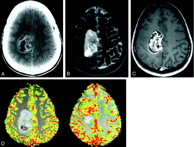
Images from the case of a 28-year-old woman with a 6-month history of headaches and left arm tingling and numbness that had gradually progressed to involve the left half of her face. A, Axial contrast-enhanced CT scan shows a 6-cm, lobulated, heterogeneously enhancing mass in the right parafalcine parietal region, extending into the underlying brain parenchyma. B, Axial T2-weighted image (3400/119/1 [TR/TE/number of excitations]) shows the mass to have high signal intensity relative to cortex, with scattered foci of low signal intensity. The surrounding brain parenchyma appears normal, with no evidence of vasogenic edema. C, Axial contrast-enhanced T1-weighted image (600/14/1) shows enhancement with linear areas of extremely low signal intensity, consistent with islands of cartilaginous tissue. D, Axial color overlay perfusion MR imaging map (1000/54). The image is a perfusion color overlay of a chondrosarcoma (left) that shows hypoperfusion in the region of the mass relative to normal white matter. The image on the right is a perfusion color overlay of a meningioma (right), which in comparison shows increased perfusion.
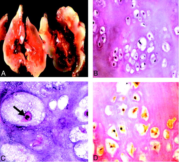
Histopathologic findings. A, Gross specimen shows a bluish white, pearly, glistening lobulated tumor. B, Low power slide shows a hyaline tumor of low to moderate cellularity with mild variation in size and shape of the tumor cells with mostly small and dark nuclei, showing mild nuclear pleomorphism (hematoxylin and eosin; original magnification, ×40). C, High power slide reveals mitotic figures (arrow). Findings consistent with the appearance of a conventional chondrosarcoma (hematoxylin and eosin; original magnification, ×200). D, Nuclear and, to a lesser degree, cytoplasmic immunoreactivity for S-100 protein are present (immunoperoxidase; original magnification, ×100), indicative of a cartilaginous tumor.
Similar articles
-
Primary falcine chondrosarcoma.J Clin Neurosci. 2008 Dec;15(12):1406-9. doi: 10.1016/j.jocn.2007.08.005. Epub 2008 Oct 7. J Clin Neurosci. 2008. PMID: 18842412
-
Intracranial Nonskull-Based Chondrosarcoma Arising from the Sagittal Sinus: A Case Report and Review of the Literature.World Neurosurg. 2018 Dec;120:234-239. doi: 10.1016/j.wneu.2018.08.239. Epub 2018 Sep 8. World Neurosurg. 2018. PMID: 30205213 Review.
-
Intracranial extraskeletal myxoid chondrosarcoma: case report and review of the literature.Acta Neurochir (Wien). 2002 Jul;144(7):735-40. doi: 10.1007/s00701-002-0949-y. Acta Neurochir (Wien). 2002. PMID: 12181708
-
Preoperative imaging of superficially located glioma resection using short inversion-time inversion recovery images in high-field magnetic resonance imaging.Clin Neurol Neurosurg. 2007 May;109(4):327-34. doi: 10.1016/j.clineuro.2007.01.005. Epub 2007 Feb 2. Clin Neurol Neurosurg. 2007. PMID: 17275995
-
[Corpus callosum. Landmark of the origin of cerebral diseases].Radiologe. 2010 Feb;50(2):152-64. doi: 10.1007/s00117-009-1945-5. Radiologe. 2010. PMID: 20012004 Review. German.
Cited by
-
Primary intracranial extra-skeletal myxoid chondrosarcoma of right lateral ventricle with EWSR1 gene fusion: a case report and review of literature.Ecancermedicalscience. 2021 Jun 30;15:1257. doi: 10.3332/ecancer.2021.1257. eCollection 2021. Ecancermedicalscience. 2021. PMID: 34567242 Free PMC article.
-
Characterization of Skull Base Lesions Using Pseudo-Continuous Arterial Spin Labeling.Clin Neuroradiol. 2019 Mar;29(1):75-86. doi: 10.1007/s00062-017-0623-7. Epub 2017 Sep 11. Clin Neuroradiol. 2019. PMID: 28894884
-
Para-Falcine Chondroma: An Entity of Unacquaintance-A Case Report and Review of Literature.Brain Tumor Res Treat. 2025 Jan;13(1):29-33. doi: 10.14791/btrt.2025.0001. Brain Tumor Res Treat. 2025. PMID: 39924714 Free PMC article.
-
Intracranial chondrosarcoma: a case report and review of the literature.J Neurooncol. 2004 May;68(1):33-9. doi: 10.1023/b:neon.0000024728.72998.7d. J Neurooncol. 2004. PMID: 15174519 Review.
-
Endoscopic transsphenoidal approach in resection of intracranial clivus chondrosarcoma: A case report.Oncol Lett. 2023 Oct 3;26(5):498. doi: 10.3892/ol.2023.14085. eCollection 2023 Nov. Oncol Lett. 2023. PMID: 37854870 Free PMC article.
References
-
- Oruckaptan HH, Berker M, Soylemezoglu F, et al. Parafalcine chondrosarcoma: an unusual localization for a classical variant: case report and review of the literature. Surg Neurol 2001;55:174–179 - PubMed
-
- Hassounah M, Al-Mefty O, Akhtar M, et al. Primary cranial and intracranial chondrosarcoma: a survey. Acta Neurochir (Wien) 1985;78:123–132 - PubMed
-
- Cianfriglia F, Pompili A, Occhipinti E. Intracranial malignant cartilaginous tumours: report of two cases and review of literature. Acta Neurochir (Wien) 1978;45:163–175 - PubMed
-
- Gerszten PC, Pollack IF, Hamilton RL. Primary parafalcine chondrosarcoma in a child. Acta Neuropathol (Berl) 1998;95:111–114 - PubMed
Publication types
MeSH terms
LinkOut - more resources
Full Text Sources
Medical