. 2003 Feb 17;197(4):403-11.
doi: 10.1084/jem.20021633.

Regulatory T cells selectively express toll-like receptors and are activated by lipopolysaccharide

Affiliations

Regulatory T cells selectively express toll-like receptors and are activated by lipopolysaccharide

Iris Caramalho et al. J Exp Med. .

Abstract

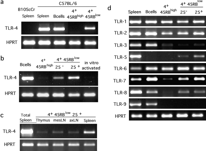
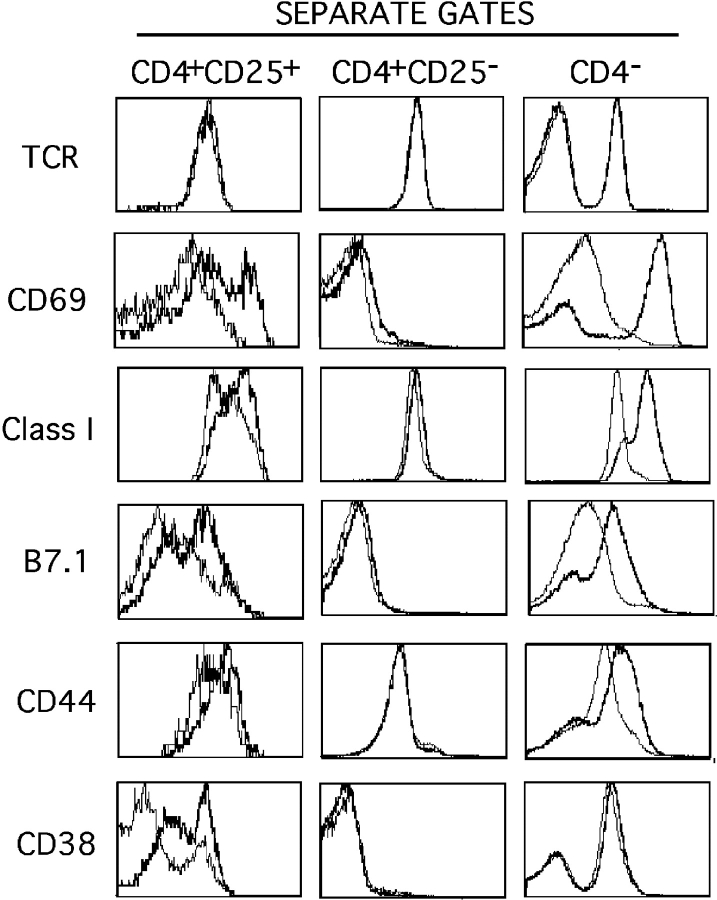
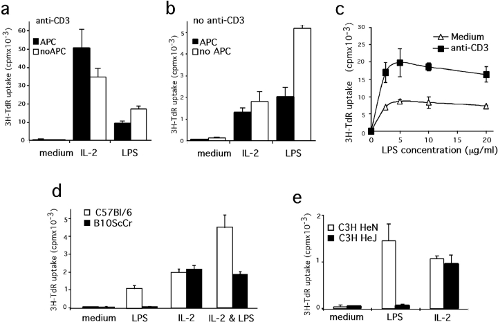
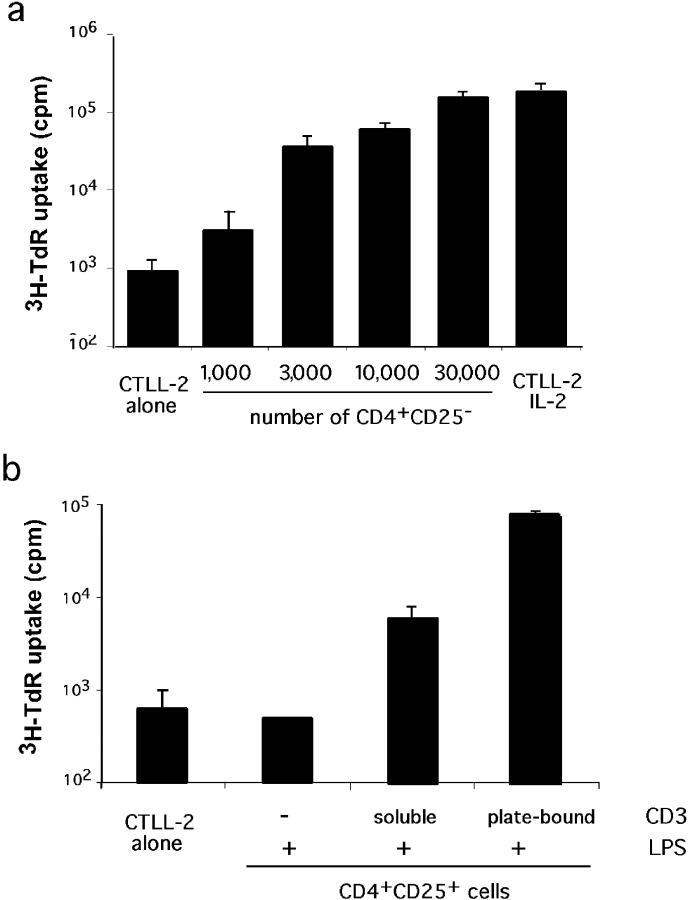
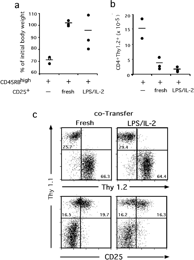
Regulatory CD4 T cells (Treg) control inflammatory reactions to commensal bacteria and opportunist pathogens. Activation of Treg functions during these processes might be mediated by host-derived proinflammatory molecules or directly by bacterial products. We tested the hypothesis that engagement of germline-encoded receptors expressed by Treg participate in the triggering of their function. We report that the subset of CD4 cells known to exert regulatory functions in vivo (CD45RB(low) CD25(+)) selectively express Toll-like receptors (TLR)-4, -5, -7, and -8. Exposure of CD4(+) CD25(+) cells to the TLR-4 ligand lipopolysaccharide (LPS) induces up-regulation of several activation markers and enhances their survival/proliferation. This proliferative response does not require antigen-presenting cells and is augmented by T cell receptor triggering and interleukin 2 stimulation. Most importantly, LPS treatment increases CD4(+) CD25(+) cell suppressor efficiency by 10-fold and reveals suppressive activity in the CD4(+) CD45RB(low) CD25(-) subset that when tested ex-vivo, scores negative. Moreover, LPS-activated Treg efficiently control naive CD4 T cell-dependent wasting disease. These findings provide the first evidence that Treg respond directly to proinflammatory bacterial products, a mechanism that likely contributes to the control of inflammatory responses.


Figures

Figure 1.
Figure 1.
Specific expression of TLR genes by Treg. RNA from sort-purified CD19+ (B cells) and various subpopulations of CD4 cells (4+) from B6 mice, either pooled LNs (a, b, and d) or specific lymphoid organs (c) were submitted to RT-PCR. CD4+ CD45RBlow or CD4+ CD45RBhigh (4+ 45RBlow or 4+ 45RBhigh) were sorted as negative for CD11c, CD11b, B220, pan-NK, and MHC II in addition to positive for CD4 and CD45RB (a) or simply according to CD4, CD45RB, and CD25 expression (b, c, and d). Erythrocyte-lysed splenocytes from B6 or TLR-4–deficient B10ScCr animals and in vitro–activated B6 CD4+ CD45RBhigh served as controls. All RT-PCR were performed at least twice on independent samples.
Figure 2.
Figure 2.
Activation of CD4+ CD25+ cells upon LPS treatment of splenocytes in vitro. Erythrocyte-depleted splenocytes were cultured for 18 h in the presence of LPS (bold) or medium alone (plain) and stained for CD4, CD25, and various other surface molecules. Histograms correspond to FACS® analyses for the indicated molecules inside the three independent gates. Representative analysis out of four independent assessments is shown. A minimum of 5,000 events was acquired in the CD4+ CD25+ gate.
Figure 3.
Figure 3.
LPS directly induces CD4+ CD25+ proliferation through TLR-4. (a and b) Proliferative responses of CD4+ CD25+ cells to LPS are APC independent. Sort-purified CD4+ CD25+ from B6 animals were maintained in culture for 3 d in the presence of anti-CD3 (a) or not (b) and supplemented or not with APC (solid or open bars). For each culture condition, proliferative responses induced by either LPS or IL-2 are compared. (c) Purified CD4+ CD25+ cells were cultured for 3 d in the absence of APC in medium supplemented with various doses of LPS. (d and e) TLR-4–deficient CD4+ CD25+ cells do not proliferate in response to LPS. CD4+ CD25+ were sort purified from TLR-4–competent animals B6 (d) and C3H/HeN (e; open bars) or TLR-4–deficient B10ScCr (d) and C3H/HeJ (e) mice (solid bars) and maintained for 3 d in culture without APC nor anti-CD3. The medium was supplemented with LPS, IL-2, or both, as indicated. Each assay has been performed at least twice on independent cell samples and with similar results.
Figure 4.
Figure 4.
IL-2 production by CD4+ CD25+ cells upon LPS stimulation. (a) Sensitivity of the assay. Supernatant of various numbers of CD4+ CD25 cells cultured for 48 h in the presence of APC and anti-CD3 were used as a source of IL-2 to support CTLL-2 cell proliferation. Proliferation of CTLL-2 cells in the absence of supernatant (alone) or when they were provided with saturating amount of IL-2 (IL-2) are shown. (b) CTLL-2 proliferation induced by supernatants of 2.105 CD4+ CD25+ cells maintained in culture for 3 d in the presence of LPS and anti-CD3 as indicated. Background proliferation is shown on the left. Each assay has been performed twice, each in triplicates.
Figure 5.
Figure 5.
LPS treatment enhances the suppressor function of Treg. (a) Suppressor efficiency of CD4+ CD25+ is greatly enhanced upon exposure to LPS. Sort-purified CD4+ CD25+ T cells were maintained in culture in medium supplemented with anti-CD3 and either LPS for 3 d followed by 3 d with IL-2 (LPS/IL-2) or with IL-2 for 6 d (IL-2). The same cell population not submitted to culture was used as reference control (fresh). Proliferation of CD4+ CD25 stimulated with anti-CD3 and APC in the presence of increasing numbers of these CD4+ CD25+ cells was monitored on day 3. The percent of inhibition ([cpm in control − cpm in experiment]/cpm in control) is plotted versus the ratio of CD4+ CD25+/CD4+CD25 cell number at the origin of the culture. (b and c) LPS treatment of CD4+ CD45RBlow CD25 cells reveals their suppressor functions. Sort-purified CD4+ CD45RBlow CD25 cells were tested in suppression assays after 6 d culture (b) or after only 3 d exposure to LPS and anti-CD3. Suppression by CD4+ CD25+ treated similarly is also shown. (c) Freshly isolated CD4+ CD25+ and CD4+ CD45RBlow CD25 served as control. Nomenclatures are as described in a. Each measurement has been performed at least twice on independent cell samples and resulted in similar curves.
Figure 6.
Figure 6.
LPS-treated CD4+ CD25+ are efficient regulatory cells in vivo. RAG-1–deficient animals (three per experimental group) received 105 CD4+ CD45RBhigh Thy1.2 cells alone or together with the same number of CD4+ CD25+ Thy1.1 cells, either freshly isolated or maintained in culture sequentially with LPS and IL-2 for a total of 6 d (as described in Fig. 4). Mice were analyzed at 21 d after transfer. (a) Control of wasting disease. The white dot represents an animal found dead on day 17 after transfer. Its weight monitored on day 16 was used. (b) Control of lymphocyte expansion. Numbers of CD4+ Thy1.2+ cells (originally CD45RBhigh) recovered in the mesenteric LNs. (c) Viability and phenotype of the transferred CD4+ CD25+ cells. Representative staining of axillary LNs from recipients of cotransfers.

Comment in

References

    1. Singh, B., S. Read, C. Asseman, V. Malmstrom, C. Mottet, L.A. Stephens, R. Stepankova, H. Tlaskalova, and F. Powrie. 2001. Control of intestinal inflammation by regulatory T cells. Immunol. Rev. 182:190–200. - PubMed
    1. Hori, S., T.L. Carvalho, and J. Demengeot. 2002. CD25+ CD4+ regulatory T cells suppress CD4+ T cell-mediated pulmonary hyperinflammation driven by Pneumocystis carinii in immunodeficient mice. Eur. J. Immunol. 32:1282–1291. - PubMed
    1. Zuany-Amorim, C., E. Sawicka, C. Manlius, A. Le Moine, L.R. Brunet, D.M. Kemeny, G. Bowen, G. Rook, and C. Walker. 2002. Suppression of airway eosinophilia by killed Mycobacterium vaccae-induced allergen-specific regulatory T-cells. Nat. Med. 8:625–629. - PubMed
    1. McGuirk, P., C. McCann, and K.H. Mills. 2002. Pathogen-specific T regulatory 1 cells induced in the respiratory tract by a bacterial molecule that stimulates interleukin 10 production by dendritic cells: a novel strategy for evasion of protective T helper type 1 responses by Bordetella pertussis. J. Exp. Med. 195:221–231. - PMC - PubMed
    1. Coutinho, A., S. Hori, T. Carvalho, I. Caramalho, and J. Demengeot. 2001. Regulatory T cells: the physiology of autoreactivity in dominant tolerance and “quality control” of immune responses. Immunol. Rev. 182:89–98. - PubMed

Publication types

MeSH terms