. 2003 Feb 17;197(4):437-49.
doi: 10.1084/jem.20020383.

Selective activation of the c-Jun NH2-terminal protein kinase signaling pathway by stimulatory KIR in the absence of KARAP/DAP12 in CD4+ T cells

Affiliations

Selective activation of the c-Jun NH2-terminal protein kinase signaling pathway by stimulatory KIR in the absence of KARAP/DAP12 in CD4+ T cells

Melissa R Snyder et al. J Exp Med. .

Abstract

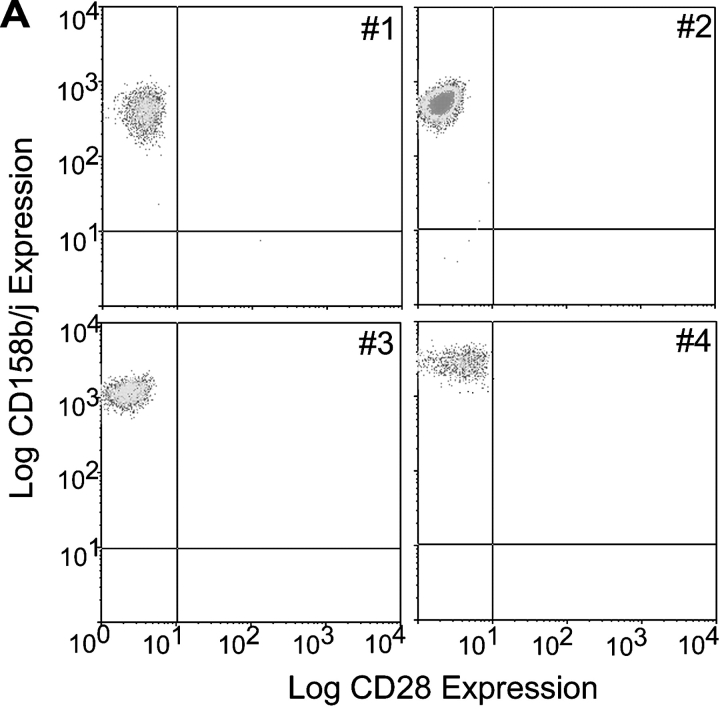
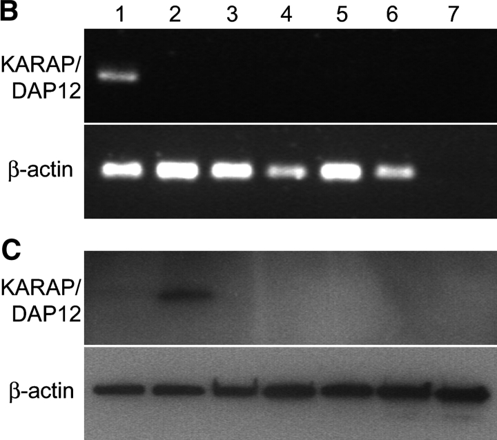
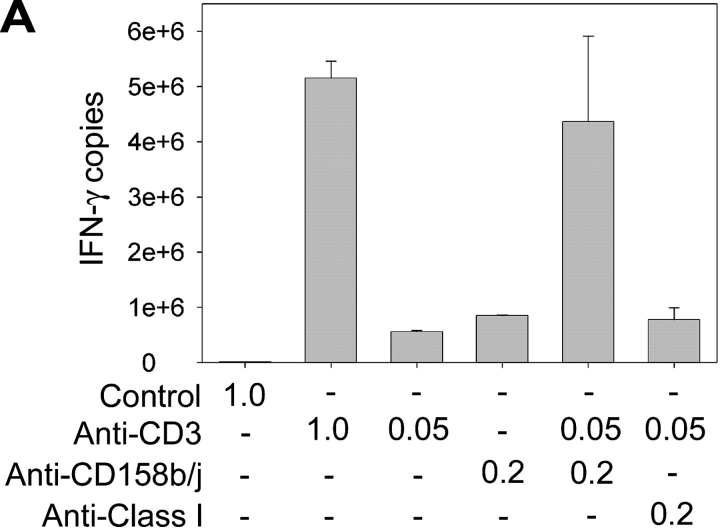
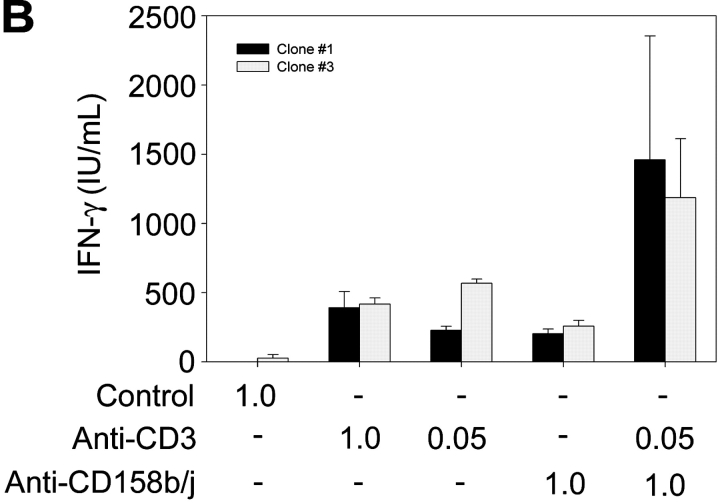
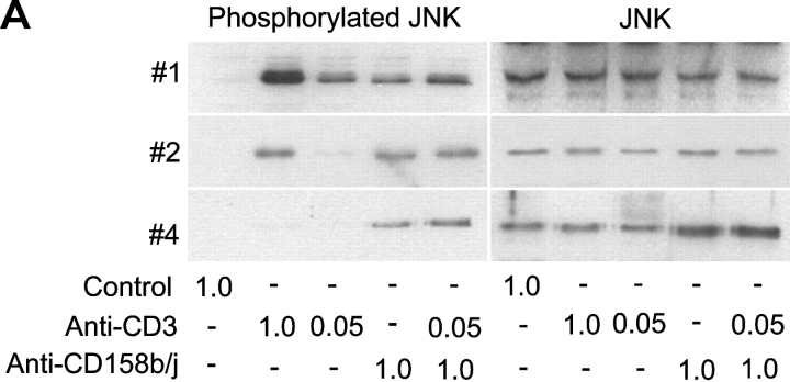
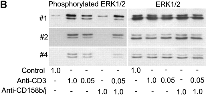
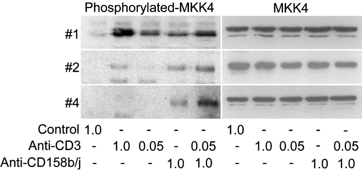
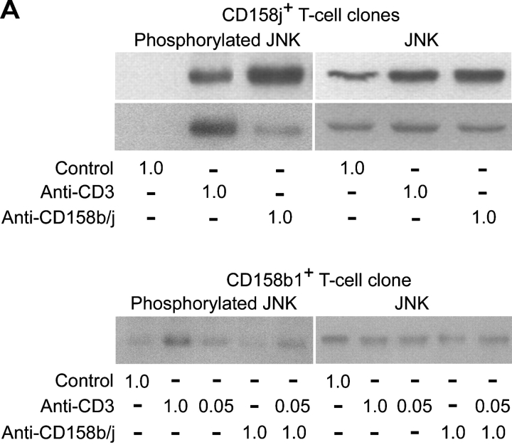
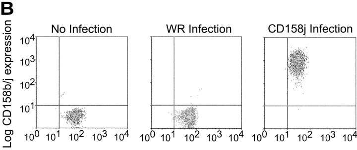
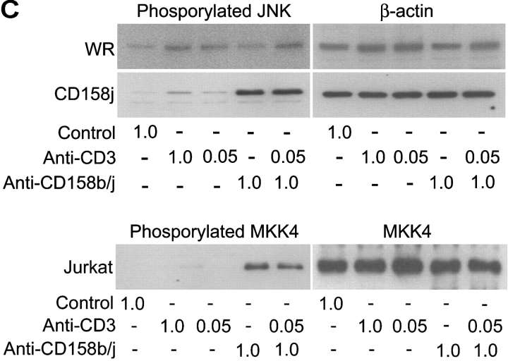
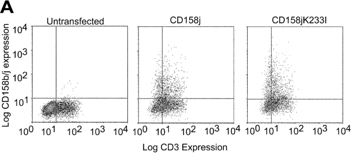
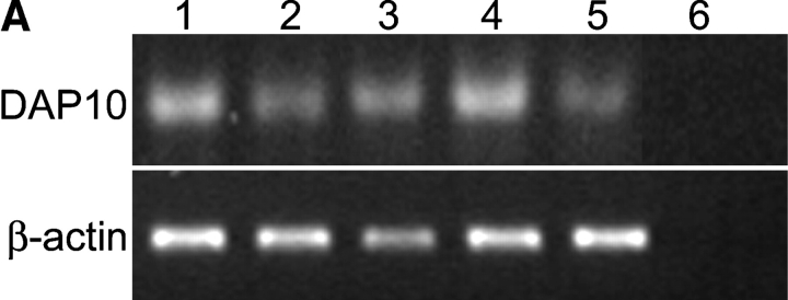
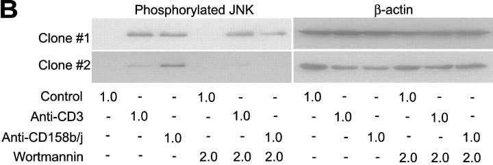
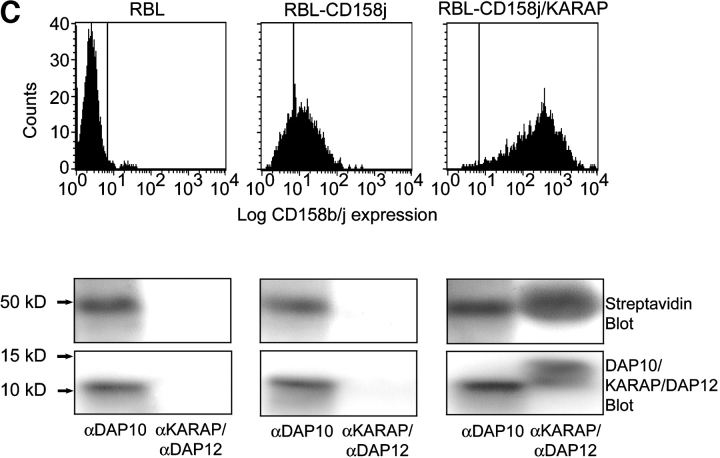
Activation of CD4(+) T cells is governed by interplay between stimulatory and inhibitory receptors; predominance of stimulatory signals favors autoimmune reactions. In patients with rheumatoid arthritis, expression of the critical costimulatory molecule, CD28, is frequently lost. Instead, CD4(+)CD28(null) T cells express killer immunoglobulin-like receptors (KIRs) with a preferential expression of the stimulatory receptor, CD158j. The frequency of CD4(+)CD28(null) T cells in rheumatoid arthritis (RA) correlates with the risk for more severe disease. Moreover, the KIR2DS2 gene, which encodes for CD158j, is a genetic risk factor for rheumatoid vasculitis. CD158j signals through the adaptor molecule, KARAP/DAP12, to positively regulate cytotoxic activity in NK cells. However, the majority of CD4(+)CD28(null) T cell clones lacked the expression of KARAP/DAP12. Despite the absence of KARAP/DAP12, CD158j was functional and augmented interferon-gamma production after T cell receptor stimulation. Cross-linking of CD158j resulted in selective phosphorylation of c-Jun NH(2)-terminal protein kinase (JNK) and its upstream kinase, MKK4 that led to the expression of ATF-2 and c-Jun, all in the absence of extracellular signal-regulated kinase (ERK)1/2 phosphorylation. Mutation of the lysine residue within the transmembrane domain of CD158j abolished JNK activation, suggesting that an alternate adaptor molecule was being used. CD4(+)CD28(null) T cells expressed DAP10 and inhibition of phosphatidylinositol 3-kinase, which acts downstream of DAP10, inhibited JNK activation; however, no interaction of DAP10 with CD158j could be detected. Our data suggest that CD158j in T cells functions as a costimulatory molecule through the JNK pathway independent of KARAP/DAP12 and DAP10. Costimulation by CD158j may contribute to the autoreactivity of CD4(+)CD28(null) T cells in RA.


Figures

Figure 1.
Figure 1.
CD4+CD28null T cell clones express CD158b/j, but do not express KARAP/DAP12. CD4+CD28null T cells were sorted from patients with RA, and clones were established by limiting dilution. Clones were analyzed by flow cytometry for expression of CD28 and CD158b/j. Four representative clones (#1 through #4) are shown. All clones expressed CD4 (unpublished data; A). RT-PCR was used to amplify transcripts for KARAP/DAP12 and β-actin from PBMCs (lane 1), Jurkat T cells (lane 2), and CD4+CD28null T cell clones #1–#4 (lanes 3–6, respectively). cDNA was omitted for the negative control (lane 7) (B). Western blotting was used to detect KARAP/DAP12 and β-actin protein (bottom panels) in Jurkat T cells (lane 1), Jurkat T cells transfected with KARAP/DAP12+ vaccinia virus (lane 2), and CD4+CD28null T cell clones (lanes 3–7) (C).
Figure 1.
Figure 1.
CD4+CD28null T cell clones express CD158b/j, but do not express KARAP/DAP12. CD4+CD28null T cells were sorted from patients with RA, and clones were established by limiting dilution. Clones were analyzed by flow cytometry for expression of CD28 and CD158b/j. Four representative clones (#1 through #4) are shown. All clones expressed CD4 (unpublished data; A). RT-PCR was used to amplify transcripts for KARAP/DAP12 and β-actin from PBMCs (lane 1), Jurkat T cells (lane 2), and CD4+CD28null T cell clones #1–#4 (lanes 3–6, respectively). cDNA was omitted for the negative control (lane 7) (B). Western blotting was used to detect KARAP/DAP12 and β-actin protein (bottom panels) in Jurkat T cells (lane 1), Jurkat T cells transfected with KARAP/DAP12+ vaccinia virus (lane 2), and CD4+CD28null T cell clones (lanes 3–7) (C).
Figure 2.
Figure 2.
CD158b/j costimulates expression of IFN-γ in CD4+ CD28null T cells. CD4+CD28nullCD158b/j+ T cell clones were stimulated using immobilized mouse IgG and anti-CD3 mAb alone or in combination with anti-CD158b/j mAb (shown as μg/ml). The number of IFN-γ transcripts was quantified using real-time PCR. Anti-MHC mAb is included as a negative control (A). Cells were stimulated as above and IFN-γ cytokine production was quantified by ELISA (B).
Figure 2.
Figure 2.
CD158b/j costimulates expression of IFN-γ in CD4+ CD28null T cells. CD4+CD28nullCD158b/j+ T cell clones were stimulated using immobilized mouse IgG and anti-CD3 mAb alone or in combination with anti-CD158b/j mAb (shown as μg/ml). The number of IFN-γ transcripts was quantified using real-time PCR. Anti-MHC mAb is included as a negative control (A). Cells were stimulated as above and IFN-γ cytokine production was quantified by ELISA (B).
Figure 3.
Figure 3.
Stimulation through CD158b/j results in an up-regulation of ATF-2 and HSP27 transcripts. The PathwayFinder cDNA Array is spotted in duplicate with 23 cDNAs. Represented on the membrane are the ERK (egr-1 and c-fos), JNK (ATF-2, hsf1, HSP27, and HSP90), NF-κB (iNos, NF-κB, and IκBα), NFAT (IL-2, Fas, and CD5), TGF-β (p16, p21, and p57Kip2), Wnt (c-myc), p53 (p21, gadd45, pig7, pig8, mdm2, and bax), and CREB pathways (egr-1, CYP19, and c-fos). The membrane also included a negative control (pUC18) and two positive controls (β-actin and GAPDH) (A). A CD4+CD28nullCD158b/j+ T cell clone was stimulated with control mouse IgG or anti-CD158j mAb and cross-linked with rabbit anti–mouse IgG Ab. Total RNA was harvested and used to probe the PathwayFinder cDNA Array (B).
Figure 3.
Figure 3.
Stimulation through CD158b/j results in an up-regulation of ATF-2 and HSP27 transcripts. The PathwayFinder cDNA Array is spotted in duplicate with 23 cDNAs. Represented on the membrane are the ERK (egr-1 and c-fos), JNK (ATF-2, hsf1, HSP27, and HSP90), NF-κB (iNos, NF-κB, and IκBα), NFAT (IL-2, Fas, and CD5), TGF-β (p16, p21, and p57Kip2), Wnt (c-myc), p53 (p21, gadd45, pig7, pig8, mdm2, and bax), and CREB pathways (egr-1, CYP19, and c-fos). The membrane also included a negative control (pUC18) and two positive controls (β-actin and GAPDH) (A). A CD4+CD28nullCD158b/j+ T cell clone was stimulated with control mouse IgG or anti-CD158j mAb and cross-linked with rabbit anti–mouse IgG Ab. Total RNA was harvested and used to probe the PathwayFinder cDNA Array (B).
Figure 4.
Figure 4.
Stimulation of CD158b/j in CD4+CD28null T cells leads to JNK, but not ERK, phosphorylation. CD4+CD28nullCD158j+ T cell clones shown in Figure 1 (clones #1, #2, and #4) were stimulated with anti-CD3 and/or anti-CD158b/j mAbs and cross-linked with rabbit anti–mouse IgG Ab. After SDS-PAGE and transfer to a nitrocellulose membrane, the cell lysates were analyzed for phosphorylation of JNK (left panels), after which the blots were stripped and reprobed with Abs against total JNK (right panels) (A). After SDS-PAGE and transfer to a nitrocellulose membrane, the cell lysates were analyzed for phosphorylation of ERK (left panels), after which the blots were stripped and re-probed with Abs against total ERK (right panels) (B).
Figure 4.
Figure 4.
Stimulation of CD158b/j in CD4+CD28null T cells leads to JNK, but not ERK, phosphorylation. CD4+CD28nullCD158j+ T cell clones shown in Figure 1 (clones #1, #2, and #4) were stimulated with anti-CD3 and/or anti-CD158b/j mAbs and cross-linked with rabbit anti–mouse IgG Ab. After SDS-PAGE and transfer to a nitrocellulose membrane, the cell lysates were analyzed for phosphorylation of JNK (left panels), after which the blots were stripped and reprobed with Abs against total JNK (right panels) (A). After SDS-PAGE and transfer to a nitrocellulose membrane, the cell lysates were analyzed for phosphorylation of ERK (left panels), after which the blots were stripped and re-probed with Abs against total ERK (right panels) (B).
Figure 5.
Figure 5.
Stimulation of CD158b/j in CD4+CD28null T cells leads to MKK4 phosphorylation. CD4+CD28nullCD158j+ T cell clones (clones #1, #2, and #4) were stimulated with anti-CD3 and/or anti-CD158b/j mAbs and cross-linked with rabbit anti–mouse IgG Ab. After SDS-PAGE and transfer to a nitrocellulose membrane, the cell lysates were analyzed for phosphorylation of MKK4 (left panels). The blots were stripped and reprobed with Ab against total MKK4 (right panels).
Figure 6.
Figure 6.
Phosphorylation of JNK is initiated by stimulation specifically through CD158j. Two CD4+CD28nullCD158j+ T cell clones (top panels) and a CD4+CD28nullCD158b1+ T cell clone (bottom panels) were stimulated with anti-CD3 and/or anti-CD158b/j mAbs and cross-linked with rabbit anti–mouse IgG Ab. After SDS-PAGE and transfer to a nitrocellulose membrane, the cell lysates were analyzed for phosphorylation of JNK (left panels). The blots were then stripped and reprobed with Abs against total JNK (right panels; A). Jurkat T cells were infected with either wild-type vaccinia virus (WR) or vaccinia virus containing CD158j cDNA and were analyzed for expression of CD158j by flow cytometry (B). Jurkat T cells infected with WR vaccinia virus or CD158j+ vaccinia virus were stimulated with anti-CD3 and/or anti-CD158b/j mAbs and cross-linked with rabbit anti–mouse IgG Ab. After SDS-PAGE and transfer to a nitrocellulose membrane, the cell lysates were analyzed for phosphorylation of JNK (top left panels) and MKK4 (bottom left panel). The blots were stripped and reprobed with Abs against β-actin (top right panels) or MKK4 (bottom right panel) (C).
Figure 6.
Figure 6.
Phosphorylation of JNK is initiated by stimulation specifically through CD158j. Two CD4+CD28nullCD158j+ T cell clones (top panels) and a CD4+CD28nullCD158b1+ T cell clone (bottom panels) were stimulated with anti-CD3 and/or anti-CD158b/j mAbs and cross-linked with rabbit anti–mouse IgG Ab. After SDS-PAGE and transfer to a nitrocellulose membrane, the cell lysates were analyzed for phosphorylation of JNK (left panels). The blots were then stripped and reprobed with Abs against total JNK (right panels; A). Jurkat T cells were infected with either wild-type vaccinia virus (WR) or vaccinia virus containing CD158j cDNA and were analyzed for expression of CD158j by flow cytometry (B). Jurkat T cells infected with WR vaccinia virus or CD158j+ vaccinia virus were stimulated with anti-CD3 and/or anti-CD158b/j mAbs and cross-linked with rabbit anti–mouse IgG Ab. After SDS-PAGE and transfer to a nitrocellulose membrane, the cell lysates were analyzed for phosphorylation of JNK (top left panels) and MKK4 (bottom left panel). The blots were stripped and reprobed with Abs against β-actin (top right panels) or MKK4 (bottom right panel) (C).
Figure 7.
Figure 7.
Stimulation of CD158j in CD4+CD28null T cells leads to expression of ATF-2 and c-Jun. CD4+CD28nullCD158j+ T cell clones (clones #2, #3, #4), and Jurkat T cells expressing CD158j were stimulated with anti-CD3 and/or anti-CD158b/j mAbs and cross-linked with rabbit anti–mouse IgG Ab. After SDS-PAGE and transfer to a nitrocellulose membrane, the cell lysates were analyzed for expression of ATF-2 and c-Jun. The membranes were stripped and reprobed with Ab against β-actin.
Figure 8.
Figure 8.
Mutation of transmembrane lysine residue in CD158j abolishes ability to induce JNK phosphorylation. Jurkat T cells were transiently transfected with constructs containing the CD158j cDNA or the CD158j233I cDNA. Cell-surface expression was confirmed by flow cytometry (A). Jurkat T cells transfected with either CD158j or CD158jK233I were stimulated with anti-CD3 or anti-CD158b/j mAb and cross-linked with rabbit anti–mouse IgG Ab. After SDS-PAGE and transfer to a nitrocellulose membrane, the cell lysates were analyzed for phosphorylation of JNK (left panels). The blots were then stripped and reprobed with Abs against total JNK (right panels) (B).
Figure 8.
Figure 8.
Mutation of transmembrane lysine residue in CD158j abolishes ability to induce JNK phosphorylation. Jurkat T cells were transiently transfected with constructs containing the CD158j cDNA or the CD158j233I cDNA. Cell-surface expression was confirmed by flow cytometry (A). Jurkat T cells transfected with either CD158j or CD158jK233I were stimulated with anti-CD3 or anti-CD158b/j mAb and cross-linked with rabbit anti–mouse IgG Ab. After SDS-PAGE and transfer to a nitrocellulose membrane, the cell lysates were analyzed for phosphorylation of JNK (left panels). The blots were then stripped and reprobed with Abs against total JNK (right panels) (B).
Figure 9.
Figure 9.
CD158j and DAP10 do not associate. RT-PCR was used to amplify transcripts for DAP10 from PBMCs (lane 1) and CD4+CD28null T cell clones (lanes 2–5). cDNA was omitted for the negative control (lane 6) (A). CD4+CD28nullCD158b/j+ T cell clones were stimulated with anti-CD3 or anti-CD158b/j in the presence or absence of 2.0 μM wortmannin. After SDS-PAGE and transfer to a nitrocellulose membrane, the cell lysates were analyzed for phosphorylation of JNK (left panels). The blots were then stripped and reprobed with Abs against β-actin (right panels). Results from two T cell clones are shown (B). DAP10-expressing RBL cells (left panel) were stably transfected with CD158j alone (middle panel) or with CD158j and KARAP/DAP12 (right panel). Cell surface expression of CD158j was confirmed by flow cytometry (top panels). DAP10 or KARAP/DAP12 was immunoprecipitated from lysates of biotinylated transfected RBL cells. After SDS-PAGE and transfer to nitrocellulose membranes, coimmunoprecipitated cell-surface proteins were detected by streptavidin-HRP (middle panels). Immunoprecipitation of DAP10 and KARAP/DAP12 was confirmed by immunoblot with anti-DAP10 or anti-KARAP/DAP12 Ab (bottom panels) (C). DAP10 or KARAP/DAP12 was immunoprecipitated from Jurkat T cells (lanes 1–3) or RBL cells (lanes 4–6). After SDS-PAGE and transfer to a nitrocellulose membrane, samples (protein-G preclear, lanes 1 and 4; DAP10 immunoprecipitate, lanes 2 and 5; KARAP/DAP12 immunoprecipitate, lanes 3 and 6) were analyzed by Western blot using DAP10 Ab (D).
Figure 9.
Figure 9.
CD158j and DAP10 do not associate. RT-PCR was used to amplify transcripts for DAP10 from PBMCs (lane 1) and CD4+CD28null T cell clones (lanes 2–5). cDNA was omitted for the negative control (lane 6) (A). CD4+CD28nullCD158b/j+ T cell clones were stimulated with anti-CD3 or anti-CD158b/j in the presence or absence of 2.0 μM wortmannin. After SDS-PAGE and transfer to a nitrocellulose membrane, the cell lysates were analyzed for phosphorylation of JNK (left panels). The blots were then stripped and reprobed with Abs against β-actin (right panels). Results from two T cell clones are shown (B). DAP10-expressing RBL cells (left panel) were stably transfected with CD158j alone (middle panel) or with CD158j and KARAP/DAP12 (right panel). Cell surface expression of CD158j was confirmed by flow cytometry (top panels). DAP10 or KARAP/DAP12 was immunoprecipitated from lysates of biotinylated transfected RBL cells. After SDS-PAGE and transfer to nitrocellulose membranes, coimmunoprecipitated cell-surface proteins were detected by streptavidin-HRP (middle panels). Immunoprecipitation of DAP10 and KARAP/DAP12 was confirmed by immunoblot with anti-DAP10 or anti-KARAP/DAP12 Ab (bottom panels) (C). DAP10 or KARAP/DAP12 was immunoprecipitated from Jurkat T cells (lanes 1–3) or RBL cells (lanes 4–6). After SDS-PAGE and transfer to a nitrocellulose membrane, samples (protein-G preclear, lanes 1 and 4; DAP10 immunoprecipitate, lanes 2 and 5; KARAP/DAP12 immunoprecipitate, lanes 3 and 6) were analyzed by Western blot using DAP10 Ab (D).

Similar articles

Cited by

References

    1. Vales-Gomez, M., H.T. Reyburn, R.A. Erskine, and J. Strominger. 1998. Differential binding to HLA-C of p50-activating and p58-inhibitory natural killer cell receptors. Proc. Natl. Acad. Sci. USA. 95:14326–14331. - PMC - PubMed
    1. Burshtyn, D.N., A.M. Scharenberg, N. Wagtmann, S. Rajagopalan, K. Berrada, T. Yi, J.P. Kinet, and E.O. Long. 1996. Recruitment of tyrosine phosphatase HCP by the killer cell inhibitor receptor. Immunity. 4:77–85. - PMC - PubMed
    1. Olcese, L., P. Lang, F. Vely, A. Cambiaggi, D. Marguet, M. Blery, K.L. Hippen, R. Biassoni, A. Moretta, L. Moretta, et al. 1996. Human and mouse killer-cell inhibitory receptors recruit PTP1C and PTP1D protein tyrosine phosphatases. J. Immunol. 156:4531–4534. - PubMed
    1. Binstadt, B.A., K.M. Brumbaugh, C.J. Dick, A.M. Scharenberg, B.L. Williams, M. Colonna, L.L. Lanier, J.P. Kinet, R.T. Abraham, and P.J. Leibson. 1996. Sequential involvement of Lck and SHP-1 with MHC-recognizing receptors on NK cells inhibits FcR-initiated tyrosine kinase activation. Immunity. 5:629–638. - PubMed
    1. Ljunggren, H.G., and K. Karre. 1990. In search of the ‘missing self’: MHC molecules and NK cell recognition. Immunol. Today. 11:237–244. - PubMed

Publication types

MeSH terms