. 2003 Feb 17;197(4):475-87.
doi: 10.1084/jem.20021765.

In vitro evidence that cytokine receptor signals are required for differentiation of double positive thymocytes into functionally mature CD8+ T cells

Affiliations

In vitro evidence that cytokine receptor signals are required for differentiation of double positive thymocytes into functionally mature CD8+ T cells

Qing Yu et al. J Exp Med. .

Abstract

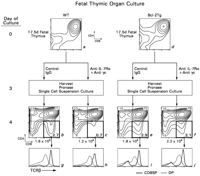
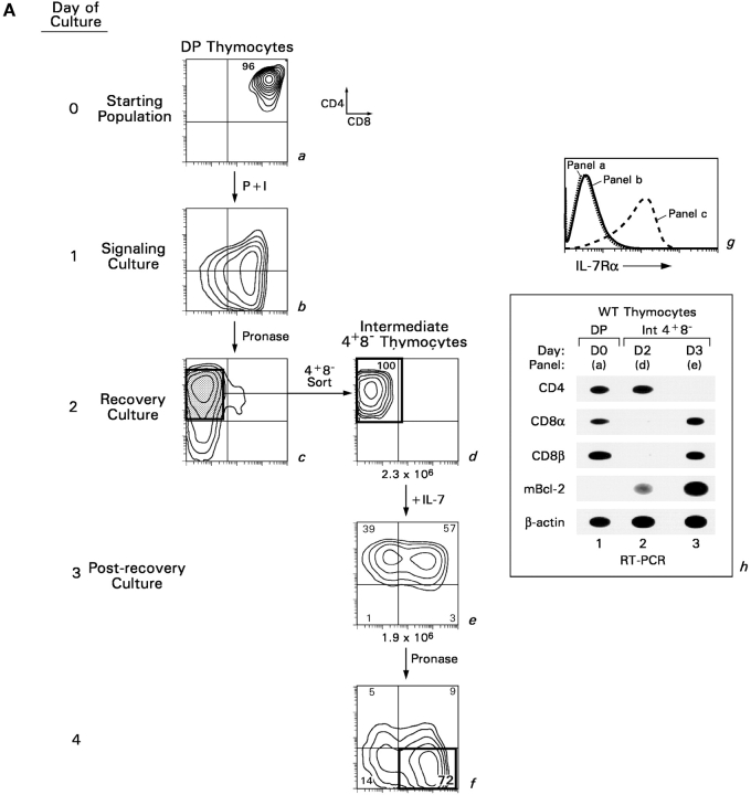
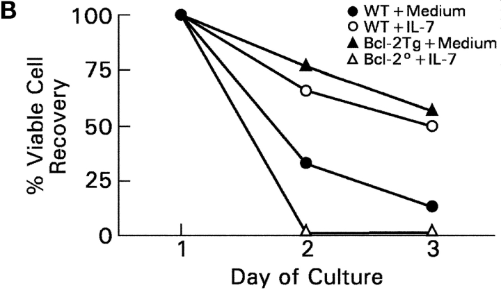
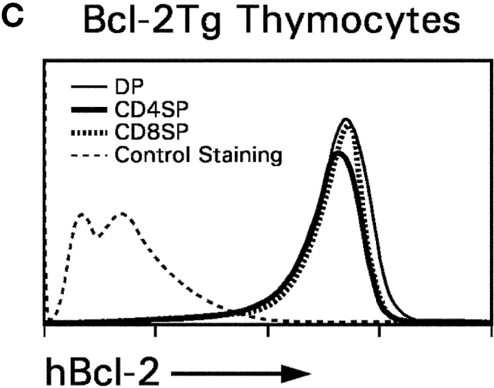
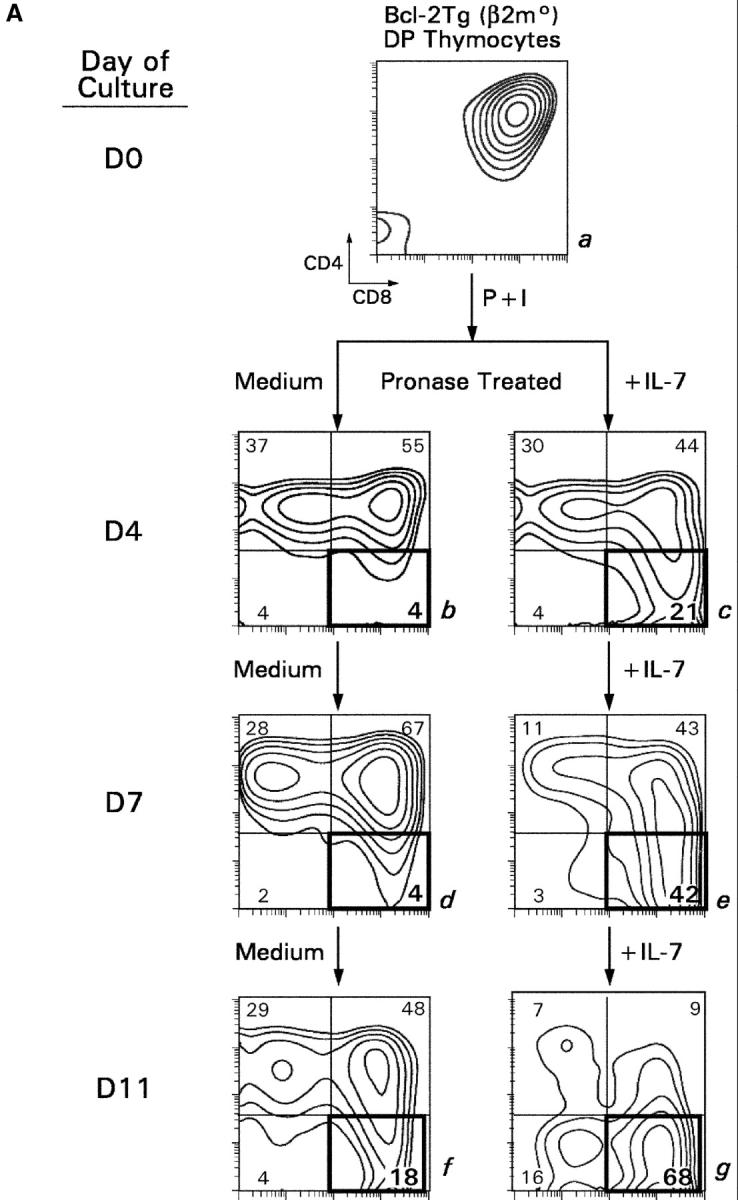
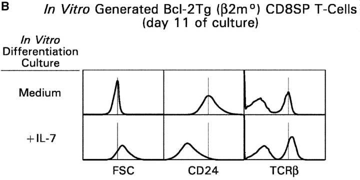
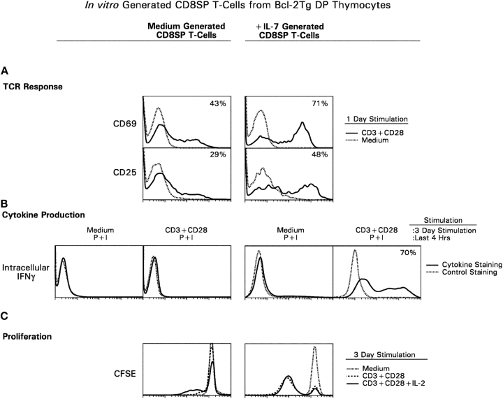
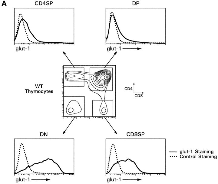
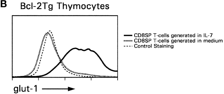
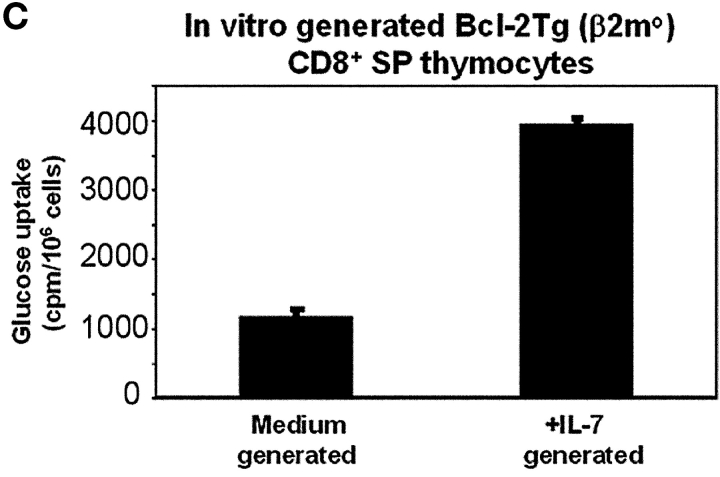
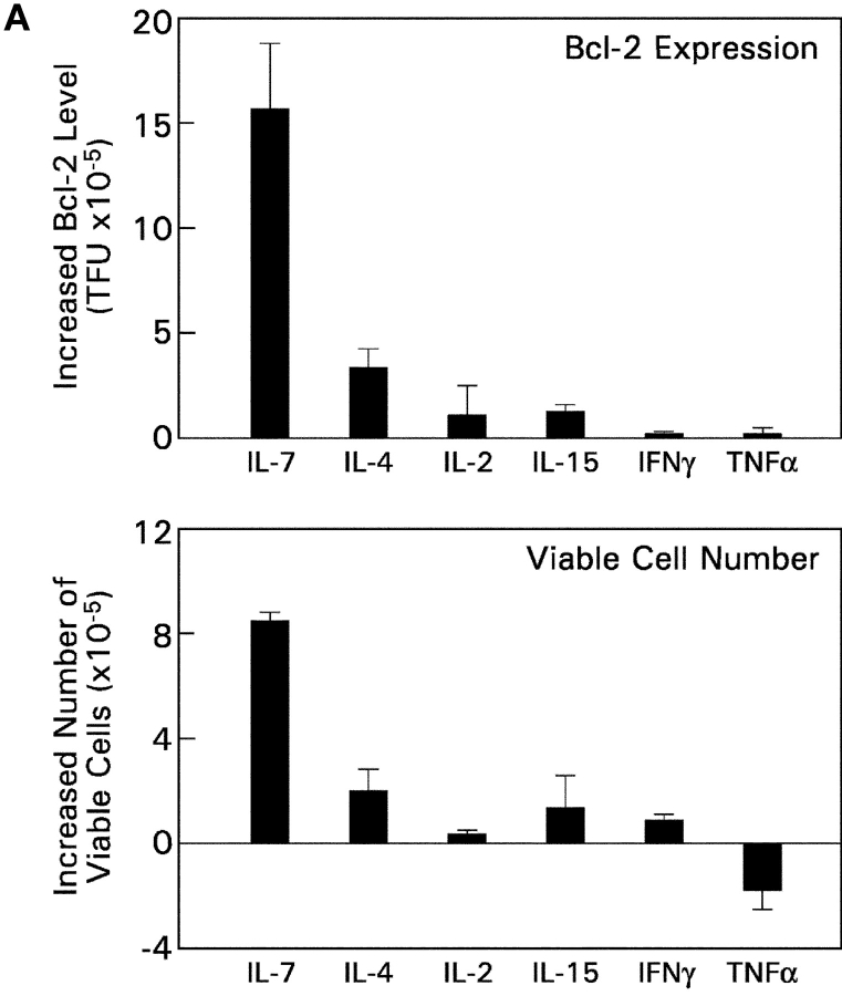
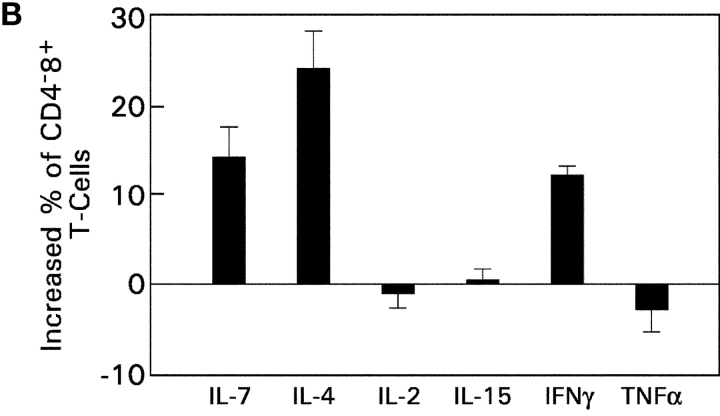
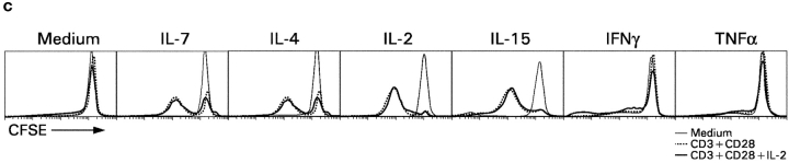
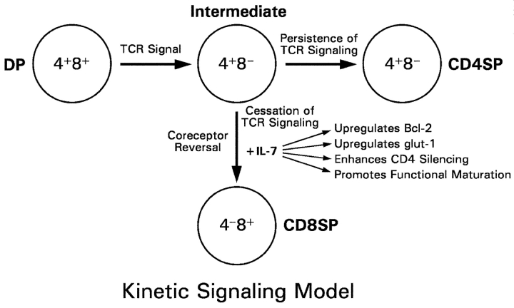
CD4(+)8(+) double positive (DP) thymocytes differentiate into CD4(+) and CD8(+) mature T cells in response to TCR signals. However, TCR signals that are initiated in DP thymocytes are unlikely to persist throughout all subsequent differentiation steps, suggesting that other signals must sustain thymocyte differentiation after TCR signaling has ceased. Using an in vitro experimental system, we now demonstrate that cytokine receptor signals, such as those transduced by IL-7 receptors, are required for differentiation of signaled DP thymocytes into functionally mature CD8(+) T cells as they: (a) up-regulate Bcl-2 expression to maintain thymocyte viability; (b) enhance CD4 gene silencing; (c) promote functional maturation;and (d) up-regulate surface expression of glucose transporter molecules, which improve nutrient uptake and increase metabolic activity. IL-7Rs appear to be unique among cytokine receptors in maintaining the viability of newly generated CD4(-)8(+) thymocytes, whereas several different cytokine receptors can provide the trophic/differentiative signals for subsequent CD8(+) thymocyte differentiation and maturation. Thus, cytokine receptors provide both survival and trophic/differentiative signals with varying degrees of redundancy that are required for differentiation of signaled DP thymocytes into functionally mature CD8(+) T cells.


Figures

Figure 1.
Figure 1.
Effect of antibody-mediated blockade of IL-7R signaling on intrathymic T cell development. Fetal thymic lobes from B6 and Bcl-2Tg (β2mο) mice (embryonic day 17.5) were placed in organ cultures to which anti–IL-7R or control rat antibodies were added. On day 3 of culture, the thymocytes were harvested into single cell suspensions, treated with extracellular pronase to strip preexisting CD4/CD8 coreceptor proteins from cell surfaces, and cultured overnight so that they would reexpress the CD4/CD8 coreceptor molecules they were actively synthesizing. Panels a–f depict CD4 versus CD8 expression, whereas panels g–j depict TCRβ expression on DP (dotted lines) and CD8SP (solid lines) thymocytes. Note that most CD8SP thymocytes at this time point were mature as their surface expression of TCRβ was high relative to that of DP thymocytes. Although not shown, similar results were obtained with anti–IL-7Rα and anti-γc individual mAbs as with both together.
Figure 2.
Figure 2.
In vitro differentiation of DP thymocytes into CD48+ cells: coreceptor reversal and requirement for Bcl-2 expression. (A) Stimulated DP thymocytes differentiate in vitro into intermediate CD4+8 cells and subsequently into CD48+ cells. Purified DP thymocytes from MHC° mice (a) were stimulated with PMA + ionomycin (P + I) for 16 h (b, signaling culture) and then allowed to recover in nonstimulatory cultures containing only medium (c, recovery culture). The cells were pronased before placement in recovery culture so that only newly synthesized coreceptor molecules would be expressed on their cell surfaces. In this way, nearly all the cells in recovery culture were CD4+8, which were purified to homogeneity by electronic cell sorting (d). In vitro–generated CD4+8 cells were referred to as “intermediate thymocytes” because they continued to differentiate. After 1 d of culture in 6 ng/ml murine IL-7 (e), the cells were pronased to identify the coreceptor molecules being actively synthesized, revealing that over 70% were now CD48+ (f). IL-7Rα surface expression was examined by flow cytometry (g), and coreceptor gene expression was examined by RT-PCR (h). (B) Survival requirements of signaled DP thymocytes in vitro. DP thymocytes from WT, Bcl-2°, and Bcl-2Tg mice were transiently signaled with P + I and cultured in medium or IL-7. Cell viability for each group was set as 100% at the beginning of recovery culture on day 1, and measured on days 2 and 3. The survival curves of Bcl-2° thymocytes cultured in medium or IL-7 were identical. (C) Bcl-2Tg expression in thymocyte subpopulations. Thymocytes from Bcl-2Tg mice were stained for intracellular human Bcl-2Tg and surface CD4 and CD8. The anti–human Bcl-2 antibody only detects transgenic Bcl-2 protein and does not detect endogenous mouse Bcl-2 protein.
Figure 2.
Figure 2.
In vitro differentiation of DP thymocytes into CD48+ cells: coreceptor reversal and requirement for Bcl-2 expression. (A) Stimulated DP thymocytes differentiate in vitro into intermediate CD4+8 cells and subsequently into CD48+ cells. Purified DP thymocytes from MHC° mice (a) were stimulated with PMA + ionomycin (P + I) for 16 h (b, signaling culture) and then allowed to recover in nonstimulatory cultures containing only medium (c, recovery culture). The cells were pronased before placement in recovery culture so that only newly synthesized coreceptor molecules would be expressed on their cell surfaces. In this way, nearly all the cells in recovery culture were CD4+8, which were purified to homogeneity by electronic cell sorting (d). In vitro–generated CD4+8 cells were referred to as “intermediate thymocytes” because they continued to differentiate. After 1 d of culture in 6 ng/ml murine IL-7 (e), the cells were pronased to identify the coreceptor molecules being actively synthesized, revealing that over 70% were now CD48+ (f). IL-7Rα surface expression was examined by flow cytometry (g), and coreceptor gene expression was examined by RT-PCR (h). (B) Survival requirements of signaled DP thymocytes in vitro. DP thymocytes from WT, Bcl-2°, and Bcl-2Tg mice were transiently signaled with P + I and cultured in medium or IL-7. Cell viability for each group was set as 100% at the beginning of recovery culture on day 1, and measured on days 2 and 3. The survival curves of Bcl-2° thymocytes cultured in medium or IL-7 were identical. (C) Bcl-2Tg expression in thymocyte subpopulations. Thymocytes from Bcl-2Tg mice were stained for intracellular human Bcl-2Tg and surface CD4 and CD8. The anti–human Bcl-2 antibody only detects transgenic Bcl-2 protein and does not detect endogenous mouse Bcl-2 protein.
Figure 3.
Figure 3.
In vitro differentiation of intermediate thymocytes from Bcl-2 transgenic mice in the presence and absence of IL-7. (A) Differentiation of intermediate thymocytes in the absence of IL-7. DP thymocytes were obtained from embryonic day 18 Bcl-2Tg (β2mo) mice, stimulated with P + I for 16 h, pronased, and electronically sorted as in Fig. 2 A to obtain purified intermediate CD4+8 thymocytes (a). 1-d culture in medium alone was sufficient for many of these intermediate thymocytes to reexpress surface CD8 (b). These cells were pronased to reveal the coreceptors being actively synthesized (c). Gene expression was assessed by RT-PCR (d). (B) IL-7R signaling increases CD48+ T cell number without cell proliferation. DP thymocytes from BCl-2Tg (β2mo) mice (a) were transiently signaled with P + I and labeled with CFSE on day 1 before placement in recovery cultures with either IL-7 or medium. On day 3, the cells were pronased and on day 4 assessed for CD4/CD8 expression (b, c) and CFSE fluorescence intensity (d). CFSE intensity of thymocytes cultured in medium (solid line) was compared with that of thymocytes cultured in IL-7 (dotted line). As a positive control, B6 lymph node T cells were labeled with CFSE in parallel on day 1 and stimulated by 1 μg/ml anti-CD3 mAb for 3 d (dashed line).
Figure 3.
Figure 3.
In vitro differentiation of intermediate thymocytes from Bcl-2 transgenic mice in the presence and absence of IL-7. (A) Differentiation of intermediate thymocytes in the absence of IL-7. DP thymocytes were obtained from embryonic day 18 Bcl-2Tg (β2mo) mice, stimulated with P + I for 16 h, pronased, and electronically sorted as in Fig. 2 A to obtain purified intermediate CD4+8 thymocytes (a). 1-d culture in medium alone was sufficient for many of these intermediate thymocytes to reexpress surface CD8 (b). These cells were pronased to reveal the coreceptors being actively synthesized (c). Gene expression was assessed by RT-PCR (d). (B) IL-7R signaling increases CD48+ T cell number without cell proliferation. DP thymocytes from BCl-2Tg (β2mo) mice (a) were transiently signaled with P + I and labeled with CFSE on day 1 before placement in recovery cultures with either IL-7 or medium. On day 3, the cells were pronased and on day 4 assessed for CD4/CD8 expression (b, c) and CFSE fluorescence intensity (d). CFSE intensity of thymocytes cultured in medium (solid line) was compared with that of thymocytes cultured in IL-7 (dotted line). As a positive control, B6 lymph node T cells were labeled with CFSE in parallel on day 1 and stimulated by 1 μg/ml anti-CD3 mAb for 3 d (dashed line).
Figure 4.
Figure 4.
Restimulation of intermediate thymocytes blocks coreceptor reversal, even in the absence of IL-7. Intermediate CD4+8 thymocytes (a) were generated from P + I-signaled Bcl-2Tg (β2mo) DP thymocytes as in Figs. 2 and 3. In the absence of restimulation (left), over 40% of these intermediate thymocytes reexpressed CD8 (b and c) with many also terminating CD4 expression to become CD48+ T cells (f and g, left). However, in the presence of P + I restimulation (right), intermediate thymocytes remained CD4+8 and neither reexpressed CD8 (d and e) nor terminated CD4 (h and i), regardless of the presence or absence of IL-7.
Figure 5.
Figure 5.
In vitro differentiation of signaled DP thymocytes in longer term cultures. (A) DP thymocytes from Bcl-2Tg (β2mo) mice (a) were signaled with P + I and placed in longer term cultures with either medium (b, d, and f) or IL-7 (c, e, and g). Cells were assessed for CD4/CD8 expression on the days indicated. (B) Effect of IL-7 on phenotypic maturation. Using day 11 cells from A, we determined their forward light scatter (FSC), which is a reflection of cell size, as well as their surface expression of CD24 and TCRβ.
Figure 5.
Figure 5.
In vitro differentiation of signaled DP thymocytes in longer term cultures. (A) DP thymocytes from Bcl-2Tg (β2mo) mice (a) were signaled with P + I and placed in longer term cultures with either medium (b, d, and f) or IL-7 (c, e, and g). Cells were assessed for CD4/CD8 expression on the days indicated. (B) Effect of IL-7 on phenotypic maturation. Using day 11 cells from A, we determined their forward light scatter (FSC), which is a reflection of cell size, as well as their surface expression of CD24 and TCRβ.
Figure 6.
Figure 6.
IL-7R signals promote differentiation of thymocytes into functional CD8+ T cells that can respond to TCR signals. Intermediate thymocytes were generated by P + I stimulation of Bcl-2Tg (β2mo) DP thymocytes, placed in short term cultures with either medium or IL-7, and pronased on day 3 to confirm the presence of CD48+ T cells. On day 4, cells that had been cultured in either medium (left) or IL-7 (right) were stimulated with immobilized anti-CD3 + anti-CD28 mAbs and assessed for TCR responses (A), intracellular cytokine production (B), and proliferation (C). (A) TCR responses. Cultured cells were stimulated with anti-CD3 + anti-CD28 mAbs for 1 d and assessed for surface expression of CD25 and CD69. Shown are CD25 and CD69 surface expression on gated CD48+ T cells (solid line, anti-CD3 + anti-CD28 stimulation; dotted line, medium). (B) Cytokine production. CD48+ T cells were purified from in vitro differentiation cultures and stimulated for 3 d with immobilized anti-CD3 + anti-CD28. 4 h before harvest, they were restimulated with P + I in the presence of monensin and assessed for intracellular IFNγ (solid line, intracellular IFNγ staining; dotted line, control IgG staining). (C) Proliferation. Cultured cells were labeled with CFSE and stimulated with anti-CD3 + anti-CD28 for 3 d either with or without additional hIL-2. CFSE intensity on gated CD48+ cells was assessed by three color flow cytometry (dotted line, no stimulation; dashed line, stimulation with anti-CD3 + anti-CD28; solid line, stimulation with anti-CD3 + anti-CD28 in the presence of 50 U/ml hIL-2). Comparable results were obtained with P + I stimulation instead of antibody stimulation (not depicted).
Figure 7.
Figure 7.
glut-1 surface expression and glucose uptake by developing CD8SP thymocytes. (A) glut-1 surface expression on thymocyte subpopulations from WT mice. Thymocytes from WT mice were stained for surface glut-1, CD4, and CD8 and analyzed by multicolor flow cytometry. Staining for glut-1 was performed by incubating thymocytes with goat anti–mouse glut-1 antibody, followed by incubation with FITC-conjugated sheep anti–goat IgG. The cells were incubated with purified rat IgG to saturate any free-binding sites on the sheep IgG before incubation with anti-CD4 and anti-CD8 mAbs. Surface expression of glut-1 is indicated relative to control staining with the secondary sheep anti–goat IgG in the absence of primary antibody. (B) IL-7R signals induce glut-1 surface expression on in vitro–generated CD8SP thymocytes. CD8SP thymocytes were generated in vitro by stimulation of Bcl-2Tg (β2mo) DP thymocytes followed by culture in either medium or IL-7. On day 7, cells were assessed for surface glut-1 expression by immuno-fluorescence and multicolor flow cytometry. (C) IL-7R signals promote glucose take. CD8SP thymocytes were generated in vitro by stimulation of Bcl-2Tg (β2mo) DP thymocytes, followed by culture in either medium or IL-7. On day 7, the cells were assayed for glucose uptake with 2-[14C(U)]-deoxy-d-glucose.
Figure 7.
Figure 7.
glut-1 surface expression and glucose uptake by developing CD8SP thymocytes. (A) glut-1 surface expression on thymocyte subpopulations from WT mice. Thymocytes from WT mice were stained for surface glut-1, CD4, and CD8 and analyzed by multicolor flow cytometry. Staining for glut-1 was performed by incubating thymocytes with goat anti–mouse glut-1 antibody, followed by incubation with FITC-conjugated sheep anti–goat IgG. The cells were incubated with purified rat IgG to saturate any free-binding sites on the sheep IgG before incubation with anti-CD4 and anti-CD8 mAbs. Surface expression of glut-1 is indicated relative to control staining with the secondary sheep anti–goat IgG in the absence of primary antibody. (B) IL-7R signals induce glut-1 surface expression on in vitro–generated CD8SP thymocytes. CD8SP thymocytes were generated in vitro by stimulation of Bcl-2Tg (β2mo) DP thymocytes followed by culture in either medium or IL-7. On day 7, cells were assessed for surface glut-1 expression by immuno-fluorescence and multicolor flow cytometry. (C) IL-7R signals promote glucose take. CD8SP thymocytes were generated in vitro by stimulation of Bcl-2Tg (β2mo) DP thymocytes, followed by culture in either medium or IL-7. On day 7, the cells were assayed for glucose uptake with 2-[14C(U)]-deoxy-d-glucose.
Figure 8.
Figure 8.
Assessment of cytokine receptor redundancy in developing CD8SP thymocytes. (A) Ability of other cytokines to up-regulate Bcl-2 expression and promote thymocyte survival. In vitro–generated intermediate CD4+8 thymocytes from MHC° mice were cultured overnight in medium or one of the indicated cytokines. (top) Cells were assessed for expression of Bcl-2 by intracellular staining, which was quantitated into total fluorescence units (TFU) and is displayed relative to that of cells cultured in medium alone (TFU of medium cultured cells was 4.6 × 10⁵). (bottom) Total number of viable cells in each culture was determined and is displayed relative to that of cells cultured in medium alone (viable cell number of medium cultured cells was 2.5 × 10⁵). (B) Ability of various cytokines to promote extinction of CD4 gene expression during coreceptor reversal. In vitro–generated intermediate CD4+8 thymocytes from Bcl-2Tg (β2mo) mice were cultured overnight in either medium or one of the indicated cytokines. The next day, cells were pronase-stripped to determine the percentage of CD48+ cells that had extinguished CD4 gene expression and undergone coreceptor reversal. The frequency of CD48+ cells in each cytokine culture is displayed relative to that of medium alone (frequency of CD48+ cells in medium alone was 13.4%). (C) Ability of various cytokines to promote functional maturation of CD8SP thymocytes. In vitro–generated intermediate CD4+8 thymocytes from Bcl-2Tg (β2mo) mice were cultured overnight in either medium or one of the indicated cytokines. The next day, cells were pronase-stripped to identify CD48+ cells, labeled with CFSE, and stimulated for 3 d with anti-CD3 + anti-CD28, either with or without additional IL-2 as indicated. Proliferation of CD48+ cells was assessed by CFSE intensity.
Figure 8.
Figure 8.
Assessment of cytokine receptor redundancy in developing CD8SP thymocytes. (A) Ability of other cytokines to up-regulate Bcl-2 expression and promote thymocyte survival. In vitro–generated intermediate CD4+8 thymocytes from MHC° mice were cultured overnight in medium or one of the indicated cytokines. (top) Cells were assessed for expression of Bcl-2 by intracellular staining, which was quantitated into total fluorescence units (TFU) and is displayed relative to that of cells cultured in medium alone (TFU of medium cultured cells was 4.6 × 10⁵). (bottom) Total number of viable cells in each culture was determined and is displayed relative to that of cells cultured in medium alone (viable cell number of medium cultured cells was 2.5 × 10⁵). (B) Ability of various cytokines to promote extinction of CD4 gene expression during coreceptor reversal. In vitro–generated intermediate CD4+8 thymocytes from Bcl-2Tg (β2mo) mice were cultured overnight in either medium or one of the indicated cytokines. The next day, cells were pronase-stripped to determine the percentage of CD48+ cells that had extinguished CD4 gene expression and undergone coreceptor reversal. The frequency of CD48+ cells in each cytokine culture is displayed relative to that of medium alone (frequency of CD48+ cells in medium alone was 13.4%). (C) Ability of various cytokines to promote functional maturation of CD8SP thymocytes. In vitro–generated intermediate CD4+8 thymocytes from Bcl-2Tg (β2mo) mice were cultured overnight in either medium or one of the indicated cytokines. The next day, cells were pronase-stripped to identify CD48+ cells, labeled with CFSE, and stimulated for 3 d with anti-CD3 + anti-CD28, either with or without additional IL-2 as indicated. Proliferation of CD48+ cells was assessed by CFSE intensity.
Figure 9.
Figure 9.
Kinetic signaling model of T cell development: role of IL-7. The kinetic signaling model postulates that TCR signals drive DP thymocytes to first differentiate into intermediate CD4+8 cells, and it is at the intermediate CD4+8 stage that CD4/CD8 lineage direction is determined by whether TCR signals are present or have ceased. Persistence of TCR signals in intermediate thymocytes maintains CD4+8 gene expression and results in differentiation into CD4+ T lineage cells; cessation of TCR signals in intermediate thymocytes results in coreceptor reversal (i.e., reinitiation of CD8 gene expression and silencing of CD4 gene expression) and differentiation into CD8+ T lineage cells. As a consequence of absent TCR signals, IL-7R signals are required to maintain Bcl-2 expression and viability of intermediate thymocytes during coreceptor reversal and differentiation into CD8+ T cells. In addition, cytokine receptor signaling promotes CD8+ T cell differentiation by providing trophic signals that support CD4 gene silencing and functional maturation in developing CD8SP thymocytes.

References

    1. Jameson, S.C., K.A. Hogquist, and M.J. Bevan. 1995. Positive selection of thymocytes. Annu. Rev. Immunol. 13:93–126. - PubMed
    1. Linette, G.P., M.J. Grusby, S.M. Hedrick, T.H. Hansen, L.H. Glimcher, and S.J. Korsmeyer. 1994. Bcl-2 is upregulated at the CD4+ CD8+ stage during positive selection and promotes thymocyte differentiation at several control points. Immunity. 1:197–205. - PubMed
    1. Moore, N.C., G. Anderson, G.T. Williams, J.J. Owen, and E.J. Jenkinson. 1994. Developmental regulation of bcl-2 expression in the thymus. Immunology. 81:115–119. - PMC - PubMed
    1. Singer, A., R. Bosselut, and A. Bhandoola. 1999. Signals involved in CD4/CD8 lineage commitment: current concepts and potential mechanisms. Semin. Immunol. 11:273–281. - PubMed
    1. Rothenberg, E.V. 1992. The development of functionally responsive T cells. Adv. Immunol. 51:85–214. - PubMed

MeSH terms