. 2003 Feb 17;197(4):515-26.
doi: 10.1084/jem.20021713.

Transgenic expression of the activating natural killer receptor Ly49H confers resistance to cytomegalovirus in genetically susceptible mice

Affiliations

Transgenic expression of the activating natural killer receptor Ly49H confers resistance to cytomegalovirus in genetically susceptible mice

Seung-Hwan Lee et al. J Exp Med. .

Abstract

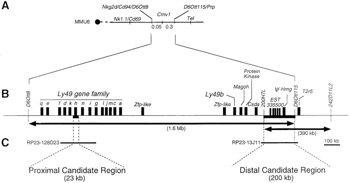
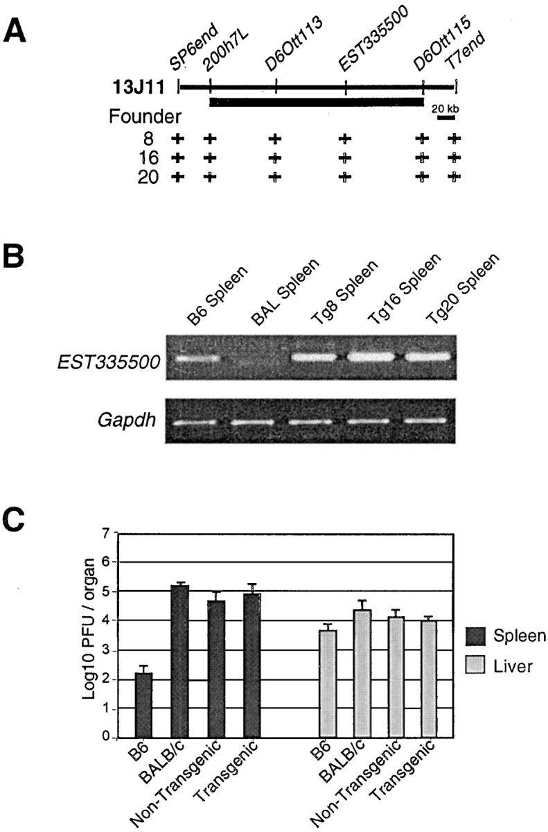
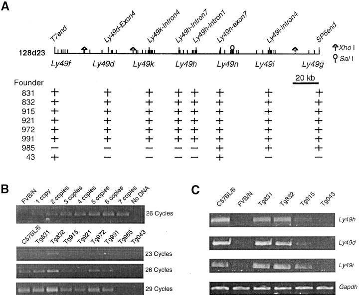
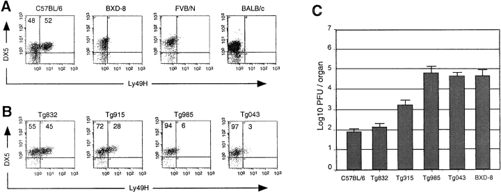
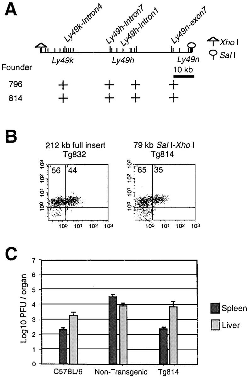
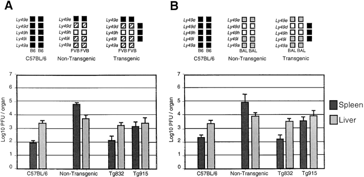
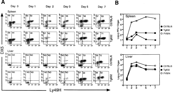
Natural resistance to infection with mouse cytomegalovirus (MCMV) is controlled by a dominant locus, Cmv1. Cmv1 is linked to the Ly49 family of natural killer receptors on distal chromosome 6. While some studies localized Cmv1 as distal to the Ly49 gene cluster, genetic and functional analysis identified Ly49h as a pivotal factor in resistance to MCMV. The role of these two independent genomic domains in MCMV resistance was evaluated by functional complementation using transgenesis of bacterial artificial chromosomes (BAC) in genetically susceptible mice. Phenotypic and genetic characterization of the transgenic animals traced the resistance gene to a single region spanning the Ly49h gene. The appearance of the Ly49H protein in NK cells of transgenic mice coincided with the emergence of MCMV resistance, and there was a threshold Ly49H protein level associated with full recovery. Finally, transgenic expression of Ly49H in the context of either of the two independent susceptibility alleles, Cmv1(sBALB) or Cmv1(sFVB), conferred resistance to MCMV infection. These results demonstrate that Ly49h is necessary and sufficient to confer MCMV resistance, and formally demonstrate allelism between Cmv1 and Ly49h. This panel of transgenic animals provides a unique resource to study possible pleiotropic effect of Cmv1.


Figures

Figure 1.
Figure 1.
Genomic targets for transgenic analysis of the Cmv1 locus. (A) Composite genetic linkage map of mouse chromosome 6 in the vicinity of Cmv1. The order and distances of the loci was determined by pedigree analysis. The centromere is represented by a black circle. Recombination frequencies in centimorgan (cM) are shown below the chromosome. Nk1.1, Cd69, Cd94, and Nkg2d code for C-lectin type receptors. Prp, prolin-rich protein; Tel, translocation-ets-leukemia (reference 7). (B) Blow-up of the minimal genetic interval between D6Ott8 and D6Ott115 and transcriptional map showing candidate genes for Cmv1. Arrows indicate the minimal Cmv1 interval as defined by Depatie et al. (references and 11) and Brown et al. (reference 9) (C). Localization of BAC clones used for transgenesis (reference 11).
Figure 2.
Figure 2.
Characterization of BAC clone 13J11 transgenic lines. (A) Physical map of BAC clone 13J11 with the localization of markers used for identification of transgenic lines by PCR. The black bar indicates the physical domain common to two independent mapping efforts for Cmv1 (references and 10). The (+) indicates a positive result in STS content analysis of transgenic founder animals. (B) RT-PCR for EST335500 and Gapdh control RNA from spleens of wild-type strains and transgenic lines mice. (C) Replication of MCMV in the spleen and liver of inbred and transgenic lines. Viral titers in the organs of mice per group were determined by plaque assay 3 d after infection with 5 × 10³ PFU of MCMV.
Figure 3.
Figure 3.
Characterization of BAC clone 128D23 transgenic lines. (A) Physical map of BAC clone 128D23 with the localization of markers used for identification of transgenic lines by PCR. The localization of genes present in the BAC clone is also indicated. The (+) indicates a positive result in STS content analysis of transgenic founder animals. Restriction sites of XhoI and SalI were shown. (B) Semiquantitative PCR to determine transgene copy number on founders. Ly49h-specific primers were used to amplify 1–7 copies of BAC DNA. Results were compared with those obtained from transgenic lines in 26 cycles of amplification. (C) RT-PCR for genes contained in BAC clone 128D23 and Gapdh control RNA from spleens of wild-type mice and transgenic lines. Ly49h, Ly49d, and Ly49i oligonucleotide primer pairs were designed to amplify specifically the C57BL/6 alleles (see Materials and Methods).
Figure 4.
Figure 4.
Acquisition of MCMV-resistance in BAC clone 128D23 transgenic lines. (A) Enriched NK cell preparations from spleens of wild-type strains were stained with the mAb DX5 and the rabbit polyclonal antibody against the cytoplasmic domain of the Ly49H peptide reported here. The numbers in the density plots indicate the percentage of DX5+ lymphocytes either Ly49H or Ly49H+. (B) Enriched NK cell preparations from spleens of BAC 128D23 transgenic lines were stained with the monoclonal antibody DX5 and the rabbit polyclonal antibody against the cytoplasmic domain of the Ly49H. The numbers in the density plots indicate the percentage of DX5+ lymphocytes either Ly49H or Ly49H+. (C) Replication of MCMV in the spleen of wild-type and transgenic lines. Viral titers in the spleen of five mice per group were determined by plaque assay 3 d after infection with 5 × 10³ PFU of MCMV. Statistically significant differences were observed between transgenic (Tg832, Tg915) and nontransgenic mice at P values of < 0.0005.
Figure 5.
Figure 5.
A 79-kb genomic construct containing Ly49H confers resistance to MCMV infection. (A) Physical map of a 79-kb fragment with the localization of markers used for identification of transgenic lines by PCR. The localization of genes present in the BAC clone is also indicated. Restriction sites of XhoI and SalI used to generate the 79-kb fragment were shown. The (+) indicates a positive result in STS content analysis of transgenic founder animals. (B) Enriched NK cell preparations from spleens of lines Tg832 and Tg814 were stained with the mAb DX5 and the rabbit polyclonal antibody against the cytoplasmic domain of the Ly49H. The numbers in the density plots indicate the percentage of DX5+ lymphocytes either Ly49H or Ly49H+. (C) Replication of MCMV in the spleen and liver of wild-type and transgenic lines. Viral titers in the spleen of five mice per group were determined by plaque assay 3 d after infection with 5 × 10³ PFU of MCMV.
Figure 6.
Figure 6.
Ly49H confers resistance to MCMV infection independently of the genetic background. Replication of MCMV in wild-type and transgenic Tg832 and Tg915 mice in the context of homozygous FVB/N (Cmv1 sFVB; A) and BALB/c (Cmv1 sBALB; B) was shown. Haplotypes of Ly49 family for Cmv1 r (C57BL/6), Cmv1 sFVB and Cmv1 sBALB are shown; black boxes, B6 alleles; hatched boxes, FVB alleles; gray boxes, BALB/c alleles; white boxes indicate the absence of alleles. Viral titers in the spleen and liver of five mice per group were determined by plaque assay 3 d after infection with 5 × 10³ PFU of MCMV.
Figure 7.
Figure 7.
Kinetics of trans-Ly49H expression. (A) Ly49H expression was followed during a 1-wk period in the transgenic line Tg832 and C57BL/6 by staining with the mAb DX5 and a rabbit polyclonal antibody against the cytoplasmic domain of the Ly49H. The numbers in the density plots indicate the percentage of DX5+ lymphocytes either Ly49H or Ly49H+. Enriched NK cell preparations were obtained from spleen and liver tissues after 0, 1, 2, 3, 5, and 7 d after infection. The proportion of NK cells in liver until 3 d after infection was not determined owing to the insignificant number of DX5+ cells on samples. (B) A fraction of the organ homogenates used in panel A were used to determine the course of MCMV infection at the indicated time points in transgenic Tg832 mice and wild-type C57BL/6 (Cmv1 r) and FVB/N (Cmv1 s) controls. Viral titers were determined by plaque after infection with 5 × 10³ PFU of MCMV.

References

    1. Chalmer, J.E., J.S. Mackenzie, and N.F. Stanley. 1977. Resistance to murine cytomegalovirus linked to the major histocompatibility complex of the mouse. J. Gen. Virol. 37:107–114. - PubMed
    1. Grundy, J.E., J.S. Mackenzie, and N.F. Stanley. 1981. Influence of H-2 and non-H-2 genes on resistance to murine cytomegalovirus infection. Infect. Immun. 32:277–286. - PMC - PubMed
    1. Allan, J.E., and G.R. Shellam. 1984. Genetic control of murine cytomegalovirus infection: virus titres in resistant and susceptible strains of mice. Arch. Virol. 81:139–150. - PubMed
    1. Scalzo, A.A., N.A. Fitzgerald, A. Simmons, A.B. La Vista, and G.R. Shellam. 1990. Cmv-1, a genetic locus that controls murine cytomegalovirus replication in the spleen. J. Exp. Med. 171:1469–1483. - PMC - PubMed
    1. Scalzo, A.A., N.A. Fitzgerald, C.R. Wallace, A.E. Gibbons, Y.C. Smart, R.C. Burton, and G.R. Shellam. 1992. The effect of the Cmv-1 resistance gene, which is linked to the natural killer cell gene complex, is mediated by natural killer cells. J. Immunol. 149:581–589. - PubMed

Publication types